Rev Bras Anestesiol. 2013;63(2):193-196
Offi cial Publication of the Brazilian Society of Anesthesiology www.sba.com.br
REVISTA
BRASILEIRA DE
ANESTESIOLOGIA
Abstract
Background and objectives: Skin fragments during lumbar punctures may develop intraspinal epidermoid tumors. The aim of this study was to determine the incidence of epithelial cells that refl ow along with the fi rst and third drops of CSF of patients undergoing spinal anesthesia. Methods: Samples of the fi rst and third drops of cerebrospinal fl uid were collected from 39 adult patients undergoing spinal anesthesia with a 25G Quincke needle. Four microscope slides were prepared: one for the fi rst drop, one for third drop, one for the needle, and one with a drop of saline for control. A pathologist examined the slides randomly.
Results: Squamous epithelial cells were identifi ed in 35 (89.7%) samples from the fi rst drop, 34 (87.2%) from the third drop, and 24 (61.5%) from spinal needle. The third drop showed a mean number of cells larger than the fi rst drop (p = 0.046). Nucleated epithelial cells were found in a sample of the fi rst drop (2.56%), in four samples of third drop (10.25%), and in one spinal needle (2.56%). Third drop showed a mean number of nucleated cells higher than fi rst drop with no statistical difference (p = 0.257).
Conclusions: High percentage of epithelial cells was found in the fi rst (89.7%) and third (87.2%) drops of CSF refl ow and in used needles (61.5%). Skin cells were found even using small gauge disposable needles with well-adapted mandrel,
© 2013 Sociedade Brasileira de Anestesiologia. Published by Elsevier Editora Ltda. All rights reserved.
There is High Incidence of Skin Cells in the First and Third
Drops of Cerebrospinal Fluid in Spinal Anesthesia
Mário Humberto Curado Taveira*
1, Antonio Fernando Carneiro
2, Gustavo Gabriel Rassi
3,
Marise Amaral Rebouças Moreira
4, Simone de Andrade Curado Taveira
51. MD, Anesthesia Study Center, Goiânia, GO, Brazil
2. TSA (Superior Title in Anesthesiology)/SBA (Brazilian Society of Anesthesiology); PhD in Medicine, Santa Casa de São Paulo, Head of Department of Surgery, Universidade Federal de Goiás; Director of Professional Defense, SBA; Specialist in Intensive Care Medicine, Goiânia, GO, Brazil
3. Clinical Pathologist, Hospital Anis Rassi de Goiânia; Director, Hospital Anis Rassi de Goiânia, GO, Brazil 4. Professor of Pathological Anatomy, Universidade Federal de Goiás, Goiânia, GO, Brazil
5. MD, Anesthesiology Department, Hospital das Clinicas, UFG, Anesthesia Study Center, Goiania, GO, Brazil Received from Anesthesia Study Center, Goiania, GO, Brazil.
Submitted on March 5, 2012. Approved on April 13, 2012.
Keywords: Anesthesia, Spinal; Cerebrospinal Fluid; Needles;
Epithelial Cells; Neoplasms.
SCIENTIFIC ARTICLE
*Corresponding author:
E-mail: [email protected]
194 M. H. C. Taveira et al.
Introduction
Epidermoid tumors of the central nervous system and
spi-nal caspi-nal are very rare 1,2. These tumors’ etiology may be
congenital or iatrogenic, being generated by epidermal cells implantation into the spinal canal. Iatrogenic spinal epidermoid tumors derive from epidermal tissue implanta-tion inside the spinal canal during lumbar puncture per-formed with needles without mandrel or with inappropriate
or maladaptive mandrel 3. In 1962, a review of 90 cases of
spinal epidermoid tumors reported that 41% of tumors had
iatrogenic origin for different reasons 4. More than 40 years
ago, two different research groups reproduced epidermoid tumors experimentally by implanting autologous skin
frag-ments into the medullary canal 5-7.
In skin puncture during spinal anesthesia or even caudal anesthesia, the needle tip without mandrel acts as a lancet, produces a biopsy and introduces these fragments within the
spinal canal, resulting in the onset of epidermoid tumors 8,9.
An evaluation of the presence of skin cells and debris in small caliber deposable needles with well-adapted man-drels showed that such fragments may be readily detected
in needles most used in spinal anesthesia 10,11. Some authors
recommend letting a few drops of cerebrospinal fl uid (CSF) drip by the hub of spinal needles in order to clean them of
any debris or other contaminants 11.
Knowledge of CSF cellularity, ignored in subarachnoid punctures, may suggest the number of drops required to wash the mandrel and if this procedure is effective or not in reducing the incidence of epidermoid tumors. The objec-tive of this randomized, double-blind study was to evaluate the incidence of skin cells in CSF inside spinal needle hubs (Quincke 25G) and if there is any difference between the amounts of epidermal material refl owing with the fi rst and third drops of CSF.
Method
After receiving approval by the Research Ethics Committee and obtaining signed informed consent, we collected samples of CSF from 25G Quincke spinal needles used in 39 patients, male and female, ASA I-II, aged between 20-80 years, un-dergoing surgery (gynecologic, urologic, orthopedic, and general) under spinal anesthesia. Anesthesiologists blinded to the study administered the anesthesia.
All needles used were 25G Quincke (BD-Becton, Dickinson and Company) with mandrels. Before puncture, the mandrel was checked for proper adaptation to the set. Patients were
anesthetized in the sitting position after intravenous ad-ministration of midazolam (2.0 mg) and fentanyl (25 mcg). After asepsis with alcohol solution, anesthetic button was performed with lidocaine (20 mg) without vasoconstrictor, using an insulin needle. After subarachnoid puncture with 25G Quincke needle and presence of CSF returning in the needle’s hub, the fi rst and third drops were collected in separate slides. The number of punctures for obtaining CSF was registered. Then, the local anesthetic previously chosen for the surgery was injected. The needle was withdrawn without the mandrel, its interior washed with injection of 0.2 mL alcohol 70% and stored in a test tube, from which a third slide would be made by centrifugation in the institu-tion laboratory.
The two slides containing the fi rst and third drops were identifi ed and registered. A fourth slide was identifi ed as control with a drop of 0.9% saline solution, and the fi xer used in the others. The slides used contained a field of 1 cm² to delimit the drops of the CSF. All material was stored in a suitable container and sent for cytological analysis by a pathologist who was blinded to the study. Analysis included assessment for the presence and amount of cells and/or epithelial tissues or other biological material (tissue or fatty tissue) unsuitable to CSF.
For statistical analysis, we used Excel program for creat-ing a database and SPSS for analysis; nonparametric Wilcoxon test was used to compare the number of cells found in every drop and test the relationship between the number of punc-tures and number of cells found; in addition to regression. The tests were performed with 95% confi dence interval and 5% standard error, with p < 0.05 considered statistically signifi cant.
Results
Regarding gender, the sample was evenly distributed with
53.8% female and 46.2% male, mean age of 46.51 ±18.40
years (range 20-80 years).
195 There is High Incidence of Skin Cells in the First and Third Drops of Cerebrospinal Fluid in Spinal Anesthesia
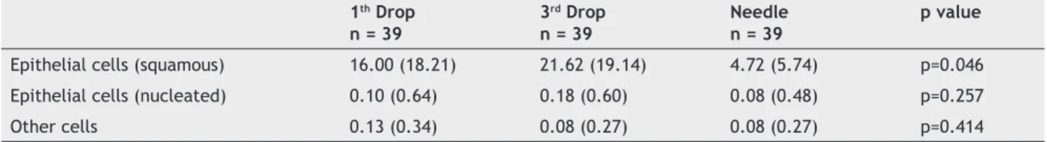
There was a signifi cant difference (p = 0.046) between the mean squamous epithelial cells in the fi rst and third drops and no difference between the fi rst and third drops regarding nucleated epithelial cells. Likewise, there was no signifi cant difference regarding other cells in the fi rst and third drops (Table 2).
Regarding the number of punctures, there was incidence of 69.2% with one puncture, 15.4%, with two punctures, 5.6% with three punctures, 2.6% with four punctures, and 7.7% with fi ve punctures. Linear regression correlating the number of punctures with the amount of cells showed no signifi cant difference at all time points (p < 0.05).
Discussion
In this study, a high percentage of epithelial cells was found in the fi rst and third drops, as well as in the needle (89.7%, 87.2%, and 61.5%, respectively).
In 1944, during an assessment of the CSF of meningitis pa-tients undergoing multiple subarachnoid punctures, research-ers found the presence of squamous epithelial cells together with staphylococci and occasionally small cylindrical skin
fragments from spinal needles 12. Twelve years later, there
were reports of increased incidence of epidermoid tumors in fi ve patients with history of meningitis that had undergone
multiple punctures 13. The ratio of iatrogenicity in the onset
of these tumors occurred with the publication of four cases of adult patients with previous normal myelography that developed squamous cell tumor at the site of subarachnoid puncture 14-17.
A comparison of 25G disposable spinal needles, in which it was not possible to identify the subarachnoid space, showed tissue fragments in 80% of the Quincke needles compared
to 41% of the Whitacre needles 11. In 27% of Quincke needles
and 12% of Whitacre needles, fragments larger than the needles’ diameter were found. In this study, in which the subarachnoid space was identifi ed in all patients, the pres-ence of epithelial cells occurred in 89.7% in the fi rst drop, 87.2% in the third drop, and 61.5% in the needle hub, while no cells were found in the control group.
Due to the high incidence of tissue fragments in 25G spinal needles (Quincke and Whitacre), letting CSF drip from the needle before local anesthetic injection was recommended believing that a few drops of CSF would wash away any
frag-ments of tissues 11. However, the high incidence of cells found
on the fi rst (89.7%) and third (87.2%) drops of CSF from the needle contradicts this theory.
This study showed the high incidence of epithelial cells found in CSF outfl ows both in the fi rst and third drops, as well as in the 25G sharp tip needles. We cannot correlate the morbidity caused by these cells, as it was not the object of the study.
References
1. Preston-Martin S - Descriptive epidemiology of primary tumors of the spinal cord and spinal meninges in Los Angeles County 1972-1985. Neuroepidemiology. 1990;9:106-111.
2. Visciani A, Savoiardo M, Balesirini MR, Solero CL - Iatrogenic intraspinal epidermoid tumors. Neuroradiology. 1989;31:273-275.
3. Reina MA, López-García A, Dittmann M, de Andrés A, Blázquez MG - Tumores epidermóides espinales iatrogénicos. Una complicación tardía de la punción espinal. Rev Esp Anestesiol Reanim. 1996;43:142-146.
4. Manno NJ, Uihiein A, Kemohan J - Intraspinal epidermoids. J Neurosurg. 1962:19:754 765.
5. Van Gilder JC, Schwartz HG - Growth at dermoids from skin implants to the nervous system and surrounding spaces al the newborn rat. J Neurosurgery. 1967:26:14-20.
6. Shaywity BA - Epidermoid spinal cord limners and previous lumbar punctures. J Pediat. 1972:80:638-640.
7. Bainitzky S, Keucher TR, Mealey J, Campbell RL - Iatrogenic intraspinal epidermoid tumours. JAMA. 1977:237:148-150. 8. Lanterburg W - Ein epidermoid frei im Wirbel kanal und siene
kombination mit himlãsionen. Virchours Arch. 1922:24:328-352.
9. Critchley M, Ferguson FR - The cerebrospinal epidermoids (Cholestealoma). Brain. 1928:51:334-384.
10. Reina MA, López A, Manzarbeitia F, Amador V, Goxencia L, Olmedilia MC - Arrastre de fragmentos epidérmicos mediante agujas espinales en cadáveres. Rev Esp Anestesiol Reanim. 1995:42:383-385.
Table 1 Epithelial Cells and Other Debris Found in the First and Third Drops, Needle, and Control. 1st Drop
n = 39
3rd Drop n = 39
Needle n = 39
Control n = 39
Epithelial cells (squamous) 35 (89.7%) 34 (87.2%) 24 (61.5%) 0 (0%)
Epithelial cells (nucleated) 1 (2.56%) 4 (10.25%) 1 (2.56%) 0 (0%)
Fragments (connective and fat tissue) 5 (12.8%) 3 (7.69%) 3 (7.69%) 0 (0%)
Table 2 Mean and SD of Different Cells Found in the First and Third Drops and Needle.
1th Drop n = 39
3rd Drop n = 39
Needle n = 39
p value
Epithelial cells (squamous) 16.00 (18.21) 21.62 (19.14) 4.72 (5.74) p=0.046
Epithelial cells (nucleated) 0.10 (0.64) 0.18 (0.60) 0.08 (0.48) p=0.257
196 M. H. C. Taveira et al.
11. Campbell DC, Douglas MJ, Taylor G - Incidence of tissue coring with the 25-Gauge Quincke and Whitacre spinal needles. Reg Anesth. 1996;21:582-585.
12. Dickson WEC - The cerebrospinal fl uid in meningitis. Postgrad Med J. 1944;20:69-74.
13. Choremis C, Ecônomos D, Papadatos C, Gargoulas A - Intraspinal epidermoid tumors (cholesteatomas) in patients treated for tuberculous meningitis. Lancet. 1956;2:437-439.
14. Mac Donald JV, Klump TE - Intraspinal epidermoid tumors caused by lumbar puncture. Arch Neurol. 1986;43:936-939.
15. Boyd HR - Iatrogenic intraspinal epidermoid. J Neurosurg. 1966;24:105-107.
16. Tabbaddor K, Lamorgese JR - Lumhar epidermoid cyst following single spinal puncture: case report. J Bone Joint Surg (Am). 1975;57:1168-1169.
17. Kudo M, Okawara S - Iatrogenic intraspinal epidermoid cyst. No-Shinkei-Geka. 1980;8:583-86.
18. Gibson T, Norris W - Skin fragmentis removed by injection needles. Lancet. 1958;2:963-985.