184
Rev Bras Ter Intensiva. 2012; 24(2):184-187
he correlation between plasma lactate concentrations
and early neonatal mortality
Correlação entre a concentração de lactato plasmático e a
mortalidade neonatal precoce
ORIGINAL ARTICLE
INTRODUCTION
Due to the diiculty in establishing a simple, sensitive, and speciic marker to predict the occurrence of neonatal death, researchers have developed scores such as Apgar,(1) Clinical Risk Index for Babies (CRIB),(2) Score for Neonatal Acute Physiology (SNAP), and Score for Neonatal Acute Physiology Perinatal Extension (SNAP-PE).(3,4) However, these scores do not fully accomplish their intended purposes.
Historically, plasma lactate measurements are used to assess early tissue injury before the appearance of speciic clinical signs.(5-9) Although reports in the medical literature have attempted to correlate plasma lactate concentrations with the progression of newborns (NBs) to death,(10-12) the results were poor when the plasma lactate levels (PLLs) were measured during the irst hours of life.
herefore, the aim of the present study was to assess the correlation between plasma lactate concentrations measured in the arterial blood during the irst 6 hours of life and neonatal mortality during the irst 3 days of life.
Herminia Guimarães Couto Fernandez1, Alan Araújo Vieira2, Adauto Dutra Moraes Barbosa2
1. Department of Neonatology, Universidade Federal Fluminense - UFF - Niterói (RJ), Brazil. 2. Department of Pediatrics, Universidade Federal Fluminense - UFF - Niterói (RJ), Brazil.
ABSTRACT
Objective: To assess the correlation between plasma lactate concentrations in the irst 6 hours of life and early neonatal mortality.
Methods: he patients were divided in 2 groups based on the cutof point, obtained from a receiver operating characteristic (ROC) curve, of the plasma lactate concentration that best predicted neonatal mortality during the irst 3 days of life. he diferences between groups and the correlations between the investigated variables and the plasma lactate concentrations measured in the irst 6 hours of life were analyzed using the Chi-square, Student’s t, or Mann-Whitney tests and logistic regression.
Results: he best cutof point of the plasma lactate concentration as determined by the ROC curve for death during the irst 3 days of life was 4.2 mmol/L. he
investigated groups difered with regards to the average birth weight, which was lower in the group with serum lactate levels > 4.2 mmol/L, and the match between birth weight and gestational age, where the group with serum lactate levels > 4.2 mmol/L exhibited a higher number of newborns small for their gestational age. Seizures, intracranial hemorrhage, and death during the irst 3 days of life occurred more frequently in the group with serum lactate levels > 4.2 mmol/L.
Conclusion: In the investigated samples, the presence of plasma lactate concentrations > 4.2 mmol/L in the irst 6 hours of life correlated with neonatal death during the irst 3 days of life, a higher frequency of neurologic morbidity, and newborns that were small for their gestational age.
Keywords: Lactic acid; Asphyxia neonatorum; Neonatal mortality (Public Health)
This study was conducted at the Universidade Federal Fluminense - UFF - Niterói (RJ), Brazil.
Conflict of interest: None. Submitted on May 18, 2012 Accepted on June 23, 2012
Corresponding author:
Plasma lactate levels and early neonatal mortality 185
Rev Bras Ter Intensiva. 2012; 24(2):184-187
METHODS
In this observational study using a historical cohort, the data were collected from the clinical records of all NBs admitted to a neonatal intensive care unit at Niterói County (RJ) (NICU) between June 2005 and February 2007. he patients transferred to other hospital units were excluded, as well as the NBs admitted after the age of 6 hours, those from whom arterial blood could not be harvested during the period of interest, and those with congenital malformations. he present study was approved by the Research Ethics Committee of the Faculty of Medicine of Fluminense Federal University/ Antônio Pedro University Hospital, number 026/07, Certiication of Presentation of Ethical Assessment nº 0648.0.000.258-07. Informed consent was waived as blood sampling was a routine practice in the institution.
One milliliter of arterial blood was harvested by puncture or umbilical catheter within the irst 6 hours of life at the time of admission to the neonatal intensive care unit (NICU). he samples were placed in vials with sodium luoride and potassium oxalate, preserved in a cooled environment, and sent immediately for processing with the kit Lactate/Rolf Greiner BioChemica using the automatized device Selecta 1 (spectrophotometric method).(13,14)
he following antenatal and neonatal data described in the clinical records were investigated: prenatal alterations (centralization on obstetric ultrasound, oligohydramnios, pregnancy-induced hypertension), type of delivery, need
for resuscitation, Apgar at 5 minutes,(1) gender, birth
weight (BW), gestational age (GA; based on the Ballard score),(15) adequacy of fetal intrauterine growth according to Alexander,(16) CRIB score,(2) presence of intracranial hemorrhage (ICH),(17) seizures after blood sample collection to measure lactate,(18) persistent pulmonary hypertension (PPHNB),(19) and death during the irst 3 days of life.
he NB were divided in 2 groups according to the plasma lactate concentration established as the cutof point to predict death during the irst 3 days of life using an ROC curve.
he qualitative variables were described as frequencies and analyzed using a Chi-square test with the Yates correction when needed. he quantitative variables were described as measures of central tendency and analyzed using a Student’s t-test (variables with normal distribution) and Mann-Whitney test (variables without criteria of normality). Logistic regression analysis was performed with all the variables that exhibited a signiicant diference between the investigated groups. he level of signiicance was established at 5%, and the data were analyzed using the MedCalc 9.0.1 and Statistical Package for Social Science (SPSS) 16.0 software packages.
RESULTS
During the investigational period, 338 NBs were hospitalized, of whom 182 NBs were excluded (5 were transferred to other hospital units, 165 were admitted after the age of 6 hours, and 12 exhibited malformations). No NB in the excluded group died during hospitalization. A total of 156 NBs were therefore included, of whom 17 (10.9%) died, 9 (5.8%) died over the irst 3 days, 3 (1.9%) died between the third and sixth days, and 5 (3.2%) died after the seventh day.
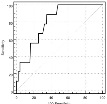
Analysis of the ROC curve showed that the PLL with the highest indexes of sensitivity and speciicity for neonatal death during the irst 3 days of life were > 4.2 mmol/L (sensitivity, 88.9%; speciicity, 64.6%; positive predictive value, 13.3%; negative predictive value, 99%), whereas the area under the curve was 0.802 (conidence interval, CI: 0.731-0.862; Figure 1).
Figure 1 - The ROC curve for the correlation of blood lactate and neonatal death during the first 3 days of life.
Transfontanellar ultrasound (TFUS) was performed in 147 of the 156 NBs. Of the 9 patients in whom TFUS was not performed, 5 died (all during the irst 3 days of life; 4 of these had PLL > 4.2 mmol/L), and 4 survived (one had PLL > 4.2 mmol/L). An echocardiogram to diagnose PPHNB was performed in 154 out of the 156 NBs. Both NBs in whom echocardiogram was not performed had PLL ≤ 4.2 mmol/L; 1 died, and the other survived.
he number of NBs who died in the irst 3 days of life was higher in the group with PLL > 4.2 mmol/L (Table 1).
186 Fernandez HGC, Vieira AA, Barbosa ADM
Rev Bras Ter Intensiva. 2012; 24(2):184-187
of NBs classiied as small for GA (SGA). All of these variables exhibited a higher frequency in the group of NB with PLL > 4.2 mmol/L. he remainder of investigated variables did not exhibit diferences (Table 1).
he group with PLL > 4.2 mmol/L exhibited a lower average BW (1,835 ± 885 g; median, 1,505 g; range, 490 - 3,760 g) compared to the group with PLL ≤ 4.2 mmol/L (2,324 ± 915 g; median, 2,400 g; range, 485 - 4,720 g; p=0.001). he CRIB score (which was measured in the 50 NB with a birth weight <1,500 g) and GA did not difer between the groups (Table 1).
he mortality during the irst 3 days did not difer as a function of the BW matched to GA. Of the 9 NBs who died, 2 were SGA, and 7 were not. Of the 147 NBs who survived the irst 3 days of life, 37 were SGA, and 110 were not (p=0.8428).
DISCUSSION
he present study used a ROC curve to deine the PLL cutof point with a better sensitivity and speciicity to predict early neonatal death in the investigated population. In addition, these indings will allow other investigators to perform the same analyses as the ones described above because there is currently no consensus for “reference values” for blood lactate concentrations in NBs.(5,10,20) In contrast, for adult patients, the reference values of PLL have already been established, and levels up to 2 mmol/L are considered normal.(6,7)
he present study found that NBs with PLL > 4.2 mmol/L during the irst 6 hours of life had higher odds of dying within the irst 3 days of life. However, the fact that the half-life of lactate is not known might explain the reason why a single measurement of PLL performed during the irst hours of life does not relect the events that occur after the third day of life.(5,12,21) Due to anaerobic cell metabolism, the PLL values are related not only to the severity of the clinical condition but also to asphyxia.(10-12)
he group of NBs with PLL > 4.2 mmol/L exhibited a higher frequency of neurologic manifestations related with the hypoxic-ischemic syndrome including a higher number of seizure episodes (OR=12.53) and ICH (OR=3.74). hese results may be due to tissue hypoperfusion and hypoxia, which induce a shift from aerobic to anaerobic metabolism. his metabolism shift results in such manifestations and might eventually culminate in patient death.(19,20,22,23)
he higher frequency of SGA NBs in the group with high lactate levels suggests that chronic intrauterine hypoxia might be one of the causes of low weight at birth.(24,25)
Although we did not correlate the CRIB scores of our groups with PLLs, Philips et al.,(26) upon performing a joint analysis of PLL and CRIB to predict mortality in extremely premature NBs, observed that the new, combined score had a good prognostic value.
Table 1 - The characteristics of the newborns, prenatal period, and labor
PLL > 4.2 mmol/L (N=60)
PLL ≤ 4.2 mmol/L (N=96)
p value
OR (CI)
NB data Gender
Male 33 (55.0) 57 (59.4) 0.591
-Female 27 (45.0) 39 (40.6)
BW 1.835.92 ± 884.71
(1505.0)
2324.37 ± 914.58 (2.400.0)
0.001
-GA 33.11 ± 4.06
(34.0)
34.32 ± 3.74 (35.0)
0.59
-CRIB 5.60 ± 4.81
(5.5)
5.80 ± 4.88 (5.5)
0.858
-BW matched to GA
SGA 23 (38.3) 16 (16.7) 0.003 3.108
(1.472-6.564)
AGA 37 (61.7) 80 (83.3)
Seizures
Yes 7 (11.7) 1 (1.0) 0.019 12.530
(1.503-104.497)
No 53 (88.3) 95 (99.0)
Death up to day 3
Yes 8 (13.3) 1 (1.0) 0.013 14.576
(1.778-119.508)
No 52 (86.7) 95 (99.0)
PPHNB (N=60)* (N=94)*
Yes 11 (18.3) 11 (11.7) 0.255
No 49 (81.7) 83 (88.3)
ICH (N=56)* (N=91)*
Yes 10 (17.9) 5 (5.5) 0.022 3.739
(1.206-11.592)
No 46 (82.1) 86 (94.5)
Prenatal data Centralization
Yes 10 (16.7) 7 (7.3) 0.075
-No 50 (83.3) 89 (92.7)
Oligohydramnios
Yes 12 (20.0) 10 (10.4) 0.099
-No 48 (80.0) 86 (89.6)
PIH
Yes 18 (30.0) 18 (18.8) 0.107
-No 42 (70.0) 78 (81.2)
Labor data Type of delivery
Vaginal 5 (8.3) 8 (8.3) 1.000
Cesarean 55 (91.7) 88 (91.7) Resuscitation
Yes 28 (46.7) 40 (41.7) 0.540
-No 32 (53.3) 56 (58.3)
Apgar < 5
Yes 9 (15.0) 9 (9.4) 0.289
-No 51 (85.0) 87 (90.6)
Plasma lactate levels and early neonatal mortality 187
Rev Bras Ter Intensiva. 2012; 24(2):184-187
CONCLUSIONS
In the investigated samples, a PLL > 4.2 mmol/L correlated with a higher frequency of SGA NBs, neurologic morbidity, and death during the irst 3 days of life.
RESUMO
Objetivo: Avaliar a correlação entre a concentração do nível plasmático de lactato, nas primeiras 6 horas de vida, e a mortalida-de neonatal precoce.
Métodos: Os pacientes foram separados em dois grupos, a par-tir do melhor ponto de corte do nível plasmático de lactato para predição da mortalidade neonatal nos 3 primeiros dias de vida, obtido por meio da construção de curva ROC. Os grupos foram separados e analisados quanto às diferenças e correlações entre as va-riáveis estudadas e nível plasmático de lactato dosado nas primeiras
6 horas de vida, por meio dos testes qui-quadrado, t de Student ou Mann-Whitney, e regressão logística.
Resultados: O melhor ponto de corte do nível plasmático de lactato determinado pela curva ROC para óbito nos 3 pri-meiros dias de vida foi 4,2mmol/L. Os grupos estudados foram diferentes em relação à média de peso de nascimento (menor no grupo com nível plasmático de lactato >4,2mmol/L), adequação entre peso de nascimento/idade gestacional, com maior número de recém-nascidos pequenos para idade gestacional nesse grupo. A ocorrência de convulsões, hemorragia intracraniana e óbito nos primeiros 3 dias de vida foi mais freqüente no grupo com nível plasmático de lactato >4,2mmol/L.
Conclusão: Para a amostragem estudada, a presença de nível plasmático de lactato > 4,2mmol/L, nas primeiras 6 horas de vida, foi correlacionada ao óbito neonatal nos 3 primeiros dias de vida, à maior frequência de morbidade neurológica e de recém-nascidos pequenos para idade gestacional.
Descritores: Ácido láctico; Asixia neonatal; Mortalidade neonatal
REFERENCES
1. Apgar V. A proposal for a new method of evaluation of the newborn infant. Curr Res Anesth Analg. 1953;32(4):260-7.
2. The CRIB (clinical risk index for babies) score: a tool for assessing initial neonatal risk and comparing performance of neonatal intensive care units. The International Neonatal Network. Lancet. 1993;342(8865):193-8. Erratum in
Lancet. 1993;342(8871):626.
3. Richardson DK, Phibbs CS, Gray JE, McCormick MC, Workman-Daniels K, Goldmann DA. Birth weight and illness severity: independent predictors of neonatal mortality. Pediatrics. 1993;91(5):969-75.
4. Richardson DK, Gray JE, McCormick MC, Workman K, Goldmann DA. Score for Neonatal Acute Physiology: a physiologic severity index for neonatal intensive care. Pediatrics. 1993;91(3):617-23.
5. Deshpande SA, Platt MP. Association between blood lactate and acid-base status and mortality in ventilates babies.Arch Dis Child Fetal Neonatal Ed.
1997;76(1):F15-20.
6. Rashkin MC, Bosken C, Baughman RP. Oxygen delivery in critically ill patients. Relationship to blood lactate and survival. Chest. 1985;87(5):580-4. 7. Weil MH, Afifi AA. Experimental and clinical studies on lactate and pyruvate
as indicators of the severity of acute circulatory failure (shock). Circulation.
1970;41(6):989-1001.
8. Barrington KJ. Hypotension and shock in preterm infant. Semin Fetal Neonatal Med. 2008;13(1):16-23.
9. Hussain F, Gilshenan K, Gray PH. Does lactate level in the first 12 hours of life predict mortality in extremely premature infants? J Paediatr Child Health. 2009;45(5):263-7.
10. Cheung PY, Etches PC, Weardon M, Reynolds A, Finner NN, Robertson CM. Use of plasma lactate to predict early mortality and adverse outcome after neonatal extracorporeal membrane oxygenation: a prospective cohort in early childhood. Crit Care Med. 2002;30(9):2135-9.
11. Groenendaal F, Lindemans C, Uiterwaal CS, Vries LS. Early arterial lactate and prediction of outcome in preterm neonates admitted to a neonatal intensive care unit. Biol Neonate. 2003;83(3):171-6.
12. Cheung PY, Chui N, Joffe AR, Rebeyka IM, Robertson CM; Western Canadian Complex Pediatric Therapies Project, Follow-up Group. Postoperative lactate concentrations predict the outcome of infants aged 6 weeks or less after intracardiac surgery: a cohort follow-up to 18 months. J Thorac Cardiovasc Surg. 2005;130(3):837-43.
13. Sinn JK, Lloyd J, Todd DA, Lazarus R, Maesel A, John E. Umbilical cord blood lactate in normal infants: comparison between two methods of measurement. J Paediatr Child Health. 2001;37(1):24-7.
14. Cohen RD, Woods HF. Lactic acidosis revisited. Diabetes. 1983;32(2):181-91.
15. Ballard JL, Khoury JC, Wedig K, Wang L, Eilers-Walsman BL, Lipp R. New Ballard Score, expanded to include extremely premature infants. J Pediatr. 1991;119(3):417-23.
16. Alexander GR, Himes JH, Kaufman RB, Mor J, Kogan M. A United States national reference for fetal growth. Obstet Gynecol. 1996;87(2):163-8. 17. Goddard-Finergold J. The nervous system during birth. Intraventricular
hemorrhage. In: Taeusch HW, Ballard RA. Avery’s diseases of the newborn. 7th ed. Philadelphia: Saunders; 1998. p. 859-64.
18. Goddard-Finergold J. The nervous system during birth. Seizures and other paroxysmal disorders. In: Taeusch HW, Ballard RA. Avery’s diseases of the newborn. 7th ed. Philadelphia: Saunders; 1998. p. 871-7.
19. Hansen T, Corbet A. Disorders of the transition, persistent pulmonary hypertension of the newborn. In: Taeusch HW, Ballard RA. Avery’s diseases of the newborn. 7th ed. Philadelphia: Saunders; 1998. p. 615-9.
20. Jung D, Lun A, Zinsmeyer J, Grauel EL, Gross J. The concentration of hypoxanthine and lactate in the blood of healthy and hypoxic newborns. J Perinat Med. 1985;13(1):43-50.
21. Koliski A, Cat I, Giraldi DJ, Cat ML. Lactato sérico como marcador prognóstico em crianças gravemente doentes. J Pediatr (Rio J). 2005;81(4):287-92. 22. Nguyen HB, Rivers EP, Knoblich BP, Jacobsen G, Muzzin A, Ressler JA, et al.
Early lactate clearance is associated with improved outcome in severe sepsis and septic shock. Crit Care Med. 2004;32(8):1637-42.
23. Procianoy RS, Silveira RC. Síndrome hipóxico-isquêmica. J Pediatr (Rio J). 2001;77(Supl 1):S63-70.
24. Soothill PW, Nicolaides KH, Campbell S. Prenatal asphyxia, hyperlacticaemia, hypoglycaemia, and erythroblastosis in growth retarded fetuses. Br Med J (Clin Res Ed). 1987;294(6579):1051-3.
25. Marconi AM, Paolini CL, Zerbe G, Battaglia FC. Lactacidemia in intrauterine growth restricted (IUGR) pregnancies: relationship to clinical severity, oxygenation and placental weight. Pediatr Res. 2006;59(4 Pt
1):570-4.