SOCIEDADE BRASILEIRA DE ORTOPEDIA E TRAUMATOLOGIA
w w w . r b o . o r g . b r
Original
article
Anatomical
variations
of
pronator
teres
muscle:
predispositional
role
for
nerve
entrapment
夽
Edie
Benedito
Caetano
∗,
Luiz
Ângelo
Vieira,
Fábio
Antonio
Anversa
Sprovieri,
Guilherme
Camargo
Petta,
Maurício
Tadeu
Nakasone,
Bárbara
Lívia
Correa
Serafim
PontifíciaUniversidadeCatólicadeSãoPaulo,FaculdadedeCiênciasMédicasedaSaúde,DisciplinadeOrtopediaeTraumatologia, Sorocaba,SP,Brazil
a
r
t
i
c
l
e
i
n
f
o
Articlehistory:
Received15February2016 Accepted18April2016 Availableonline2March2017
Keywords: Pronation Mediannerve
Nervecompressionsyndromes
a
b
s
t
r
a
c
t
Objective:Toassesstheanatomicalvariationsofthepronatorteresmuscle(PTM)andits implicationinthecompressionofthemediannerve,whichpassesthroughthehumeral andulnarheadsofthePTM.
Methods:Forthepresentstudy,100upperlimbsfromhumancadaversfromtheanatomy laboratoryweredissected.Forty-sixspecimensweremaleandfour,female,whoseaged rangedfrom28to77years;27werewhiteand23,non-white.Apilotstudyconsistingofsix handsfromthreefreshcadaverdissectionswasconductedtofamiliarizetheauthorswith thelocalanatomy;thesewerenotincludedinthepresentstudy.
Results:ThehumeralandulnarheadsofPTMwerepresentin86limbs.In72outofthe86 limbs,themediannervewaspositionedbetweenthetwoheadsofthePTM;in11,itpassed throughthemusclebellyofulnarheadofthePTM,andinthree,posteriorlytobothheads ofthePTM.Whenbothheadswerepresent,themediannervewasnotobservedaspassing throughthemusclebellyofthehumeralheadofPTM.In14outofthe100dissectedlimbs,the ulnarheadofthePTMwasnotobserved;inthissituation,themediannervewaspositioned posteriorlytothehumeralheadin11limbs,andpassedthroughthehumeralheadinthree. In17limbs,theulnarheadofPTMwaslittledeveloped,withafibrousbandoriginatingfrom theulnarcoronoidprocess,associatedwithadistalmusclecomponentneartheunionwith thehumeralhead.Infourlimbs,theulnarheadoftheMPRwasrepresentedbyafibrous band.Inbothlimbsofonecadaver,afibrousbandwasobservedbetweenthesupinator muscleandthehumeralheadofthePTM,passingovermediannerve.
Conclusion: Theresultssuggestthattheseanatomicalvariationsinrelationship median nerveandPTMarepotentialfactorsformediannervecompression,astheynarrowthe spacethroughwhichthemediannervepasses.
©2017PublishedbyElsevierEditoraLtda.onbehalfofSociedadeBrasileiradeOrtopedia eTraumatologia.ThisisanopenaccessarticleundertheCCBY-NC-NDlicense(http:// creativecommons.org/licenses/by-nc-nd/4.0/).
夽
StudyconductedatthePontifíciaUniversidadeCatólicadeSãoPaulo,FaculdadedeCiênciasMédicasedaSaúde,DisciplinadeOrtopedia eTraumatologia,Sorocaba,SP,Brazil.
∗ Correspondingauthor.
E-mail:[email protected](E.B.Caetano). http://dx.doi.org/10.1016/j.rboe.2017.02.003
Variac¸ões
anatômicas
do
músculo
pronador
redondo
e
sua
importância
nas
síndromes
compressivas
Palavras-chave: Pronac¸ão Nervomediano
Síndromesdecompressão nervosa
r
e
s
u
m
o
Objetivo: Analisarasvariac¸õesanatômicasdomúsculopronadorredondo(MPR)esuas implicac¸õesnacompressãodonervomediano,quepassaentreascabec¸asumeraleulnar doMPR.
Método: Foramdissecados100membrossuperioresdecadáveresadultospertencentesao laboratóriodeanatomia;46cadávereseramdosexomasculinoequatrodofeminino.A idadevariouentre28e77anos;27eramdaetniabrancae23,nãobranca.Umestudopiloto queincluiutrêscadáveresfrescosfoifeito,parafamiliarizac¸ãodosautorescomaanatomia regional.Essesnãoforamincluídosnoestudo.
Resultados:Em86membros,observou-seapresenc¸adascabec¸asumeraleulnardoMPR.Em 72dos86membros,onervomedianoestavaposicionadoentreascabec¸asumeraleulnar doMPR;em11,esseencontrava-seatravésdamassamusculardacabec¸aulnardoMPReem três,onervomedianoestavaposicionadoposteriormenteàsduascabec¸asdoMPR.Noscasos emqueasduascabec¸asdomúsculoestavampresentes,nãoseobservouonervomediano passandoatravésdamassamusculardacabec¸aumeraldoMPR.Em14dos100membros dissecados,acabec¸aulnardoMPRnãoestavapresente.Nessasituac¸ão,onervomediano posicionava-seposteriormenteàcabec¸aumeralem11membroseatravésdacabec¸aumeral emtrêsmembros.Em17membros,acabec¸aulnarestavamuitopoucodesenvolvida,com conformac¸ãofibrosaemsuaorigemnoprocessocoronoidedaulna,associadaaum com-ponentemusculardistal,próximoasuauniãocomacabec¸aumeral.Emquatromembros,a cabec¸aulnardoMPRestavarepresentadaapenasporumabandafibrosa.Nosdoismembros deumcadáver,observou-seumaexpansãofibrosaquesaíadomúsculosupinadorparaa cabec¸aumeraldoMPR,passandocomoumacintasobreonervomediano.
Conclusões: Essesresultadossugeremqueasvariac¸õesanatômicasnarelac¸ãonervo medi-anoeMPRrepresentamfatorespotenciaisparacompressãonervosa,porestreitaroespac¸o noqualpassaonervomediano.
©2017PublicadoporElsevierEditoraLtda.emnomedeSociedadeBrasileirade OrtopediaeTraumatologia.Este ´eumartigoOpenAccesssobumalicenc¸aCCBY-NC-ND (http://creativecommons.org/licenses/by-nc-nd/4.0/).
Introduction
Thereareseveralanatomicalstructuresthatcancompressthe mediannerveneartheelbowjoint.Fromproximaltodistal, thecompressionmaybecausedbytheStruthers’ligament1,2 withorwithoutthe supracondylarprocess ofthehumerus, byaponeuroticexpansionofthebicepsbrachiimuscle( Lacer-tusfibrosus),3,4betweenthehumeralandulnarheadsofthe pronatorteresmuscle(PTM),5,6bythevascularnetworkofthe region,7andbythearchformedbythetwoinsertionsofthe superficialflexormuscleofthefingers.8
Regardlessofthesesiteswherecompressionoccurs,this condition istermed pronator teres syndrome, because the compressionoccursmostfrequentlybetweenthetwoheads ofthismuscle.9–11Themaincausesaretheanatomic varia-tionsofthePTM.Thenormalanatomicalpatterndescribed bytheclassicalanatomystudies12–14 isthatthePTMis con-stitutedbytwo heads. Thehumeralhead, moreextensive, originatesinthesupracondylarprocessofthehumerusand adjacencies.Theulnarheadoriginatesinthe coronoid pro-cessoftheulna.Thetwoportionsuniteforinsertionintothe diaphysisoftheradius,contouringtoit.Themediannerveis positionedbetweenthetwoheadsofthePTM.However,the
relationship between the median nerve and the humeral and ulnar heads of the PTM is subject to numerous variations.4,6,15,16 This study aimed to analyze, through anatomicaldissections,therelationshipbetweenthePTMand mediannerveandthuscontributetoabetterunderstanding ofthecausesofthepronatorteressyndrome.
Material
and
methods
One hundred upper limbs of 50 adult cadavers from the anatomydepartmentofthisinstitutionweredissectedforthis study,46cadaversweremaleandfourwerefemale.Theage rangedfrom28to77years;27werewhiteand23,non-white. Cadaverswhoseforearmsweredeformedbytraumas, malfor-mations,andscarswereexcluded.Apilotstudythatincluded threefreshcadaverswasconductedsothattheauthorscould familiarizethemselveswiththelocalanatomy.Thesewerenot includedinthisstudy.
10cmproximaltotheintercondylarlineofthehumerus;at this location, it was positioned mediallyin relationto the brachialartery.Thedissectionproceededdistallyuntil reach-ingthebicipitalaponeurosis,whichwassectioned,allowing thevisualizationoftheproximalmarginofthePTM.The pres-enceofvariationsinthesurfaceheadandfibrousbandsatthe sitewasrecorded.Subsequently,thesuperficialheadofthe PTMwassectionedtransverselytoallowthevisualizationof thedeephead,whosepresentationwasvaried;itwasabsent in14ofthe100dissectedlimbs.Themediannervewasdistally dissecteduntilitpassedthroughthearchformedbythe prox-imalinsertionsoftheflexordigitorumsuperficialismuscles. Inalldissectedlimbs,thefirstbranchofthemediannervein theforearmwasalwaystowardsthesuperficialheadofthe PTM.Allforearmmusclesweredissected;theirinnervation, thepresenceofcommunicationamongtheforearmnerves (Martin-Gruber anastomosis), the relationship between the mediannerveandthebicipitalaponeurosis,andthe relation-shipofthemedianandanteriorinterosseousnerveswiththe archoforiginofthethreeheadsoftheflexordigitorum super-ficialismusclewereanalyzed.Theanatomicalvariationswere annotatedandphotographed.AKeller2.5Xmagnifyingglass wasusedformagnification.Thisstudywasapprovedbythe hospital’sethicscommitteeundertheCAAENo.1.356.351.
Results
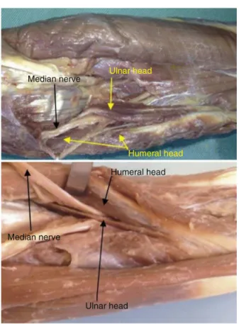
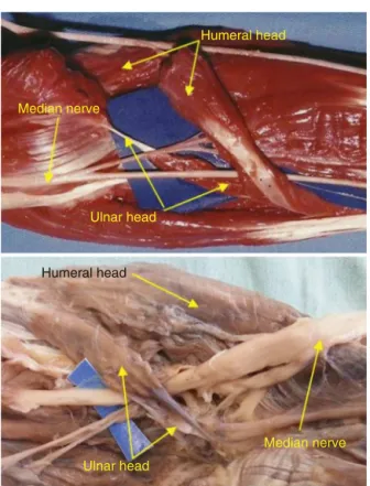
In86dissectedlimbs,thehumeralandulnarheadsofthePTM werewellindividualized,consistingoftwodistinctmuscular portionsthatjoinedtoinsertthroughanenlargedtendon, con-touringtoandinsertinginthemiddlethirdofthediaphysisof theradius(Fig.1).In72ofthe86limbs,themediannervewas positionedbetweenthehumeralandulnarheadsofthePTM (Fig.1).In11limbs(fourbilaterally),itwaspositionedthrough themuscularmassoftheulnarheadofthePTM(Fig.2).In threeforearms(onebilaterally),themediannervewas pos-itionedposteriorlytothetwoheadsofthePTM(Fig.3).When bothheadsofthemusclewerepresent,nocasesofthemedian nervepassingthroughthemusclemassofthehumeralheadof thePTMwereobserved.In14ofthe100limbs,theulnarhead ofthePTMwasabsent.Inthissituation,themediannervewas positionedposteriorlytothehumeralheadin11limbs(Fig.4) andthroughthehumeralheadinthreelimbs(Fig.5).
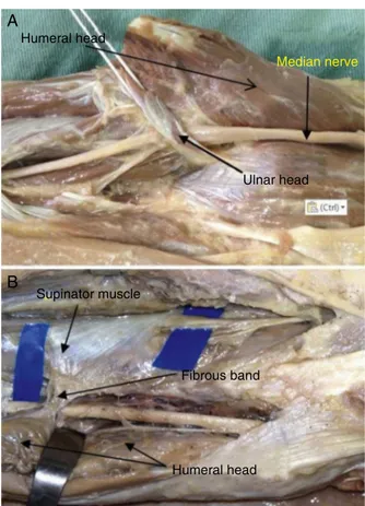
Thehumeralheadwaslargerthantheulnarhead(Fig.1). Apoorlydevelopedulnarheadwasobservedin17limbs,with afibrousconformationinitsorigininthecoronoidprocessof theulna,associatedwithadistalmuscularcomponent,close toitsunionwiththehumeralhead(Fig.6).Theulnarheadof thePTMwasrepresentedbyafibrousbandnotassociatedwith amuscularcomponentinonlyfourlimbs(Fig.7A).Onboth limbsofasinglecorpse,afibrousexpansionextendingfrom thesupinatormuscletothehumeralheadofthePTM,passing asabandoverthemediannerve,wasobserved(Fig.7B).In fivelimbs,theulnarheadwasinsertedalongsidetheGantzer muscle,inthecoronoidprocessoftheulna.Ineightlimbs,a highinsertionofthehumeralheadPTMrangingfrom2.8to 3.5cmproximaltothemedialepicondylewasobserved(Fig.7A andB;Table1).
Median nerve
Median nerve
Ulnar head
Humeral head
Humeral head
Ulnar head
Fig.1–In86limbs,thehumeralandulnarheadsofthePR
musclewerewellindividualized.
Median nerve
Median nerve
Ulnar head
Ulnar head Humeral head
Humeral head
Fig.2–Inninelimbs(threebilaterally),themediannerve
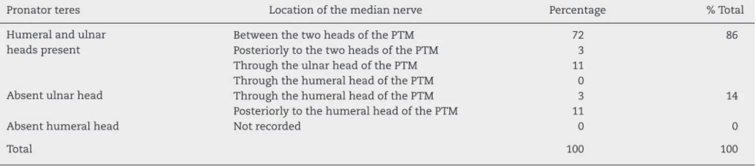
Table1–EvaluationofthehumeralandulnarheadsofPTMandtheirrelationshipswiththemediannervein100 dissectedlimbs.
Pronatorteres Locationofthemediannerve Percentage %Total
Humeralandulnar headspresent
BetweenthetwoheadsofthePTM 72 86
PosteriorlytothetwoheadsofthePTM 3
ThroughtheulnarheadofthePTM 11
ThroughthehumeralheadofthePTM 0
Absentulnarhead ThroughthehumeralheadofthePTM 3 14
PosteriorlytothehumeralheadofthePTM 11
Absenthumeralhead Notrecorded 0 0
Total 100 100
Discussion
In 86 of the 100 dissected limbs (86%), the humeral and ulnarheadsofthePTMwerepresent,whichisinagreement withtheresultsreportedbyStabilleetal.6(83.5%),Jamieson andAnson15 (81%), andHollinshead17 (91%).Other authors observeddifferentpercentages:HoferandHofer,1856%,and Nebot-Cegarraetal.,1968%.In74ofthese86limbs,themedian nervewaspositionedbetweenthehumeralandulnarheadsof thePTM;in11,themediannervepassedthroughthe muscu-latureoftheulnarheadofthePTM.Inthreelimbs,themedian nervewaspositionedposteriorlytobothPTMheads.Incases inwhichbothheadsofthe PTMwere present,nocasesof themediannervepassingthroughthemuscularmassofthe humeralheadofthePTMwereobserved.
Median nerve
Median nerve Humeral head
Humeral head
Ulnar head
Ulnar head
Fig.3–Inthreeforearms(onebilaterally),themediannerve
waspositionedposteriorlytothetwoheadsofthePTM.
ThePTMulnarheadwasabsentin14% ofthedissected limbs.InthestudybyStabilleetal.,6itwasobservedin15%; inthatbyHollinshead,17in9%,andinthatbyNebot-Cegarra et al.,19 in21.7%.Kaplan20 and Zancolli21 reportedthatthe absenceoftheulnarheadisfrequent.TestutandLatarjet12 andChiarugi14alsoobservedthisabsence,butdidnotrecord howoftenthisanatomicalvariationmayoccur.
Testut and Latarjet12 and Le Double and Anatole22 describedthatatendonsegmentmayreplacetheulnarhead ofthePTM;Spinner23considersthatthisvariationoccurs fre-quently.Stabilleetal.6observedtheulnarheadrepresentedby afibrousbandin9%ofthecases.Inthepresentstudy,17limbs hadapoorlydevelopedulnarhead,withafibrouscomponent initsproximalportioninthecoronoidprocessoftheulna,but
Humeral head
Humeral head
Median nerve
Median nerve
Fig.4–AbsenceoftheulnarheadofthePTM.Inthis
situation,themediannervewaspositionedposteriorlyto
Median nerve
Median nerve
Humeral head
Humeral head
Fig.5–AbsenceoftheulnarheadofthePTM.Inthis
situation,themediannervepassedthroughthehumeral
headinthreelimbs.
associatedwithamuscularcomponentinthedistalportion, beforejoiningthehumeralhead.Inonlyfourlimbs,theulnar headofthePTMwasrepresentedbyafibrousband,not asso-ciatedwithamuscularcomponent.Inagreementwithallthe studiesanalyzed,nocasesofabsenthumeralheadofthePTM wereobserved.
Testutand Latarjet12 and Le Double and Anatole22 also describedthatthehumeralandulnarheadofthePTMmaybe completelyseparate,withdistalinsertionsatdifferentsites. Inthepresentstudy,inagreementwithStabilleetal.,6inthe 86limbsinwhichbothheadswerepresent,thesewereunited beforetheirinsertionintothemiddle-thirdoftheradial dia-physis.
Barret,24in200dissectedcases,observedinoneofthem anadditionalportionofthehumeralhead,termingitthethird headofthePTM.HealsoreportedthatBuchananhadobserved athirdheadofthePTM, whichoriginatedfrom the supra-condylarprocessofthehumerus.Caetanoetal.2publisheda caseinwhichtherewasmediannervecompressioncausedby theinsertionofthehumeralheadofthePTMin supracondy-larhumeralprocess.Inthepresentstudy,theinsertionofthe humeralhead(2.8–3.5cm)proximaltothemedialepicondyle ofthehumeruswasobservedineightcases.
Thepositioning ofthe mediannerveposteriorly toboth headsofthePTM,describedbyJamiesonandAnson15in6% oflimbsandbyDiDioandDangelo16in2.5%,wasobservedin threeforearms(3%)inthepresentstudy.
Median nerve
Median nerve Ulnar head
Ulnar head
Humeral head
Humeral head
Fig.6–In11limbs,apoorlydevelopedulnarheadwas
observed,withafibrousconformationinitsorigininthe
coronoidprocessoftheulna,associatedwithadistal
muscularcomponent,closetoitsunionwiththehumeral
head.
Humeral head Supinator muscle Fibrous band Humeral head Ulnar head
A
B
Median nerveFig.7–(A)Infourlimbs,theulnarheadofthePTMwas
representedbyafibrousbandnotassociatedwithamuscle
component;(B)intwolimbs,afibrousexpansionextended
fromthesupinatormuscletothehumeralheadofthePTM,
passingasabandoverthemediannerve.
Surgeons should be aware ofthe anatomical variations thatcanbeobservedintheelbowregion,astheychangethe positionofthenoblestructures,puttingthematriskduring arthroscopicproceduresandopensurgicalapproachesinthe region.
Conclusion
Theanatomicalvariationsofthehumeraland ulnarheads ofthe PTM inrelation totheir constitution and frequency canalterthepositioningofthemediannerve.Themost fre-quentlyobservedvariationwasabsenceoftheulnarhead.In thepresenceoffibrousbands,especiallywhenthe median nerveextendsclosetothecoronoidprocessoftheulnawhere theulnarheadofthePTMhasafibrousconstitution,nerve compressioncanoccurduetoanarrowingofthenerve pas-sagespace.Itisclearthatthepassageofthenervethrough themusclemassofthePTMisnotonlyaplacewithgreater potentialtocausepronatorteressyndrome,butalsoaltersthe normalcourseofthemediannerve.
Conflicts
of
interest
Theauthorsdeclarenoconflictsofinterest.
r
e
f
e
r
e
n
c
e
s
1.SenerE,TakkaS,CilaE.Supracondylarprocesssyndrome. ArchOrthopTraumaSurg.1998;117(6–7):418–9.
2.CaetanoEB,BrandiS,LeeHJ.Compressãodonervomediano porprocessosupracondilardoúmero.RevBrasOrtop. 1989;24(9):323–6.
3.SpinnerRJ,CarmichaelSW,SpinnerM.Partialmediannerve entrapmentinthedistalarmbecauseofanaccessory bicipitalaponeurosis.JHandSurg.1991;16(2):236–44. 4.SeitzWHJr,MatsuokaH,McAdooJ,ShermanG,StickneyDP.
Acutecompressionofthemediannerveattheelbowbythe Lacertusfibrosus.JShoulderElbowSurg.2007;16(1):91–4. 5.HartzCR,LinscheidRL,GramseRR,DaubeJR.Thepronator
teressyndrome:compressiveneuropathyofthemedian nerve.JBoneJointSurgAm.1981;63(6):885–90.
6.StabilleSR,DuarteE,CarvalhoVC.Pronatorteresmuscle: anatomicalvariationsandpredispositionforthecompression ofthemediannerve.ActaSciBiolSci.2008;24:631–7.
7.PiresPR,AndradeRP.Síndromescompressivasnomembro superior.In:PardiniA,FreitasA,editors.Cirurgiadamão: lesõesnãotraumáticas.2a
ed.RiodeJaneiro:Medbook;2008. p.263–97.
8.DellonAL,MackinnonSE.Musculoaponeuroticvariations alongthecourseofthemediannerveintheproximal forearm.JHandSurgEdinbScotl.1987;12(3):359–63. 9.JohnsonRK,SpinnerM,ShrewsburyMM.Mediannerve
entrapmentsyndromeintheproximalforearm.JHandSurg. 1979;4(1):48–51.
10.EversmannWW.Proximalmediannervecompression.Hand Clin.1992;8(2):307–15.
11.BayerlW,FischerK.Thepronatorteressyndrome.Clinical aspects,pathogenesis,andtherapyofanon-traumatic mediannervecompressionsyndromeinthespaceofthe elbowjoint.Handchirurgie.1979;11(2):91–8.
12.TestutL,LatarjetA.Tratadodeanatomiahumana.Barcelona: Salvat;1947.
13.TandlerJ.Tratadodeanatomiasistematica.2a
ed.Barcelona: Salvat;1928.
14.ChiarugiG.Istituzionidianatomiadell’uomo.7aed.Milano:
Sovete;1949.
15.JamiesonRW,AnsonBJ.Therelationofthemediannerveto theheadsoforiginofthepronatorteresmuscle,astudyof 300specimens.QBullNorthwestUnivMedSch.
1952;26(1):34–5.
16.DiDioJA,DangeloJG.Nervusmedianuspiercingthecaput humeraleofthem.pronatorteres.AnatAnz.1963;112:385–8. 17.HollinsheadWH.Anatomyforsurgeons.NewYork:Hoeber
Harper;1958.
18.HoferK,HoferG.UeberdenVerlaufderarteriabrachialismit demnervusmedianuszwischendenbeidenkopfendes musculuspronatorteres.AnatAnzeles.1910;36:510. 19.Nebot-CegarraJ,Perez-BerruezoJ,ReinadelaTorreF.
Variationsofthepronatorteresmuscle:predispositionalrole tomediannerveentrapment.ArchAnatHistolEmbryol. 1991–1992;74:35–45.
20.KaplanEB.Anatomiafunctionalyquirurgicadelamano. BuenosAires:Artecnica;1961.
21.ZancolliE.Structuralanddynamicbasesofhandsurgery.2nd ed.Philadelphia:Lippincott;1979.
22.LeDoubleAF,AnatoleF.Traitédesvariationsdusystème musculairedel’hommeetdeleursignificationaupointde vuedel’anthropologiezoologique.Paris:SchleicherFrères; 1897.
24.BarrettJH.Anadditional(thirdandseparate)headofthe pronatorteresmuscle.JAnat.1936;70Pt4:577–8.
25.GessiniL,JandoloB,PietrangeliA.Entrapmentneuropathies ofthemediannerveatandabovetheelbow.SurgNeurol. 1983;19(2):112–6.