Larry Ching Fan Li**, Ricky Wing-Kit Wong***
introduction
Based on the incisor relationship, Class II mal-occlusion is defined as the lower incisor edges lying posterior to the cingulum plateau of the upper in-cisors resulting in an increase in overjet.1 The
prev-alence of having an overjet greater than 10 mm was
reported to be around 0.2% of the population.2
Large overjet, especially in children and ado-lescents, is associated not only with an increased risk of traumatic injury to the upper anterior teeth but also psychological distress which results in loss of self-esteem and problems with social in-teraction. Among different malocclusions, Class II malocclusion was rated as the most unattractive
* MOrthRCSEd, Membership of Orthodontics Examination of the Royal College of Surgeons of Edinburgh.
Management of severe Class II malocclusion
with sequential removable functional
and orthodontic appliances: A case for
MOrthRCSEd* examination
introduction: Functional appliance is an effective way of treating skeletal Class II
mal-occlusion in children and adolescents. A 12-month stepwise mandibular advancement protocol has been proved to enhance the condylar growth and improve the mandibular
prognathism using Herbst appliance. objective: The following case report documented a
11 year old Chinese girl with 11 mm overjet treated by a phase I 12-month growth modi-fication therapy using Twin Block appliance with Hyrax palatal expander and high-pull headgear in a stepwise mandibular advancement protocol followed by a phase II pread-justed Edgewise appliance therapy. It is one of the cases submitted for the Membership of Orthodontics Examination of the Royal College of Surgeons of Edinburgh.
Abstract
Keywords: Myofunctional therapy. Functional appliances. Angle Class II malocclusion.
** MSc in Orthodontics, MOrthRCSEd. MRACDS (Member of The Royal Australasian College of Dental Surgeons). *** Adjunct Professor of Orthodontics, Deparment of Orthodontics, School of Dentistry, University of Hong Kong.
How to cite this article: Li LCF, Wong RWK. Management of severe Class
II malocclusion with sequential removable functional and orthodontic appli-ances: A case for MOrthRCSEd examination. Dental Press J Orthod. 2011 Sept-Oct;16(5):46.e1-11.
» The authors report no commercial, proprietary, or inancial interest in the
by both orthodontists and laypersons.3 Albino4
as-sets appearance is the most common reason given for seeking treatments.
Class II malocclusion can usually be cor-rected by either extracting two upper premo-lars followed by retraction of the upper anterior teeth (camouflage) or advancing the mandible by growth modification or orthognathic surgery. There are still controversies about how effec-tive is growth modification for the correction of large overjets. Fixed functional appliances such as Herbst has been proven to effectively enhance condylar growth and improve mandibular
prog-nathism in both adolescents5 and adults6 using
a 12 month stepwise mandibular advancement
protocol.7 The following case report
document-ed a 11 year and 2 month old Chinese girl with 11 mm overjet treated by a phase I growth modi-fication using Twin Block appliance with the 12 month stepwise protocol followed by a phase II preadjusted Edgewise appliance therapy. It is one of the cases submitted to the Membership of Or-thodontics Examination of the Royal College of Surgeons of Edinburgh (MOrthRCSEd).
cAse PresentAtion
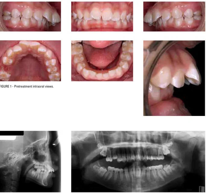
clinical examination and diagnostic summary A 10 year and 10 month old Chinese girl at-tended our clinic complaining of crooked and sticking out upper anterior teeth. Extraorally, she had no apparent facial asymmetry. The lips were incompetent at rest due to an increase in overjet and also unfavorable muscle tone. The upper lip length was 18 mm, which was consid-ered shorter than the average. There was an ac-ceptable amount of incisal and gingival display on smiling and at rest, with the upper and lower dental midlines coinciding with the midfacial line. The nasolabial angle was acute, reflecting the protrusive upper lip, which together with the retruded mandible and chin, contributed to her convex profile. The temporomandibular joints were normal.
Intraorally, she presented a early permanent dentition with a Class II Division 1, incisor rela-tionship and increased overjet of 11 mm. The over-bite was increased at 4 mm (57%), complete and traumatic. The molar and canine relationships were full unit Class II on both sides. There was scissor-bite between teeth #14 and #44. There was mild crowding in the upper arch. The curve of Spee was increased at 3 mm. She also had a reduced anterior Bolton tooth size ratio of 71.3% (77.7% for normal
Class II Southern Chinese females8) due to
rela-tively smaller teeth in the lower anterior segment. The oral hygiene needed to be improved (Fig 1).
Radiographically, the increased ANB (6°) and Wits appraisal (+6 mm) confirmed that the
pa-tient had a Class II skeletal pattern.9 The normal
SNA and reduced SNB and SNPg indicated a normal maxilla, receding mandible and chin. The SN-mandibular plane angle and the lower facial proportion were normal. The upper incisors were proclined while the lower incisors were normally angulated. The lower incisors were far behind the A-Pogonion line and the lower lip was retrusive to the Ricketts E plane by 2.1 mm. The cervical ver-tebrae maturation (CVM) stage was CVS3, which
was around the peak of growth spurt10 (Fig 2).
Aims of treatment
1. Improve oral hygiene.
2. Enhance forward growth of the mandible to improve facial profile and mandible/cranial base relationship.
3. Reduce overjet and overbite and achieve Class I incisor and buccal segment relationships.
4. Relieve crowding and align teeth.
5. Eliminate lip trap and improve lip compe-tency.
FIGURE 1 - Pretreatment intraoral views.
FIGURE 2 - Pretreatment lateral cephalometric and panoramic radiographs.
treatment progress
Phase I: Growth modification therapy
The patient was referred to a dental hygienist for oral hygiene instruction and scaling and pro-phylaxis. After achieving a satisfactory oral hy-giene level, orthodontic treatment commenced. An acrylic Twin Block appliance was issued for full-time wear with an initial mandibular ad-vancement of 5 mm and 7 mm vertical opening at
week for 12 weeks to achieve a palatal expansion of 6 mm (Fig 3). Maxillary canine to canine were bonded with 0.014-in nickel-titanium (NiTi) arch-wire with laceback between teeth #11 and #21 to align the teeth.
Phase II: Fixed appliance
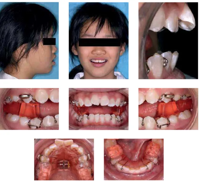
The Twin Block appliance was removed af-ter 12 months into treatment. The overjet was reduced to 1 mm. Crowding was relieved in the
upper arch due to distalizing effect of the den-tition as well as the palatal expansion. An up-per Twin Block retainer providing a positive incisal stop was issued to be worn full-time for 3 months (Fig 4). Both upper and lower arches were bonded using 0.022 x 0.028-in slot pread-justed Edgewise appliance with Roth’s prescrip-tion and aligned with 0.014-in nickel-titanium wires. The archwires were subsequently changed to 0.017 x 0.025-in NiTi for further alignment
FIGURE 4 - Twin Block retainer. Space developed distal to upper canines due to headgear effect of Twin Block appliance and pala-tal expansion.
and for torque control. After three months, 0.017 x 0.025-in stainless steel archwire was placed on the upper arch while 0.019 x 0.025-in stainless steel archwire was used in the lower arch for arch coordination. Class II elastics were worn full time to maintain the buccal relationships and the overjet. The treatment was completed in 26 months. Fixed 0.018-in Twisflex fixed lingual re-tainers were delivered on both arches. Upper and lower Hawley’s retainers were also issued as an additional protection measure against unnoticed debonding of the fixed lingual retainers.
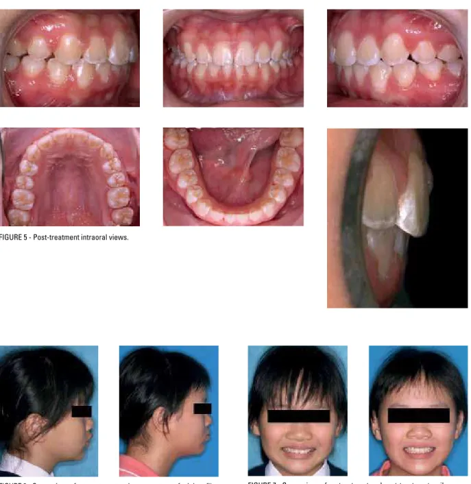
treatment changes
The total treatment time was 26 months. Overjet and overbite were reduced to 3 mm and 2.5 mm respectively. Super Class I buccal relationships were achieved on both sides. Ca-nine guidance was present on the left and right sides during lateral excursions and incisal guid-ance was present on protrusion. There were no non-working side interferences during function. Protrusive movements were also normal. Good buccal interdigitation was achieved despite buc-cal overjet was slightly increased due to the tooth size discrepancy. A 97.8% reduction in PAR (Peer Assessment Rating) score was achieved with the
initial PAR score of 45 points reduced to 1 point post-treatment. This can be categorized as great-ly improved (Figs 5, 6 and 7).
An appreciable amount of sagittal and vertical mandibular growth was observed during the treat-ment period. The facial profile, measured as facial convexity angle, improved 8°. Mandible/cranial base relationship improved to 3.6° and Wits ap-praisal value was at -2 mm indicating a Class I
skel-etal base.9 Vertically, the mandibular plane angle
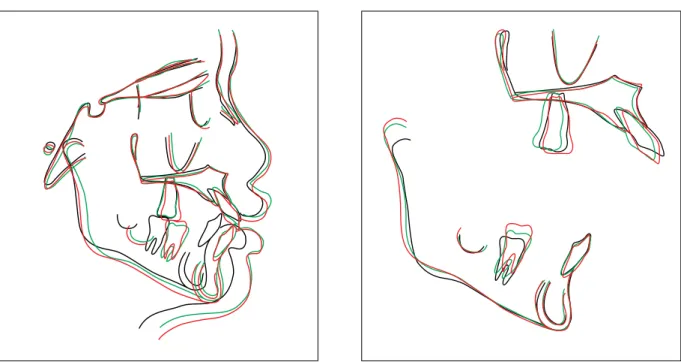
was unchanged when compared to pretreatment despite the lower facial height increased by 1.8%. From the superimposition, maxillary growth was restrained during headgear Twin Block treatment, and resumed to a forward and downward pattern during the phase II treatment (Table 1).
Maxillary cephalometric tracings superimpo-sition (Fig 10) indicates that there has been ret-roclination of upper incisors. Eruption of upper molars was restrained during the headgear/Twin Block treatment, but they were extruded during phase II with fixed appliance treatment.
Mandibular cephalometric tracings superim-position (Fig 10) indicates that there has been very slight lower incisor proclination and main-tenance of vertical position, and lower molars ex-truded and moved mesially during headgear/Twin Block treatment. This explained the development of mild crowding in the lower anterior region af-ter headgear/Twin Block treatment. During the fixed appliance phase, there was continued extru-sion of lower molars to level the curve of Spee. The lower incisors were retroclined as a result of rounding off the molar and premolar area. Up-ward and backUp-ward condylar growth was obvious during the overall treatment (Figs 8, 9 and 10).
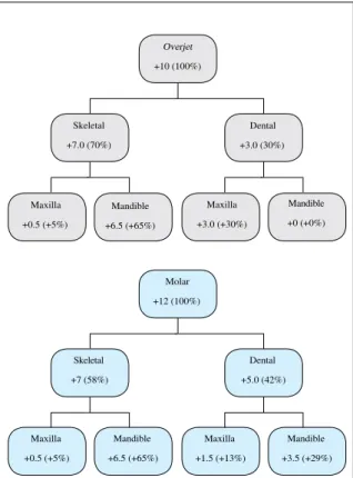
Sagittal-Occlusion Analysis
From the Sagittal-Occlusion Analysis (SO-analy-sis)11 immediately after 12 months of headgear/Twin
FIGURE 5 - Post-treatment intraoral views.
FIGURE 6 - Comparison of pretreatment and post-treatment facial profile. FIGURE 7 - Comparison of pretreatment and post-treatment smile.
a maxillary restraint (ss/RLp’ - ss/RLp) of 0.5 mm. Maxillary growth restraint was not maintained after phase I, as sagittal maxillary growth caught up by 1 mm during phase II of treatment. For the mandible, pg-RLp continued to increase although at a much slower rate during phase II, and relapse was minimal according to the analysis.
From the SO-analysis during the headgear/Twin
FIGURE 8 - Post-Twin Block lateral cephalometric and panoramic radiographs.
FIGURE 9 - Post-treatment lateral cephalometric and panoramic radiographs.
discussion treatment rationale
In many respects the patient was an ideal candidate for functional appliance treatment. She presented with a mild to moderate Class II skeletal discrepancy, average vertical dimensions, mild crowding and proclined upper incisors, with the lower incisors presenting average inclination. The functional appliance was used to correct the skeletal discrepancy, and correct incisor and buc-cal segment relationships to Class I. As a result of the potential skeletal and dentoalveolar changes produced by the functional appliance, a more fa-vorable soft tissue environment was created with elimination of the lip trap and the lower lip acting labially on the upper incisors.
Orthodontic camouflage by extraction of up-per premolars could have been another treatment option but was not considered for a number of reasons. The patient and her mother were keen to avoid extractions due to concerns about removing healthy teeth. Extraction of upper premolar teeth might be able to retract the upper protrusive lip and improve facial convexity to certain extent, but would not improve mandibular retrogna-thism. Extraction approach also required careful anchorage management, which in her case, might involve headgear or bone anchorage, and thus car-ry other potential problems such as incompliance and patient discomfort.
Removable functional appliance was used in this patient because her premolars were not fully
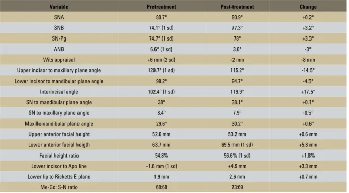
TABLE 1 - Cephalometric assessment.
Note that the norm values were based on a sample of 12 year old children. Note: All values above have been adjusted to the SN-Frankfort Horizontal plane. Sources of normal values for chinese:
Cooke MS and Wei SHY(1988) Eur J Orthod. 10(3):264-72
Cooke MS (1986) Ph. D Thesis. the University of Hong Kong. Cephalometric analyses based on natural head posture of children in Hong Kong (1 sd) = One standard deviation.
(2 sd) = Two standard deviation.
Variable Pretreatment Post-treatment Change
SNA 80.7° 80.9° +0.2°
SNB 74.1° (1 sd) 77.3° +3.2°
SN-Pg 74.7° (1 sd) 78° +3.3°
ANB 6.6° (1 sd) 3.6° -3°
Wits appraisal +6 mm (2 sd) -2 mm -8 mm
Upper incisor to maxillary plane angle 129.7° (1 sd) 115.2° -14.5°
Lower incisor to mandibular plane angle 98.2° 94.7° -4.5°
Interincisal angle 102.4° (1 sd) 119.9° +17.5°
SN to mandibular plane angle 38° 38.1° +0.1°
SN to maxillary plane angle 8,4° 7.9° -0,5°
Maxillomandibular plane angle 29.6° 30.2° +0.6°
Upper anterior facial height 52.6 mm 53.2 mm +0.6 mm
Lower anterior facial heigth 63.7 mm 69.5 mm (1 sd) +5.8 mm
Facial height ratio 54.8% 56.6% (1 sd) +1.8%
Lower incisor to Apo line +1.6 mm (1 sd) +4.9 mm +3.3 mm
Lower lip to Ricketts E plane 1.9 mm 2.6 mm +0.7 mm
Overjet +10 (100%) Maxilla +3.0 (+30%) Mandible +0 (+0%) Skeletal +7.0 (70%) Dental +3.0 (30%) Maxilla +0.5 (+5%) Mandible +6.5 (+65%) Maxilla +1.5 (+13%) Mandible +3.5 (+29%) Maxilla +0.5 (+5%) Mandible +6.5 (+65%) Molar +12 (100%) Skeletal +7 (58%) Dental +5.0 (42%)
FIGURE 11 - Sagittal Occlusion Analysis.
erupted yet when the treatment started. Other-wise, Herbst appliance would be another option because we could minimize any potential
com-pliance problem12 and maximize treatment
effi-ciency. The success of the treatment was largely dependent on the patient’s compliance which was very well accomplished in this case. Mandibular advancement every six months in a stepwise man-ner has been proved more effective in
stimulat-ing condylar growth13 and improving mandibular
prognathism.14 The use of high-pull headgear
dur-ing the functional appliance stage helped restrain-ing the maxillary growth and also prevented clock-wise rotation of the maxilla which might cause backward and downward rotation of the mandible thus jeopardizing the treatment effects.15
Lower incisor proclination is one of the major side effects of functional appliances. In this case, the proclination of the lower inci-sors was minimized to only 0.6°. This common side effect in using functional appliance was avoided by trimming away the acrylic lingual to the lower anterior teeth. The fact that pa-tient had smaller lower incisors and also the pressure from the lower lip might also have helped maintaining the angulations of the lower incisors.
two phase vs. one phase treatment
The total treatment time was 26 months in-cluding 12 months of growth modification. The main aim of the two phase orthodontic treat-ment was to enhance the patient’s potential for favourable mandibular growth and improve her skeletal and soft tissue profile by growth modifi-cation. It was also planned to avoid over-retrac-tion of her upper incisors with respect to the incisor angulations, future nasal growth and her existing smile line. The decision to start treat-ment with a first phase of functional appliance was proved to be appropriate.
The patient had her menarche in the sixth month during the Twin Block phase treatment, in-dicating that she was around the peak of her
puber-tal growth spurt,16 which was corresponding to her
CVM stage.17 The treatment effect by the functional
appliance could be maximized during this period.18
The lip competency improved although was not fully corrected due to unfavourable muscle tone. There was increased buccal overjet as a result of tooth size discrepancy between upper and lower arches. It was decided not to change the size and shape of the teeth by stripping or building-up with resin because of esthetic concerns. There was mild generalized gingival swelling at the interdental papilla area, despite fair oral hygiene had been maintained by the patient during treatment. The gingival swelling resolved greatly one week after debonding.
Long term prognosis
The mandibular intercanine width has been maintained at its original while the upper was expanded during the functional appliance phase. The prognosis for stability is good pro-vided the patient’s growth pattern is favour-able and the mandible will not rotate down-ward and backdown-ward. Good buccal interdigita-tion and incisal contact also helped to stabilize the occlusal stability, as well as the fixed and removable retainers.
concLusion
1. Mitchell L, Carter NE, Doubleday B. An introduction to Orthodontics. 2nd ed. Oxford: Oxford University Press; 2001.
2. Profit WR, Fields HW Jr, Sarver DM. Contemporary
Orthodontics. 4th ed. Missouri: Elsevier; 2007.
3. Cochrane SM, Cunningham SJ, Hunt NP. Perceptions of facial appearance by orthodontists and the general public. J Clin Orthod. 1997 Mar;31(3):164-8.
4. Albino JE. A psychologist’s guide to oral diseases and disorders and their treatments. Prof Psychol Res Pract. 2002;33(2):176-82.
5. Hägg U, Du X, Rabie AB. Initial and late treatment effects of headgear-Herbst appliance with mandibular step-by-step advancement. Am J Orthod Dentofacial Orthop. 2002;122(5):477-85.
6. Purkayastha SK, Rabie AB, Wong R. Treatment of skeletal Class II malocclusion in adults: stepwise vs single-step advancement with the Herbst appliance. World J Orthod. 2008 Fall;9(3):233-43.
7. Rabie AB, She TT, Hägg U. Functional appliance therapy accelerates and enhances condylar growth. Am J Orthod Dentofacial Orthop. 2003 Jan;123(1):40-8.
8. Ta TA, Ling JY, Hägg U. Tooth-size discrepancies among different occlusion groups of southern Chinese children. Am J Orthod Dentofacial Orthop. 2001;120(5):556-8.
9. Cooke MS, Wei SHY. Cephalometric standards for the southern Chinese. Eur J Orthod. 1988;10:264-72.
10. Baccetti T, Franchi L, James A, McNamara JA Jr. The cervical vertebral maturation (CVM) method for assessment of optimal treatment timing in dentofacial orthopaedic. Semin Orthod. 2004;11:119-29.
references
11. Pancherz H. The mechanism of Class II correction in Herbst appliance treatment. A cephalometric investigation. Am J Orthod. 1982;82(2):104-13.
12. O’Brien K, Wright J, Conboy F, Sanjie Y, Mandall N, Chadwick S, et al. Effectiveness of treatment for Class II malocclusion with the Herbst or Twin-Block appliances: a randomized, controlled trial. Am J Orthod Dentofacial Orthop. 2003;124(2):128-37.
13. Bakr A, Rabie AB, Al-Kalaly A. Does the degree of advancement during functional appliance therapy matter? Eur J Orthod. 2008;30(3):274-82.
14. Hägg U, Rabie AB, Bendeus M, Wong RW, Wey MC, Du X, et al. Condylar growth and mandibular positioning with stepwise vs maximum advancement. Am J Orthod Dentofacial Orthop. 2008 Oct;134(4):525-36.
15. Du X, Hägg U, Rabie AB. Effects of headgear Herbst and mandibular step-by-step advancement versus conventional Herbst appliance and maximal jumping of the mandible. Eur J Orthod. 2002;24(2):167-74.
16. Hägg U, Taranger J. Maturation indicators and the pubertal growth spurt. Am J Orthod. 1982;82(4):299-309.
17. Al Khal HA, Wong RW, Rabie AB. Elimination of hand-wrist radiographs for maturity assessment in children needing orthodontic therapy. Skeletal Radiol. 2008;37(3):195-200. Epub 2007 Oct 3.
18. Hägg U, Pancherz H. Dentofacial orthopaedics in relation to chronological age, growth period and skeletal development. An analysis of 72 male patients with Class II division 1 malocclusion treated with the Herbst appliance. Eur J Orthod. 1988;10(1):169-76.
Submitted: August 26, 2010
Revised and accepted: December 29, 2010
contact address Larry Ching Fan Li