Arq. NeuroPsiquiatr. vol.68 número5
Texto
Imagem
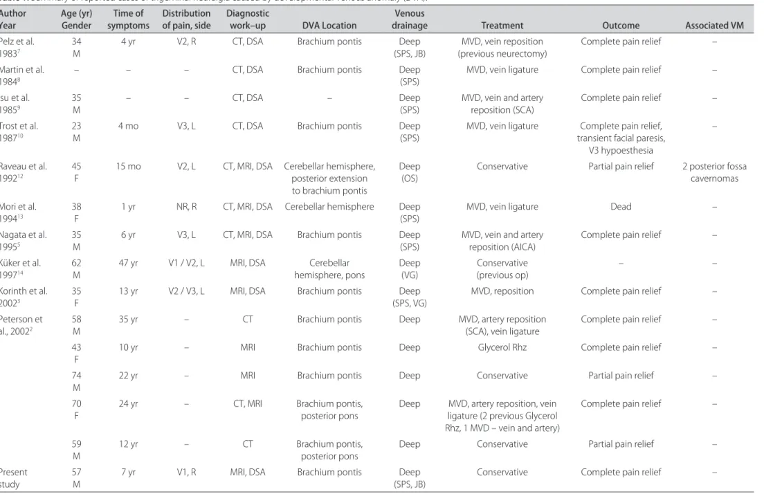
![Fig 1. Sequential axial 3D CISS (three-dimensional constructive interference in steady state) MRI (1 mm width) reveals a large right transparenchymal vein (white arrow) and caput medusae (white ar-row heads) [A] as well as the course of the trigeminal ne](https://thumb-eu.123doks.com/thumbv2/123dok_br/15432808.595101/1.955.272.851.759.1051/sequential-dimensional-constructive-interference-reveals-transparenchymal-medusae-trigeminal.webp)

Documentos relacionados
Computed tomography scan and magnetic resonance imaging revealed a pituitary tumor invading the left sphenoidal and cavernous sinuses.. Laboratory data excluded
A computed tomography (CT) scan ( Figure 1 ) and magnetic resonance imaging (MRI) scan of the chest ( Figure 2 ) revealed the presence of a nodular lesion in the right pulmonary
This finding was confirmed by magnetic resonance angiography of the cervical vessels, and axial computed tomography angiography showed agenesis of the right carotid canal..
Amongst the available imaging methods (Doppler ultrasonography, computed tomography, magnetic resonance imaging and digital subtraction angiography), angiography is considered the
The diagnosis of mature cystic ter- atoma by computed tomography (CT) and magnetic resonance imaging (MRI) is fairly straightforward as such modalities are more fat sensitive.. At
Inclusion criteria for selecting articles were: » Studies based on magnetic resonance imag- ing (MRI), computed tomography (CT) and/ or volumetric cone-beam tomography, which assessed
However, the appearance of this cyst on cone beam computed tomography (CBCT) and magnetic resonance imaging (MRI) have received relatively little attention.. CBCT and
(C) Control MRI six months after surgery showing recurrent tumor in the left cerebellar hemisphere invading the superior cerebellar peduncle and brainstem.... Arq
