High frequency of silent brain infarcts associated with
cognitive deficits in an economically disadvantaged
population
Paula Squarzoni,IJaqueline H. Tamashiro-Duran,IFabio L.S. Duran,IClaudia C. Leite,IIMauricio Wajngarten,III Marcia Scazufca,IPaulo R. Menezes,IVPaulo A. Lotufo,VTania C.T.F. Alves,IGeraldo F. BusattoI,*
IDepartamento de Psiquiatria, Instituto de Psiquiatria (IPQ), Hospital das Clinicas HCFMUSP, Faculdade de Medicina, Universidade de Sao Paulo, Sao Paulo,
SP, BR.IIDepartamento de Radiologia e Oncologia, Faculdade Medicina FMUSP, Universidade de Sao Paulo, Sao Paulo, SP, BR.IIIInstituto do Coracao (InCor), Hospital das Clinicas HCFMUSP, Faculdade de Medicina, Universidade de Sao Paulo, Sao Paulo, SP, BR.IVDepartamento de Medicina Preventiva, Faculdade de Medicina FMUSP, Universidade of Sao Paulo, Sao Paulo, SP, BR.VCentro de Pesquisa Clinica e Epidemiologica, Faculdade de Medicina FMUSP, Universidade de Sao Paulo, Sao Paulo, SP, BR.
OBJECTIVE:Using magnetic resonance imaging, we aimed to assess the presence of silent brain vascular lesions in a sample of apparently healthy elderly individuals who were recruited from an economically disadvantaged urban region (Sa˜o Paulo, Brazil). We also wished to investigate whether the findings were associated with worse cognitive performance.
METHODS:A sample of 250 elderly subjects (66-75 years) without dementia or neuropsychiatric disorders were recruited from predefined census sectors of an economically disadvantaged area of Sao Paulo and received structural magnetic resonance imaging scans and cognitive testing. A high proportion of individuals had very low levels of education (4 years or less, n=185; 21 with no formal education).
RESULTS:The prevalence of at least one silent vascular-related cortical or subcortical lesion was 22.8% (95% confidence interval, 17.7–28.5), and the basal ganglia was the most frequently affected site (63.14% of cases). The subgroup with brain infarcts presented significantly lower levels of education than the subgroup with no brain lesions as well as significantly worse current performance in cognitive test domains, including memory and attention (po0.002).
CONCLUSIONS:Silent brain infarcts were present at a substantially high frequency in our elderly sample from an economically disadvantaged urban region and were significantly more prevalent in subjects with lower levels of education. Covert cerebrovascular disease significantly contributes to cognitive deficits, and in the absence of magnetic resonance imaging data, this cognitive impairment may be considered simply related to ageing. Emphatic attention should be paid to potentially deleterious effects of vascular brain lesions in poorly educated elderly individuals from economically disadvantaged environments.
KEYWORDS: Framingham Coronary Heart Disease Risk; Ageing; Educational Level; Cognition; Silent Brain Infarction.
Squarzoni P, Tamashiro-Duran JH, Duran FL, Leite CC, Wajngarten M, Scazufca M, et al. High frequency of silent brain infarcts associated with cognitive deficits in an economically disadvantaged population. Clinics. 2017;72(8):474-480
Received for publication onFebruary 13, 2017;First review completed onMarch 14, 2017;Accepted for publication onApril 10, 2017
*Corresponding author. E-mail: [email protected]
’ INTRODUCTION
Brain infarcts and lacunae are common in elderly popula-tions and may be easily detected by high-resolution magnetic resonance imaging (MRI). These lesions are typically asso-ciated with poor cognitive function and a loss of the ability to
perform daily living activities; they also predict a future risk of dementia (1). According to many recent large-scale MRI stu-dies of elderly populations, a considerable proportion of these brain infarcts remains undetected due to the absence of overt clinical impairments (2-4). In contrast to the traditional view that cardiovascular diseases result from a combination of gen-etic, lifestyle and physiological risk factors, a recent trend has incorporated the social determinants of health as an addi-tional, critical dimension of cardiovascular risk (5). Education is the most frequently used indicator of socioeconomic position in the United States and was the predictor with the highest significance in determining cardiovascular disease outcomes (5). Elderly subjects with very low levels of education and dis-advantageous socioeconomic conditions, which are common DOI:10.6061/clinics/2017(08)04
Copyright&2017CLINICS–This is an Open Access article distributed under the terms of the Creative Commons License (http://creativecommons.org/licenses/by/ 4.0/) which permits unrestricted use, distribution, and reproduction in any medium or format, provided the original work is properly cited.
in many low- and middle-income countries, present increa-sed rates of cardiovascular risk factors, stroke and dementia (6-9). However, silent brain infarcts have not been specifically investigated in disadvantaged populations with very low levels of education using MRI (4). Studies evaluating the profile of cognitive deficits associated with silent cerebro-vascular lesions in populations with low levels of education are also relevant because lower education influences cog-nitive reserves to a certain degree and may lead to greater cognitive deficits associated with brain lesions, whereas indi-viduals with higher levels of education are expected to exhi-bit better sustained cognitive performance when suffering the same degree of brain damage (10,11).
In the present community-based study, we acquired struc-tural MRI scans in a population-based sample of 250 elderly (66 to 75 years old) subjects without dementia or other neuro-psychiatric disorders, including a high proportion of indi-viduals with very low levels of education (4 years or less). This sample was recruited from a circumscribed, economically disadvantaged catchment area of Sao Paulo, Brazil. We determined the frequency of silent brain infarcts in this sample population and investigated the profile of cognitive deficits associated with those brain lesions. We predicted that the frequency of silent brain infarcts would be substantially high and more prevalent in subjects with fewer years of formal education.
’ METHODS
The study received approval from the local Committee for Ethics and Research of the Faculty of Medicine, University of Sao Paulo (#0450/05). Written consent was obtained from all subjects.
Participants were selected from a community-based sam-ple of elderly individuals recruited for the Sao Paulo Ageing and Health (SPAH) study (12), an epidemiological investiga-tion aimed at determining the prevalence of dementia, other mental disorders, and their risk factors (12). Residents aged 65 or older from predefined census sectors of an econo-mically disadvantaged area of Sao Paulo were recruited to undergo a cognitive evaluation using the protocol developed by the 10/66 Dementia Research Group (12-14). This pro-tocol included a structured neurological assessment, a struc-tured cardiological evaluation, the Geriatric Mental State (GMS) (a standardized psychiatric interview), and the Com-munity Screening Instrument for Dementia (CSI-D) (12). The CSI-D consists of a 32-item cognitive test administered to the participant (approximately 20 minutes) and a 26-item informant interview that includes items inquiring about the participant’s daily functioning and general health (approxi-mately 15 minutes) (15). Three summary scores are generated from the CSI-D, including the COGSCORE, an item-weigh-ted summary score from the participant’s 32-item cognitive test (seven-item object denomination, four-item object defi-nition, two verbal category fluency tasks, word repetition, identification of a famous person, temporal and spatial orientation, three orders, three-word recall, six-chunk story recall, two drawings of intersecting circles and pentagons) that also incorporates the Consortium to Establish a Registry for Alzheimer’s Disease (CERAD) animal naming verbal fluency task and the modified CERAD 10 word-list learn-ing task with delayed recall (16); the informant score (RELSCORE), which is an unweighted total score from the informant interview; and the discriminant function score
(DFSCORE), which is a weighted score combining COG-SCORE and RELCOG-SCORE (17). The SPAH composite cognitive algorithm was used to exclude subjects with dementia.
Subjects also underwent a short (approximately 10 minutes) evaluation using the Short Cognitive Performance Test (SKT), which was validated in Brazil by Flaks et al. (18), to briefly document the cognitive performance of subjects on the day of MRI scanning. The SKT permits a rapid evaluation of overall cognitive performance and performance in specific domains, such as memory, attention and automatic inhibition, with higher scores indicating a more severe cognitive impairment (19). Thus, the measures of cognitive performance used in the present investigation included total SKT scores, SKT-based attention and memory subscores, total COGSCORE, and category verbal fluency scores. In addition, IQ estimates were obtained for all subjects who underwent MRI scanning with the Wechsler Abbreviated Scale of Intelligence (WASI) (20).
All subjects were also ranked according to their cardio-vascular risk using the 10-year Framingham Risk Score for Coronary Heart Disease Risk (FRS), which is a composite index comprising five clinical factors (age, blood pressure, diabetes mellitus, smoking status, and cholesterol levels) (21,22). Subjects were classified into three subgroups according to their degree of cardiovascular risk (10-year risk): low-risk (o10%), medium-risk (10 to 20%), and high-risk (420%) (22).
From the initial SPAH databank (n=2,072), we excluded all subjects with a diagnosis of dementia (n=105), subjects aged greater than 75 years at the time of recruitment for MRI scanning (n=996) and subjects with a history of major neuro-logical disorders (such as epilepsy and Parkinson’s disease) or lifetime diagnosis of major depressive disorder according to the International Statistical Classification-10th revision cri-teria (ICD-10) (n=52). Eight hundred twelve potentially eligible individuals were identified after excluding indivi-duals with missing clinical data (n=107). The remaining potentially eligible subjects were contacted by telephone to assess the presence of contra-indications for MRI scanning and to exclude individuals who had a previous history of severe head trauma. We failed to contact 103 subjects. Of the subjects who were contacted, we excluded 206 subjects (132 females and 74 males) who fulfilled the above exclusion criteria; thus, 503 subjects were invited to undergo the brain imaging session. After excluding illiterate subjects (i.e., indi-viduals who could not write or read a simple message/letter) and excluding additional subjects due to budget constraints, only 306 of the 503 available individuals were invited to undergo the MRI scanning session. Fifty-two of the invi-ted subjects refused to participate in the study, resulting in 254 dementia-free elderly subjects between 66–75 years old
(female/male [139/115]) who underwent MRI scanning. We excluded illiterate subjects because the SKT-based cog-nitive performance indices that we acquired on the day of MRI scanning may have limited the validity of the results obtained from these individuals (18).
No significant differences in age (t-test=1.354, p=0.176),
prevalence of hypertension (Fisheŕs exact test, p=0.480) or
prevalence of diabetes (Fisheŕs exact test, p=0.089) were
observed between the individuals who underwent MRI scanning (n=254) and the eligible individuals who were not included in the study (n=558). The gender distribution was significantly different between the two groups (Fisher’s exact test,p=0.003), with a larger proportion of males in the group
that underwent MRI scanning than the potentially eligible individuals who were not included in the study (45.3%versus
34.2%). The studied group also displayed a larger number of years of formal education (3.49±3.28 years) then the individuals who were not included in the investigation (2.15 ±2.74 years) (t-test=-6.038,po0.01).
MRI datasets were acquired using a 1.5-T General Electric Signa LX CVi scanner (Milwaukee, WI, USA) with the following acquisition protocol: a) a dual-spin echo sequence of 120 transaxial slices across the entire brain (axial PD/T2); b) a T2-weighted fast spin-echo transaxial sequence (88 slices); and c) a 3D Spoiled Gradient Recalled Acquisition sequence of 124 slices with a repetition time (TR)/echo time (TE) of 21.7/5.2 msec, a flip angle of 20 degrees, 220-mm field of view, 1.5-mm slice thickness, 1 measurement, and a 256192 matrix. All MRI scans were examined by experi-enced radiologists who were unaware of the study aims, and they identified the presence, number and location of silent brain infarcts. Infarcts were detected as low-signal-intensity lesions on the spoiled gradient echo (SPGR) sequence and hyperintense lesions on the T2-weighted images. Vascular lesions that were 3 to 15 mm in diameter were classified as lacunae (24,25)
The Statistical Package for Social Sciences (SPSS) for Windows (17.0) was used for the statistical analyses. Cog-nitive data obtained from elderly individuals with and without silent brain infarcts were compared using ANOVA (statistical significance set at po0.05). For comparisons of each individual group, subjects were automatically excluded when values for the specific variable under evaluation were missing. For the overall sample of 254 elderly subjects, data for FRS scores (n=6), IQ (n=1), and COGSCORE (n=10) were missing.
’ RESULTS
Frequency of silent brain infarcts
Four of the 254 subjects assessed with MRI were excluded from the analyses due to artefacts during MRI scanning that
prevented the accurate detection of gross brain lesions, resulting in a total of 250 individuals who were selected for the analysis. Fifty-seven individuals had at least one silent vascular-related lesion, yielding a prevalence of 22.8% (95% confidence interval: 17.7 to 28.5). Details for these lesions and their brain locations are provided in Table 1. An additional 4 subjects presented with meningioma and 2 had vascular malformations.
When we restricted the MRI data analysis to the sub-sample of individuals with very low levels of education (4 years or less, n=185), 25.41% (n=47) were presented at least one silent vascular-related lesion.
Demographic and clinical differences between subjects with and without silent vascular-related brain lesions
After excluding individuals with meningioma or vascular malformations, the subgroup with silent vascular lesions (n=57) was older (po0.001) and had lower levels of edu-cation than the subgroup with no brain infarcts (p=0.038)
(Table 2). No significant differences in the gender distri-bution, current socioeconomic status and estimated IQ were observed between individuals with and without silent infarcts (Table 2). The proportion of individuals with a high 10-year cardiovascular risk (FRS420%) was greater in the
subgroup with silent vascular lesions in the brain than that in the subgroup without silent infarcts (Table 2).
When we restricted the analysis to the subsample of individuals with very low levels of education (4 years or less, n=185; 21 with no formal education), the subgroup with silent brain infarcts (n=47) was older than individuals with-out silent brain infarcts (72.43±3.41vs70.60±2.68, t=-3.749,
po0.001), and a trend towards a greater proportion of indi-viduals with a high cardiovascular risk (55.3% vs 39.1%, p=0.079) was observed. No significant differences in gender
distribution, current socioeconomic status and estimated IQ were observed between individuals in the subsample with very low levels of education who presented with or without silent infarcts.
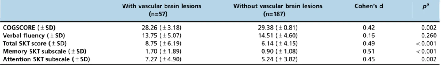
Comparison of cognitive performance between subjects with and without silent vascular-related brain lesions
The subgroup with silent vascular lesions performed sig-nificantly worse on all cognitive measures, except for the verbal fluency category (Table 3). Cohen’s d values were
Table 1-Location of silent vascular brain lesions (n=57).
Brain location Number of subjects %
Basal ganglia/thalamus 26 45.61
White matter 9 15.79
Basal ganglia/thalamus/cerebellum/brainstem 4 7.02
Cerebellum 4 7.02
Basal ganglia/thalamus/cerebellum/brainstem/frontal cortex 1 1.75
Basal ganglia/thalamus/white matter 1 1.75
Cerebellum/brainstem 3 5.26
Basal ganglia 2 3.51
Parietal cortex 2 3.51
Basal ganglia/thalamus/white matter/cerebellum/brain stem 1 1.75
Basal ganglia/thalamus/white matter/cerebellum/brain stem/occipital cortex 1 1.75
Frontal cortex 1 1.75
Occipital cortex 1 1.75
greater than 0.40 for all cognitive measures, except for the verbal fluency category (Table 3). Total raw SKT scores were not able to be transformed into norm values for 11 indi-viduals with low estimated IQ values.
Based on the between-group differences in mean current age and level of education (Table 2), we repeated the anal-yses and used age and years of education as confounding covariates. The subgroup with brain lesions again performed significantly worse, based on their COGSCORE performances (p=0.014), total SKT scores (p=0.015), SKT-based memory
subscores (p=0.004) and attention subscores (p=0.038).
Finally, we counted the number of individuals with a COGSCORE performance that was 1.5 standard deviations below the mean performance of all subjects between 66 and 75 years of age from the original SPAH sample. This analysis generated the criterion for mild cognitive impairment (MCI) based on the actual mean cognitive profile of the elderly population from which the present sample was drawn (26). Significantly more individuals (n=5) with silent vascular lesions who fulfilled the criterion (8.77%), as only 4 indi-viduals with MCI were identified in the subgroup without such brain lesions (2.14%) (Fisher’s exact test,p=0.034). Four
individuals who fulfilled the MCI criterion in the subgroup with silent brain infarcts (80%) and 4 subjects with MCI in the subgroup with no vascular brain lesions (100%) had less than four years of formal education.
’ DISCUSSION
This study investigated the presence of silent brain infarcts detected by MRI scanning and their associated cognitive deficits in a relatively large sample of apparently healthy individuals aged 66-75 years who were recruited from an economically disadvantaged elderly population in Sao Paulo, Brazil. The frequency of silent brain infarcts in our
sample was considerably high and represented approxi-mately 23% of subjects from 66-75 years old. The frequency of silent brain infarcts reported in this study is at the higher end of the range of prevalence estimates reported in MRI studies conducted on populations from high-income coun-tries (2,4). Moreover, our sample was restricted to indivi-duals aged 65-75 years, whereas many previous studies included older subjects who may have a higher frequency of these lesions (27). Prevalence estimates in community-based samples with a mean age that is comparable to the ages included in our study have ranged from 8% to 20% (2,4,28). Thus, according to our study, the prevalence of silent brain vascular lesions in elderly subjects recruited in an economic-ally disadvantaged urban centre may be substantieconomic-ally high.
The FRS has been shown to underestimate cardiovascular disease risk in socioeconomically disadvantaged individuals (29). Nevertheless, we detected a significant, direct relation-ship between silent brain infarcts and higher FRS in our economically disadvantaged cohort, which replicates find-ings from studies conducted in higher-income environments (28,30).
As predicted, the presence of silent brain infarcts was associated with significantly worse cognitive performance. Significant deficits were detected in memory and other cognitive domains, reinforcing the view that covert vascular lesions in elderly populations are related to deficits in several cognitive domains (31,32). Cohen’s d values were moderate (approximately 0.5) for the total and memory SKT scores and greater than 0.40 for the COGSCORE and the SKT attention subscore. These effect sizes were not modest, suggesting that cognitive deficits associated with silent brain infarcts in elderly subjects recruited in an economically disadvantaged urban centre should not be judged as subtle, as suggested by other researchers examining elderly subjects in higher-income environments (33,34).
Table 2-Demographics, estimated intelligence and medical characteristics of the participants in the subgroups with and without silent vascular lesions.
With vascular brain lesions (n=57)
Without vascular brain lesions (n=187)
p
Men 25 (43.9%) 88 (47.1%) 0.672a
Mean years of education (±SD) 3.46 (±2.66) 4.54 (±3.63) 0.038a
Mean age (±SD) in years 72.11 (±3.36) 70.51 (±2.57) o0.001a
Hypertension (%) 39 (68.4%) 114 (61.0%) 0.308b
Diabetes (%) 19 (33.3%) 49 (26.3%) 0.304b
High cardiovascular risk (%) * 30 (52.6%) 67 (35.8%) 0.034b
Estimated IQ (±SD) 75.84 (±11.33) 76.92 (±10.19) 0.497a
SD:standard deviation; IQ = estimated coefficient of intelligence.
* As determined by the Framingham Coronary Heart Disease Risk score (FRS). Subjects were classified as low-risk (o10%), medium-risk (10 to 20%), and high-risk (420%).
aUnpaired t-tests;bChi-square tests (Pearson).
Table 3-Comparisons of the performance of individuals with and without silent vascular brain lesions on cognitive tests.
With vascular brain lesions (n=57)
Without vascular brain lesions (n=187)
Cohen’s d pa
COGSCORE (±SD) 28.26 (±3.18) 29.38 (±0.81) 0.42 0.002
Verbal fluency (±SD) 13.75 (±5.07) 14.51 (±4.60) 0.16 0.260
Total SKT score (±SD) 8.75 (±6.19) 6.14 (±4.15) 0.49 o0.001
Memory SKT subscale (±SD) 1.70 (±1.89) 0.90 (±1.08) 0.51 o0.001 Attention SKT subscale (±SD) 7.27 (±4.90) 5.24 (±3.82) 0.45 0.002
SD:standard deviation.
Consistent with previous investigations conducted on other populations as well as populations in Brazil, we detec-ted more vascular brain lesions in less educadetec-ted individuals (35,36). The overall sample investigated in the present study predominantly had low levels of education, with a large proportion of individuals (75.82%) having less than four years of formal education. The frequency of silent vascular-related brain infarcts surpassed the level of 25% in our sample when the inspection of MRI data was restricted to individuals with very low levels of education (4 years or less). This frequency of silent brain infarcts is greater than the average values reported in a number of studies conducted on populations with higher mean levels of education (2,4,28). Therefore, the prevalence of silent brain infarcts in indivi-duals with very low levels of education detected in our study may be disproportionally large, which is consistent with the known unfavourable association between lower levels of education and cardiovascular risk factors (37). Since poten-tially eligible subjects who did not undergo MRI scanning had a significantly lower mean number of years of formal education, our prevalence rates of silent brain infarcts may have been even higher if we had examined the illiterate individuals who were not included in the study. Notably, the majority of individuals who fulfilled our MCI criterion presented silent brain infarcts and had very low levels of education (less than four years). This outcome is consistent with the hypothesis that reduced cognitive reserves increase the risk of clinically significant cognitive deficits in the pre-sence of brain damage (10).
The preferential location of silent brain infarcts in the basal ganglia in our study is highly consistent with the findings of previous studies (3) and supports the hypothesis that this brain region exhibits greater vulnerability to cerebrovascular-related damage and the associated cognitive deficits in mul-tiple domains (34). Based on recent tractography MRI studies using diffusion tension imaging, even small silent vascular basal ganglia lesions may affect the function of large-scale cortical white matter networks, providing a potential expla-nation for the emergence of the association between cogni-tive deficits in multiple domains with silent basal ganglia infarcts (32). Moreover, resting-state functional MRI investi-gations have revealed aberrant patterns of functional con-nectivity in both intra- and between brain networks in patients with vascular basal ganglia lesions, which are directly propor-tional to the degree of cognitive impairment (31). Subgroups with and without silent brain infarcts in our study did not exhibit differences in category fluency scores. This lack of dif-ference is not likely to be due to compensatory brain mecha-nisms that are recruited to sustain fluency performance in subjects who presented cerebral lesions, because other studies have reported verbal fluency deficits in elderly subjects with silent brain infarcts (33,38). Category verbal fluency scores were relatively modest in both elderly subgroups (possibly due to the influence of the overall low level of education in the sample) (39). The scores obtained in our sample are lower than the scores reported with the same version of the task applied to populations of elderly individuals without dementia who were examined in Latin-American countries, India and China (40). Therefore, floor effects may have prevented us from detecting subgroup differences in this task.
Moreover, the subgroup with silent vascular-related brain lesions in our study was older than the subgroup without these lesions. However, this age difference was not likely to have weakened our reported association between significantly
worse cognitive performance and silent vascular brain lesions for two reasons. First, both subgroups had mean current ages of greater 70 years, and the between-group difference in mean age values was less than 2 years. Second, measures of cogni-tive performance remained significantly different between groups when we repeated the statistical analyses by entering the current age as a confounding covariate.
We must acknowledge that our investigation was restric-ted to documenting the presence of sizeable brain infarcts through a visual inspection of morphological scans acqui-red with a 1.5-T scanner. We did not assess the presence of changes of potentially lesser cerebrovascular disease severity in the brain, including white matter hyperintensities (which are thought to reflect microvascular injuries in elderly popu-lations) (41-43) or microinfarcts (which may not be clearly detectable using a 1.5-T MRI scanner) (44,45). Therefore, the prevalence rates of silent cerebrovascular disease markers may have been even larger in our elderly population if we had been able to assess the full range of these markers. Another limitation is that not all eligible individuals were evaluated with MRI due to the exclusion of illiterate subjects and budget constraints. Thus, the figures reported in this study cannot be considered true population-based preva-lence estimates of silent brain infarcts.
Our results obtained from a sample of predominantly poorly educated elderly individuals from an economically disadvantaged urban region indicate that covert cerebrovas-cular disease is frequent. As suggested by previous findings, covert vascular brain damage has meaningful consequences and significantly contributes to deficits in cognitive perfor-mance that may be considered related to aging in the absence of MRI data (4). Our findings reinforce the need to carefully examine the potential deleterious effects of vascular brain lesions in poorly educated elderly individuals from econom-ically disadvantaged environments. Future longitudinal studies are needed to investigate the long-term prognosis and rates of conversion to dementia in these individuals.
’ ACKNOWLEDGMENTS
This work was supported by the São Paulo Research Foundation (FAPESP), reference numbers 2013/03231-3, 2012/50239-6 and 04/15336-5, the Well-come Trust, UK (GR066133MA) and the National Council for Scientific and Technological Development (CNPq).
’ SOURCES OF FUNDING
This research was funded by the Wellcome Trust, UK (GR066133MA) and by FAPESP (2004/15336-5, 2012/50329-6 and 2013/03231-3), Brazil. PS was supported by FAPESP-Brazil (2013/03231-3). GFB, MS, PRM, and TCTFA were sup-ported by CNPQ-Brazil.
’ AUTHOR CONTRIBUTIONS
Busatto GF conceived the study and participated in study design. Squarzoni P analyzed and interpreted the data and drafted and revised the manuscript. Tamashiro-Duran JH, Duran FL, Leite CC, Wajngarten M, Scazufca M, Menezes PR, Lotufo PA and Alves TC analyzed and interpreted the data and revised the manuscript. All authors read and approved thefinal version of the manuscript.
’ REFERENCES
Cardiovascular Health Study. Stroke. 1998;29(2):388-98, http://dx.doi. org/10.1161/01.STR.29.2.388.
2. Vermeer SE, Longstreth WT Jr, Koudstaal PJ. Silent brain infarcts: a sys-tematic review. Lancet Neurol. 2007;6(7):611-9, http://dx.doi.org/ 10.1016/S1474-4422(07)70170-9.
3. Delgado P, Riba-Llena I, Tovar JL, Jarca CI, Mundet X, López-Rueda A, et al. Prevalence and associated factors of silent brain infarcts in a Med-iterranean cohort of hypertensives. Hypertension. 2014;64(3):658-63, http://dx.doi.org/10.1161/HYPERTENSIONAHA.114.03563.
4. Fanning JP, Wong AA, Fraser JF. The epidemiology of silent brain infarction: a systematic review of population-based cohorts. BMC Med. 2014;12:119, http://dx.doi.org/10.1186/s12916-014-0119-0.
5. Havranek EP, Mujahid MS, Barr DA, Blair IV, Cohen MS, Cruz-Flores S, et al. Social Determinants of Risk and Outcomes for Cardiovascular Disease: A Scientific Statement From the American Heart Association. Circulation. 2015;132(9):873-98, http://dx.doi.org/10.1161/CIR.00000000 00000228.
6. Cox AM, McKevitt C, Rudd AG, Wolfe CD. Socioeconomic status and stroke. Lancet Neurol. 2006;5(2):181-8, http://dx.doi.org/10.1016/S1474-4422(06)70351-9.
7. Kuper H, Adami HO, Theorell T, Weiderpass E. The socioeconomic gra-dient in the incidence of stroke: a prospective study in middle-aged women in Sweden. Stroke. 2007;38(1):27-33, http://dx.doi.org/10.1161/ 01.STR.0000251805.47370.91.
8. Xu W, Tan L, Wang HF, Tan MS, Tan L, Li JQ, et al. Education and Risk of Dementia: Dose-Response Meta-Analysis of Prospective Cohort Studies. Mol Neurobiol. 2016;53(5):3113-3123, http://dx.doi.org/10.1007/s12035-015-9211-5.
9. Nitrini R, Bottino CM, Albala C, Custodio Capuñay NS, Ketzoian C, Llibre Rodriguez JJ, et al. Prevalence of dementia in Latin America: a collaborative study of population-based cohorts. Int Psychogeriatr. 2009; 21(4):622-30, http://dx.doi.org/10.1017/S1041610209009430.
10. Amieva H, Mokri H, Le Goff M, Meillon C, Jacqmin-Gadda H, Foubert-Samier A, et al. Compensatory mechanisms in higher-educated subjects with Alzheimer’s disease: a study of 20 years of cognitive decline. Brain. 2014;137(Pt 4):1167-75, http://dx.doi.org/10.1093/brain/awu035. 11. Farfel JM, Nitrini R, Suemoto CK, Grinberg LT, Ferretti RE, Leite RE,
et al. Very low levels of education and cognitive reserve: a clinicopatho-logic study. Neurology. 2013;81(7):650-7, http://dx.doi.org/10.1212/ WNL.0b013e3182a08f1b.
12. Scazufca M, Menezes PR, Vallada HP, Crepaldi AL, Pastor-Valero M, Coutinho LM, et al. High prevalence of dementia among older adults from poor socioeconomic backgrounds in São Paulo, Brazil. Int Psychogeriatr. 2008;20(2):394-405, http://dx.doi.org/10.1017/S104161020 7005625.
13. Prince M, Ferri CP, Acosta D, Albanese E, Arizaga R, Dewey M, et al. The protocols for the 10/66 dementia research group population-based research programme. BMC Public Health. 2007;7:165, http://dx.doi.org/ 10.1186/1471-2458-7-165.
14. 10/66 Dementia Research Group. Subjective memory deficits in people with and without dementia: findings from the 10/66 dementia research group pilot studies in low- and middle-income countries. J Am Geriatr Soc. 2009;57(11):2118-24, http://dx.doi.org/10.1111/j.1532-5415.2009. 02523.x.
15. Hall KS, Hendrie HH, Brittain HM, Norton JA, Rodgers DD, Prince CS, et al. The development of a dementia screening interview in two distinct languages. Int J Meth Psychiatr Res. 1993;3:1-28.
16. Copeland JR, Dewey ME, Griffiths-Jones HM. A computerized psychiatric diagnostic system and case nomenclature for elderly subjects: GMS and AGECAT. Psychol Med. 1986;16(1):89-99, http://dx.doi.org/10.1017/ S0033291700057779.
17. Prince M, Acosta D, Chiu H, Scazufca M, Varghese M. 10/66 Dementia Research Group. Dementia diagnosis in developing countries: a cross-cultural validation study. Lancet. 2003;361(9361):909-17, http://dx.doi. org/10.1016/S0140-6736(03)12772-9.
18. Flaks MK, Yassuda MS, Regina AC, Cid CG, Camargo CH, Gattaz WF, et al. The Short Cognitive Performance Test (SKT): a preliminary study of its psychometric properties in Brazil. Int Psychogeriatr. 2006;18(1):121-33, http://dx.doi.org/10.1017/S1041610205002577.
19. Lehfeld H, Erzigkeit H. The SKT--a short cognitive performance test for assessing deficits of memory and attention. Int Psychogeriatr. 1997;9 Suppl 1:115-21, http://dx.doi.org/10.1017/S104161029700478X. 20. Wechsler D. Wechsler Abbreviated Scale of Intelligence (WASI).
Psycho-logical Corporation; 1999.
21. Grundy SM, Balady GJ, Criqui MH, Fletcher G, Greenland P, Hiratzka LF, et al. Primary prevention of coronary heart disease: guidance from Fra-mingham: a statement for healthcare professionals from the AHA Task Force on Risk Reduction. American Heart Association. Circulation. 1998;97(18):1876-87, http://dx.doi.org/10.1161/01.CIR.97.18.1876.
22. Wilson PW, D0Agostino RB, Levy D, Belanger AM, Silbershatz H,
Kannel WB. Prediction of coronary heart disease using risk factor categories. Circulation. 1998;97(18):1837-47, http://dx.doi.org/10.1161/01. CIR.97.18.1837.
23. ABA/ABIPEME. Critério de classificac¸ão sócio-econômica. Rio de
Janeiro, ABA, 1978 [Portuguese]. 1978, http://dx.doi.org/10.1590/S0103-64402003000200001.
24. Wardlaw JM, Smith EE, Biessels GJ, Cordonnier C, Fazekas F, Frayne R, et al. Neuroimaging standards for research into small vessel disease and its contribution to ageing and neurodegeneration. Lancet Neurol. 2013;12 (8):822-38, http://dx.doi.org/10.1016/S1474-4422(13)70124-8.
25. Fanning JP, Wesley AJ, Wong AA, Fraser JF. Emerging spectra of silent brain infarction. Stroke. 2014;45(11):3461-71, http://dx.doi.org/10.1161/ STROKEAHA.114.005919.
26. Squarzoni P, Tamashiro-Duran J, Souza Duran FL, Santos LC, Vallada HP, Menezes PR, et al. Relationship Between Regional Brain Volumes and Cognitive Performance in the Healthy Aging: An MRI Study Using Voxel-Based Morphometry. J Alzheimers Dis. 2012;31(1):45-58, http://dx.doi. org/10.3233/JAD-2012-111124.
27. DeCarli C, Massaro J, Harvey D, Hald J, Tullberg M, Au R, et al. Measures of brain morphology and infarction in the framingham heart study: establishing what is normal. Neurobiol Aging. 2005;26(4):491-510, http://dx.doi.org/10.1016/j.neurobiolaging.2004.05.004.
28. Vermeer SE, Koudstaal PJ, Oudkerk M, Hofman A, Breteler MM. Pre-valence and risk factors of silent brain infarcts in the population-based Rotterdam Scan Study. Stroke. 2002;33(1):21-5, http://dx.doi.org/10.1161/ hs0102.101629.
29. Brindle PM, McConnachie A, Upton MN, Hart CL, Davey Smith G, Watt GC. The accuracy of the Framingham risk-score in different socioeconomic groups: a prospective study. Br J Gen Pract. 2005;55(520): 838-45.
30. Das RR, Seshadri S, Beiser AS, Kelly-Hayes M, Au R, Himali JJ, et al. Prevalence and correlates of silent cerebral infarcts in the Framingham offspring study. Stroke. 2008;39(11):2929-35, http://dx.doi.org/10.1161/ STROKEAHA.108.516575.
31. Chen Y, Wang J, Zhang J, Zhang T, Chen K, Fleisher A, et al. Aberrant functional networks connectivity and structural atrophy in silent lacunar infarcts: relationship with cognitive impairments. J Alzheimers Dis. 2014;42(3):841-50, http://dx.doi.org/10.3233/JAD-140948.
32. Tang J, Zhong S, Chen Y, Chen K, Zhang J, Gong G, et al. Aberrant white matter networks mediate cognitive impairment in patients with silent lacunar infarcts in basal ganglia territory. J Cereb Blood Flow Metab. 2015;35(9):1426-34, http://dx.doi.org/10.1038/jcbfm.2015.67.
33. Blanco-Rojas L, Arboix A, Canovas D, Grau-Olivares M, Oliva Morera JC, Parra O. Cognitive profile in patients with a first-ever lacunar infarct with and without silent lacunes: a comparative study. BMC Neurol. 2013; 13:203, http://dx.doi.org/10.1186/1471-2377-13-203.
34. Chen Y, Wang A, Tang J, Wei D, Li P, Chen K, et al. Association of white matter integrity and cognitive functions in patients with subcortical silent lacunar infarcts. Stroke. 2015;46(4):1123-6, http://dx.doi.org/10.1161/ STROKEAHA.115.008998.
35. Blomstrand A, Blomstrand C, Ariai N, Bengtsson C, Björkelund C. Stroke incidence and association with risk factors in women: a 32-year follow-up of the Prospective Population Study of Women in Gothenburg. BMJ Open. 2014;4(10):e005173, http://dx.doi.org/10.1136/bmjopen-2014-005173. 36. Bensenor IM, Goulart AC, Szwarcwald CL, Vieira ML, Malta DC, Lotufo
PA. Prevalence of stroke and associated disability in Brazil: National Health Survey - 2013. Arq Neuropsiquiatr. 2015;73(9):746-50, http://dx. doi.org/10.1590/0004-282X20150115.
37. Bruthans J, Mayer O Jr, De Bacquer D, De Smedt D, Reiner Z, Kotseva K, et al. Educational level and risk profile and risk control in patients with coronary heart disease. Eur J Prev Cardiol. 2016;23(8):881-90, http://dx. doi.org/10.1177/2047487315601078.
38. Fang M, Feng C, Xu Y, Hua T, Jin AP, Liu XY. Microbleeds and silent brain infarctions are differently associated with cognitive dysfunction in patients with advanced periventricular leukoaraiosis. Int J Med Sci. 2013;10(10):1307-13, http://dx.doi.org/10.7150/ijms.6430.
39. Brewster PW, Tuokko H, MacDonald SW. Measurement equivalence of neuropsychological tests across education levels in older adults. J Clin Exp Neuropsychol. 2014;36(10):1042-54, http://dx.doi.org/10.1080/13803395. 2014.967661.
40. Sosa AL, Albanese E, Prince M, Acosta D, Ferri CP, Guerra M, et al. Population normative data for the 10/66 Dementia Research Group cognitive test battery from Latin America, India and China: a cross-sec-tional survey. BMC Neurol. 2009;9:48, http://dx.doi.org/10.1186/1471-2377-9-48.
41. Söderlund H, Nyberg L, Adolfsson R, Nilsson LG, Launer LJ. High pre-valence of white matter hyperintensities in normal aging: relation to blood pressure and cognition. Cortex. 2003;39(4-5):1093-105, http://dx.doi.org/ 10.1016/S0010-9452(08)70879-7.
42. Gouw AA, Seewann A, van der Flier WM, Barkhof F, Rozemuller AM, Scheltens P, et al. Heterogeneity of small vessel disease: a sys-tematic review of MRI and histopathology correlations. J Neurol
Neuro-surg Psychiatry. 2011;82(2):126-35, http://dx.doi.org/10.1136/jnnp.
2009.204685.
43. Barker R, Ashby EL, Wellington D, Barrow VM, Palmer JC, Kehoe PG,
and vascular dementia. Brain. 2014;137(Pt 5):1524-32, http://dx.doi.org/ 10.1093/brain/awu040.
44. Ince PG, Minett T, Forster G, Brayne C, Wharton SB. Medical Research Council Cognitive Function and Ageing Neuropathology Study. Microinfarcts in an older population-representative brain donor cohort (MRC CFAS): Prevalence, relation to dementia and mobility, and
implications for the evaluation of cerebral Small Vessel Disease. Neuropa-thol Appl Neurobiol. 2017;43(5):409-18, http://dx.doi.org/10.1111/nan. 12363.