*Correspondence: Dr. M. Asadullah Madni. Department of Pharmacy. Khawaja Fareed Campus. The Islamia University of Bahawalpur - 63100 - Bahawalpur, Pakistan. E-mail: [email protected]
A
vol. 51, n. 3, jul./sep., 2015 http://dx.doi.org/10.1590/S1984-82502015000300012
Formulation design and characterization of a non-ionic surfactant
based vesicular system for the sustained delivery of a new
chondroprotective agent
Muhammad Imran Khan, Asadullah Madni
*, Saeed Ahmad, Muhammad Ahmad Mahmood,
Mubashar Rehman, Muhammad Ashfaq
Department of Pharmacy, The Islamia University of Bahawalpur, Bahawalpur, Pakistan
Diacerein is used for symptomatic relief and cartilage regeneration in osteoarthritis. Due to gastrointestinal
side efects, poor aqueous solubility and low bioavailability, its clinical usage has been restricted. The objective of the present study was to enhance its dissolution proile and to attain sustained release by designing a novel delivery system based on niosomes. Five niosomal formulations (F1-F5) with
non-ionic surfactant (sorbitan monostearate) and cholesterol in varying ratios of 5:5, 6:4, 7:3, 8:2 and 9:1 were developed by the reverse-phase evaporation technique. The size and polydispersivity index (PDI) were found in the range of 0.608 µm to 1.010 µm and 0.409 to 0.781, respectively. Scanning electron microscopy (SEM) of the selected formulation (F3) revealed spherical vesicles, and 79.8% entrapment
was achieved with F3 (7:3). Dissolution studies using the dialysis method showed sustained release
behaviour for all formulations. The optimized surfactant-to-cholesterol concentration (7:3) in formulation
F3 sustained the drug-release time (T50%) up to 10 hours. Kinetic modelling exhibited a zero-order release
(R2=0.9834) and the release exponent ‘n’ of the Korsmayer-Peppas model (n=0.90) conirmed non-ickian
and anomalous release. The results of this study suggest that diacerein can be successfully entrapped into niosomes using sorbitan monostearate and that these niosomes have the potential to deliver diacerein eiciently at the absorption site.
Uniterms: Chondroprotective. Diacerein/dissolution proile. Diacerein/sustained release. Niosomes. Sorbitan monostearate. Reverse-phase evaporation.
A diacereína é usada para o alívio sintomático e para a regeneração da cartilagem na osteoartrite. Devido
aos efeitos adversos gastrointestinais, baixa solubilidade aquosa e biodisponibilidade, o seu uso clínico tem sido restrito. O objetivo do presente estudo foi melhorar o peril de dissolução deste fármaco e obter liberação prolongada através do planejamento de um novo sistema de liberação designado de niossoma. Cinco formulações distintas de niossomas (F1 a F5) contendo tensoativos não iônicos (monoestearato
de sorbitano) e colesterol, em diferentes proporções, de 5:5, 6:4, 7:3, 8:2 e 9:1, foram desenvolvidas através da técnica de evaporacão de fase reversa. Os tamanhos e índices de polidispersibilidade (PDI) obtidos variam entre 0,608 e 1,01 µm e entre 0,409 e 0,7781, respectivamente. Imagens de microscopia
electrônica de varrimento (SEM) da formulação selecionada (F3) revelaram vesículas esféricas. Obteve-se
encapsulação de 79,8% com a formulação F3 (7:3). Estudos de dissolução usando o método de diálise
demonstraram padrão de liberacão prolongada para todas as formulações. A proporção de tensoativo e colesterol (7:3) na formulacão F3 prolongou o tempo de liberação do fármaco (T50%) até 10 horas.
Estudos de modelação cinética demonstraram ordem de liberacão zero (R2=0,9834) e o expoente de
liberação “n” do modelo de Korsmayer-Peppas (n=0.90) conirmou a liberação não-ickiana e anômala.
Os resultados deste estudo sugerem que a diacereína pode ser encapsulada com sucesso no interior de niossomas, utilizando monostearato de sorbitano, o qual tem potencial para liberar, eicientemente, a diacereína no local de absorção.
INTRODUCTION
Vesicular systems, including liposomes and niosomes, are spherical configurations comprised of
amphiphiles assembled into unilamellar or multilamellar structures. They are considered as cell-like bioreactors or primitive cell models and as suitable candidates for biomaterial encapsulation. Niosomes have prime importance in drug delivery as they can reduce toxicity, alter pharmacokinetics, enhance bioavailability and also sustain the release of the drug (Muzzalupo et al., 2008).
In the 1970s, niosomes were considered as a part of the cosmetic industry. The self-assembled nature of nonionic surfactants in an aqueous medium leads to the formation of a close concentric bilayer structural configuration, similar to that of liposomes but without
phospholipids (Waddad et al., 2013). Niosomes have the
ability to encapsulate hydrophilic drug moieties in the inner aqueous core and hydrophobic drugs into the bilayer structure (Hong et al., 2009). Niosomes have better stability, more convenient handling and storage conditions and are
more cost efective than liposomes. These characteristics have attracted much attention in the ield of drug delivery (Marianecci et al., 2012). Sorbitan fatty acid esters, alkyl
ethers, alkyl glyceryl ethers and polyoxyethylene (4) lauryl ether (Brij 30) are extensively employed components of niosomal formulations (Mahale et al., 2012).
Osteoarthritis is a degenerating disease of tissues and
cartilage that causes changes in biochemical properties of synovial luid due to breakdown in joints by local protease enzymes (Pearle, Warren, Rodeo, 2005). Metabolic,
developmental, traumatic and genetic diseases may elicit osteoarthritis and alter the functionality of particular joints,
such as the spine, hands, hip and knee (Shari, Bathon, 2012). Diacerein is a new anthraquinone derivative that has analgesic and anti-inlammatory properties (Hunter, Wise, 2007). Clinical investigations have demonstrated that diacerein is an efective drug for the symptomatic relief of osteoarthritis (Nguyen et al., 1994). Diacerein
is sparingly soluble in water (less than 0.01 mg/ml) (Maski et al., 2009). Several formulation strategies have
been employed to produce diacerein with an improved dissolution and hence bioavailability proile. Diacerein was complexed with PEG 6000 in solid dispersions
and there was a marked improvement in its dissolution
rate and solubility (Aggarwal, Singh, 2011). An attempt
was made to encapsulate diacerein into solid lipid
nanoparticles by modified high-shear homogenization using an ultrasonication technique and stearic acid as the main constituting agent. Stearic acid-based solid lipid nanoparticles were found to be promising carriers
for diacerein (Jain et al., 2013). Polyvinyl pyrrollidone
K30 (PVP-K30) and hydroxypropyl methylcellulose E4 (HPMC E4) have been employed to produce diacerein in a solid dispersion form to improve its solubility and dissolution rate (Deshmukh et al., 2010).
Limited data is available for the development of
niosomal delivery systems for this chondroprotective agent. Therefore, the present study aimed to prepare a
niosomal system to improve the solubility and thereby the bioavailability of diacerein by entrapping the drug in the vesicles of these systems. The added beneits are the low
cost of formulation ingredients, easily operated methods, cost effectiveness, enhanced entrapment and retarded release of the drug.
MATERIAL AND METHODS
Material
Diacerein was received as a research material
donation from Consolidated Chemical Laboratories (Pvt.) Ltd. Pakistan. Span 60 and cholesterol were purchased from Sigma Aldrich, UK. All solvents including ethanol (Sigma Aldrich), diethyl ether (Sigma Aldrich) and methanol were of HPLC grade (Sigma Aldrich). Distilled water was prepared fresh in the laboratory using distillation apparatus (Hamilton, England).
Preparation of niosomes
The niosomes were prepared using a reverse-phase
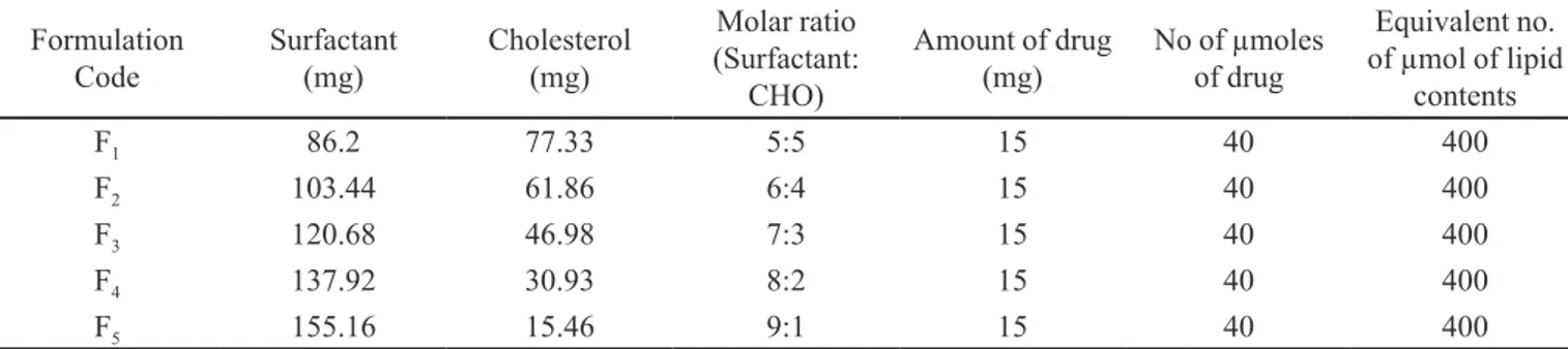
evaporation method. The mixtures of surfactant and cholesterol in different molar ratios (5:5, 6:4, 7:3, 8:2, 9:1) were accurately weighed (Table I) and dissolved in 20 mL of chloroform/ethanol mixture (2:1 v/v) separately.
The organic solvents from all formulations were slowly evaporated under reduced pressure in a rotary evaporator
(Heidolph, Germany) at 60 oC until a smooth ilm of lipid
components was formed on the wall of the rotating lask. The film was resuspended in 20 mL of ether. The drug solution (1 mg/mL) was prepared in a 15-mL mixture (2:1) of phosphate-bufered saline (PBS; pH 7.4) and ethanol. The mixture of lipid solution and drug solution was sonicated for 5 minutes and swirled by hand and then sonicated again for 2 minutes in a bath sonicator. The resulting dispersion
was immediately evaporated to disrupt the formed gel.
Then, 10 ml of PBS (pH 7.4) was added to the rotating lask
and the evaporation was continued for 15 minute to ensure the complete removal of the remaining diethyl ether. The
niosomal suspension was refrigerated at 4 oC overnight to
Characterization of vesicles
The developed niosomal formulations of diacerein
with diferent molar ratios of Span 60 and cholesterol were characterized for vesicle size, morphology, polydispersity index (PDI) and entrapment eiciency.
Fourier transform infrared (FTIR) spectroscopic studies
The compatibility of the drug and other formulation components were studied using FTIR (Bruker FTIR, Tensor 27Series, Germany). Spectra of individual
niosomal components and formulations were measured
using a pike single-bounce attenuated total relectance (ATR) cell equipped with a ZnSe single crystal. For solid samples (pure drug, cholesterol), samples were added to the ATR cell and measured directly. For liquids (niosomal
formulation), samples were placed directly on the small crystal spot and lever having concave surface is placed over it in order to prevent evaporation.
Drug entrapment studies
Diacerein-loaded niosomal formulations were
ultracentrifuged at 14,800 × g for 1.5 h to separate the unentrapped drug from the niosomes. The concentration of
free drug in the supernatant was estimated by measuring the absorbance at 258.50 nm using a UV spectrophotometer (IRMECO U2020, Germany). This process was repeated
to ensure complete removal of the free drug. The amount
of entrapped drug was determined by mathematical equation (1), as described elsewhere (Alsarra et al., 2005):
Total drug – Drug in supernatant
% Drug entrapment × 100
Total drug
−
= (1)
Morphological study
The morphological characteristics of niosomes
were investigated using a field emission scanning
electron microscope (JSM-7500F, Jeol, Japan). A drop of
optimized niosomal formulation (F3) was mounted on an
aluminium stub with adhesive silver tape. The stubs were
stored overnight under a vacuum and then sputter-coated using gold.
Size and distribution measurements
Size and polydispersivity index were measured by a dynamic light-scattering technique using a Zetasizer (ZS-90 Malvern Instruments, UK). For the measurements,
vesicle dispersions were diluted 100 times with the
same PBS (pH 7.4) used for their preparation to avoid
multiscattering of light. Buffer solutions were filtered
through 0.45-μm cellulose ilters to remove impurities. Vesicle mean size and size distribution (PDI) were determined at 25 °C while keeping the scattering angle at
90°. The instrument was operated at medium refractive index 1.330, a medium viscosity of 1.0 mPa.s and dielectric constant of 80.4. Sample mean size and PDI
were measured using the third-order cumulant fitting autocorrelation function.
Assessment of stability
The ability of niosomes to retain the drug was assessed by storing the niosomal formulations at two
different temperatures, i.e. refrigeration temperature
(4-8 oC) and ambient temperature (25oC ± 2 oC). Samples were stored in glass vials for 3 months and were withdrawn at regular time intervals. The entrapment efficiency of niosomal formulations was analysed in order to
characterize stability proile.
In vitro release studies
A dialysis method was adopted for determination
of the release proile of niosomal formulations (F1-F5).
TABLE I - Composition of diferent niosomal formulations
Formulation Code
Surfactant
(mg) Cholesterol (mg)
Molar ratio (Surfactant:
CHO)
Amount of drug
(mg) No of µmoles of drug
Equivalent no. of µmol of lipid
contents
F1 86.2 77.33 5:5 15 40 400
F2 103.44 61.86 6:4 15 40 400
F3 120.68 46.98 7:3 15 40 400
F4 137.92 30.93 8:2 15 40 400
Each formulation (1 ml) was added to a dialysis bag, clamped and placed in a beaker containing 200 mL of PBS maintained at pH 7.4 and 37 oC ± 2 ºC, which acted as the receptor compartment, and continuous stirring was
by a magnetic stirrer at 50 rpm. Samples were taken at various time-points for a period of 12 hours and assayed at 258.50 nm.
Release kinetic behaviour
The release pattern of diacerein from the niosomal
formulations (F1-F5)was evaluated by applying diferent
kinetic models to the observed release data to find the
release mechanism. The following models were applied to the release data in order to assess the release mechanism
from niosomes. The kinetic models (Dash et al., 2010) are
given in equation 2, 3, 4 and 5.
Zero order Qt = Q0 + K0 (2)
where Qt is the amount of drug released in time t, Q0 is the initial amount of the drug in the solution, and Ko is the
zero-order release constant.
Kt
log log Co
2.303
First Order C= − (3)
where Co is the initial drug concentration, t is time, and K
is the irst-order release constant.
Higuchi model Q = KH . t1/2 (4)
where Q is the amount of drug released in time t, and KH
is the Higuchi dissolution constant.
Moreover, in order to determine the release mechanism, the Korsemeyer–Peppas model was applied to all formulations:
Korsemeyer–Peppas Model Mt Ktn
M∞ = (5)
where Mt/M∞ is the amount of drug released at time t, K
is the release rate constant, and n is the release exponent. The n value is used to characterize the diferent release
mechanisms.
Statistical analysis
To compare the mean values of vesicle size and entrapment eiciencies of prepared niosomes and to assess statistical signiicance, a one-way analysis of variance (ANOVA) was performed at 95% confidence interval using SPSS 20.0.
RESULTS AND DISCUSSION
Formation of niosomes
The reverse-phase evaporation technique was found to be optimal for designing niosomes of diacerein.
Formulations were prepared containing a total lipid
mixture of 400 μmol in the order of increasing surfactant
and decreasing the cholesterol concentration. The
surfactant and cholesterol were dissolved in a mixture
of chloroform and ethanol. This organic solution was
evaporated and mixed with the drug solution following
sonication. The emulsion was again evaporated to remove traces of the organic solvent and hydration was continued at 60 oC.
Due to its inherent characteristics, Span 60 was
employed in this study. It is solid at room temperature, has
a higher phase-transition temperature (53-57 oC) and an
HLB value of 4.7. Moreover, it can form vesicles with or without cholesterol (Abdelkader et al., 2010). Surfactants
with higher solubility and HLB are not able to organize
into vesicles. Therefore, niosomes generally do not
develop between HLB values of 14 and 17 (Girigoswami, Das, De, 2006). Cholesterol has been incorporated into formulations as a vesicle stabilizer and it also abolishes the gel-liquid transition of the niosomes making these
less leaky. The finding of this study indicated that all
ratios of Span 60 and cholesterol were able to develop niosomes due to the lipophilic nature of the Span 60 and higher eiciency of encapsulating hydrophobic drugs like
diacerein.
Size and polydispersivity index
The size of the vesicles was found to be in the range
of 0.5 μm to 1 μm (Table II). The comparatively small size
of the niosomes is due to the low HLB value of Span 60 used in the present study. The size of niosomes increases with increasing values of HLB (Pardakhty, Varshosaz, Rouholamini, 2007). The smaller size of niosomes can be explained on the basis of the low HLB value of Span 60,
which results in low surface free energy, which leads to relatively less uptake of water in the core, and hence to smaller niosomes(Yoshioka, Sternberg, Florence, 1994).
The size of the vesicles was afected signiicantly (ANOVA, p<0.05) by altering the surfactant to cholesterol level in the formulations. These indings are in accordance
with the results of niosomal encapsulation of oestradiol
are multidispersed systems. Among all ive formulations,
F3 showed the lowest value of PDI (0.409), which also
favours the F3 formulation to be the more optimized.
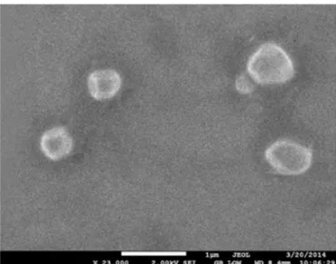
Scanning electron microscopy
The SEM image of the selected formulation (F3)
revealed the spherical shape of niosomes (Figure 1).
The shape of vesicles is predicted on the basis of the critical packing parameter (CPP), which is calculated from
the area of the hydrophilic head group and the length of
the alkyl chain of the surfactant by the following formula:
CPP = ʋ /lc.α
0 (6)
where ʋ is the lipophilic group volume, lc is the critical
hydrophobic group length, and α0 is the area of the
hydrophilic head group. Span 60 has a CPP value in the range of 0.5 to 1 and can give rise to niosomes (Uchegbu,
Vyas, 1998). The spherical shape of the niosomes obtained in our study is in agreement with previous reports of entrapping opioid antagonist naltrexone by sorbitan monostearate (Abdelkader et al., 2012) and also
entrapment of calcein in the same surfactant (Manosroi
et al., 2003).
Drug entrapment studies
The ultracentrifugation method was found to be suitable for the determination of entrapment eiciency of niosomal formulations (F1-F5) as shown in Table III. The
amount of entrapped drug was obtained by subtracting
the amount of unentrapped drug from the total drug incorporated.
The entrapment efficiency was affected mainly
by the molar concentration of cholesterol because the
total molar concentration of the lipid content was kept
constant (Table I) throughout the experiments. Previous studies have reached contrasting conclusions about the efect of the concentration of cholesterol on entrapment eiciency. Some reports describe that there was no efect
of cholesterol concentration on niosomes entrapment
efficiency (Uchegbu et al., 1995), while other studies
show that entrapment eiciency increases with increasing cholesterol concentration from 0% to 50% mol/mol (Yoshioka, Sternberg, Florence, 1994) and another report showed limited enhancement of entrapment eiciency up to 30% mol/mol (Moazeni et al., 2010).
In our studies, entrapment eiciency was enhanced up to 30% mol/mol concentration of cholesterol. In
formulations F1 and F2, entrapment eiciency was lower than in formulation F3, which might be ascribed to the fact that higher concentrations of cholesterol may cause disruption of the vesicles leading to leakage of drug from TABLE II - Average size and polydispersivity index of the
niosomal formulations
Formulation Code Size a (μm) Polydispersivity
Index F1 1010×10
-3 ± 0.142 0.476 ± 0.04
F2 987.1×10⁻³ ± 0.260 0.504 ± 0.14
F3 965.7×10
-3 ± 0.119 0.409 ± 0.17
F4 835.9×10⁻³ ± 0.188 0.632 ± 0.05
F5 608.4×10⁻³ ± 0.312 0.781 ± 0.21
Data are means ±SD; p<0.05. a Size of niosomes was signiicantly
diferent from each other according to the post-hoc test.
FIGURE 1 - SEM image of optimized formulation F3.
TABLE III - Average entrapment efficiency of the niosomal formulations
Formulation Code
Surfactant:cholesterol
ratio (μmolar ratio) % Entrapment eiciencyb
F1 5:5 47.15 ± 0.46
F2 6:4 59.65 ± 0.51
F3 7:3 79.80 ± 0.35
F4 8:2 68.54 ± 0.39
F5 9:1 61.47 ± 0.44
Data are means ± SD; p<0.05. b Entrapment eiciency of all
the microenvironment of the vesicles, thus leading to
comparatively low entrapment efficiency (Abdelkader
et al., 2010). Formulation F3 had the highest entrapment
efficiency (79.80%) among all the formulations as it contained the optimized concentration of cholesterol.
Formulations F4 and F5 had lower entrapment eiciency than F3, which might be correlated with the lower amounts of cholesterol as compared to F3 (Table III). Statistical
analysis of the data given in Table III demonstrated that percentage entrapment eiciency was signiicantly
dependent upon the surfactant to cholesterol level in the
formulations (ANOVA; p<0.05).
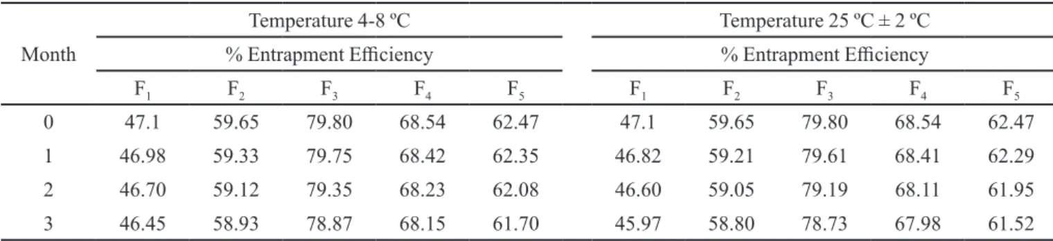
Stability of niosomes
The encapsulation efficiency of diacerein in the vesicles was determined at the end of each month. There
was no signiicant change in entrapment eiciency for
diacerein at two different temperatures after 3 months
(Table IV). Therefore, niosomes may be an effective
formulation to protect from drug leakage.
FTIR studies
The spectra of individual components and formulations of diacerein were recorded and provided
in Figure 2. The FTIR spectrum of diacerein showed the principal peaks of diacerein at 1764.56 cm-1 (C=O, ester),
1677 cm-1 (C=O stretch COOH), 1188.89 cm-1 (C—O
stretch, ester), 760.43 cm-1 (m-substituted benzene) and
703.02 cm-1 (benzene). Such peaks of diacerein have also
been reported elsewhere (Silverstein, Webster, Kiemle, 2005), which shows the purity of the diacerein sample
and its free nature without any type of pharmacophore.
In the spectrum of the Span 60, there were peaks of the
hydroxyl group at 3390.20 cm-1, strong aromatic –CH
3 group2916.75 cm-1 and the strong C=O ester bond at
1734.63 cm-1 (Li et al., 2008). Cholesterol showed peaks
of the hydroxyl group at 3429.82 cm-1, strong aromatic
stretching of CH=CH at 2931.41 cm-1, and the strong C=O
bond of carboxylic acid at 1770.20 cm-1 (Reis, Winter,
Zerda, 1996).
The FTIR spectrum of the selected niosomal
formulation (F3) demonstrated a slight shifting of the
peaks and peaks were found to be remarkably difused (Figure 2). No formation of any new peak was observed
indicating that no chemical interaction occurred, and further smoothening of peaks depicted the strong physical interaction that led to the formation of niosomes.
TABLE IV - Stability of diacerein niosomes at diferent temperatures
Month
Temperature 4-8 ºC Temperature 25 ºC ± 2 ºC % Entrapment Eiciency % Entrapment Eiciency
F1 F2 F3 F4 F5 F1 F2 F3 F4 F5
0 47.1 59.65 79.80 68.54 62.47 47.1 59.65 79.80 68.54 62.47
1 46.98 59.33 79.75 68.42 62.35 46.82 59.21 79.61 68.41 62.29 2 46.70 59.12 79.35 68.23 62.08 46.60 59.05 79.19 68.11 61.95 3 46.45 58.93 78.87 68.15 61.70 45.97 58.80 78.73 67.98 61.52
FIGURE 2 - FTIR Spectra of (A) diacerein, (B) sorbitan
Release studies of niosomal formulations
An overall sustained release behaviour was observed with all formulations (F1-F5) as shown in Figure 3.The
selected formulation (F3) expressed more prolonged
release and only ~55% of the drug was released in the 12-hour study design. The reason for this more retarded
release from F3 was due to the optimum concentration of cholesterol in its vicinity, which provided the optimal
strength and luidity to the niosomes in a more eicient
way.
Another reason is the reduction of the efflux of diacerein from niosomal vesicles by stabilizing the vesicular membrane and making it more rigid and less
leaky (Demel, Dekruyf, 1976; Korchowiec et al., 2006).
A similar phenomenon was observed with
niosome-entrapped rifampicin, where the cholesterol was found to
retard the release of the drug (Jain, Vyas, 1995). The slow release of drug from niosomes is also attributed to the use of Span 60, which has a slower rate of release than Span 20, 80 and 85, which in turn is due to the reason that Span 60 molecules remain in an ordered gel state, but other Spans are in a disordered liquid crystalline state (Attia et al., 2007).
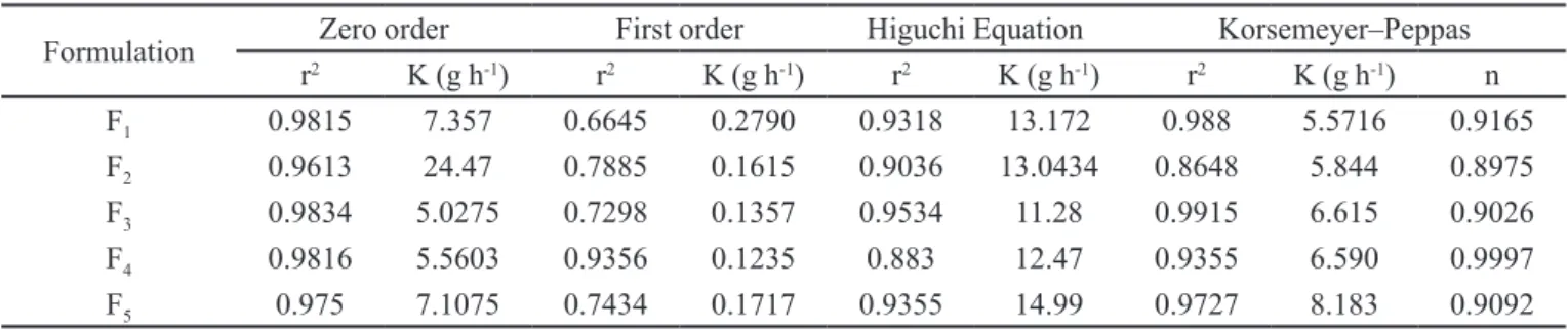
Release kinetic behaviour
The regression coefficient (r2) values were
approximated to follow zero-order release. Formulation
F3 was the more optimized as it showed r2=0.9834.
All equations including zero order, Higuchi model and Korsemeyer-Peppas equations, exhibited good linearity for the optimized selected formulation (F3). The Korsemeyer-Peppas model was applied to all formulations data and the n value was calculated. The value of n for
all formulations suggests non-ickian (anomalous) - i.e.
coupling of both diffusion and erosion - mechanisms (Costa, Sousalobo, 2001). Table V gives the values of
the rate constants and regression correlations using rate
equations for the release of diacerein from niosomes. The overall results revealed that the zero-order r2 values for all
formulations were higher than the irst-order values, which suggests that niosomal formulations (F1-F5) can release the drug in sustained manner.
CONCLUSION
Niosomal formulations of Span 60 and cholesterol were successfully prepared by a reverse-phase evaporation method. Optimizing the concentration of cholesterol to an
optimum level (F3) increased the entrapment eiciency
and sustained the release of diacerein from the niosomes.
The formulation (F3, 7:3) showed maximum encapsulation
efficiency (79.80%) and a prolonged release profile (55%) in 12 hours. The prepared niosomes can be used as
potential carrier for the controlled release and enhanced
availability of the diacerein at the site of absorption.
ACKNOWLEDGEMENTS
This study was a part of PhD thesis by Muhammad Imran Khan that was supported by the Higher Education Commission of Pakistan (Grant number: 112-23886-2BM1-354). The authors would like to thank the Chairman
TABLE V - Kinetic modelling of formulations (F1-F5)
Formulation Zero order First order Higuchi Equation Korsemeyer–Peppas r2 K (g h-1) r2 K (g h-1) r2 K (g h-1) r2 K (g h-1) n
F1 0.9815 7.357 0.6645 0.2790 0.9318 13.172 0.988 5.5716 0.9165
F2 0.9613 24.47 0.7885 0.1615 0.9036 13.0434 0.8648 5.844 0.8975 F3 0.9834 5.0275 0.7298 0.1357 0.9534 11.28 0.9915 6.615 0.9026
F4 0.9816 5.5603 0.9356 0.1235 0.883 12.47 0.9355 6.590 0.9997 F5 0.975 7.1075 0.7434 0.1717 0.9355 14.99 0.9727 8.183 0.9092
of the Department of Pharmacy and the Vice Chancellor of The Islamia University of Bahawalpur for providing all the necessary facilities during the study.
REFERENCES
AGGARWAL, A.K.; SINGH, S. Physicochemical characterization and dissolution study of solid dispersions of diacerein with polyethylene glycol 6000. Drug Dev. Ind. Pharm., v.37, n.10, p.1181-1191, 2011.
ABDELKADER, H.; ISMAIL, S.; KAMAL, A.; ALANY, R. Preparation of niosomes as an ocular delivery system for naltrexone hydrochloride: physicochemical characterization. Pharmazie,v.65, n.11, p.811-817, 2010.
ABDELKADER, H.; WU, Z.; AL-KASSAS, R.; ALANY, R. Niosomes and discomes for oculardelivery of naltrexone hydrochloride: morphological, rheological, spreading properties and photo-protective effects. Int. J. Pharm., v.433, n.1, p.142-148, 2012.
ATTIA, I.A.; EL-GIZAWY, S.A.; FOUDA, M.A.; DONIA, A.M. Influence of a niosomal formulation on the oral bioavailability of acyclovir in rabbits. AAPS Pharm. Sci. Tech., v.8, n.4, p.E106, 2007.
ALSARRA, I.A.; BOSELA, A.A.; AHMED, S.M.; MAHROUS, G.M. Proniosomes as a drug carrier for transdermal delivery of ketorolac. Eur. J. Pharm. Biopharm., v.59, n.3, p.485-490, 2005.
COSTA, P.; SOUSA LOBO, J.M. Modeling and comparison of dissolution proiles. Eur. J. Pharm. Sci., v.13, n.2, p.123-133, 2001.
DESHMUKH, D.B.; GAIKWAD, P.D.; BANKAR, V.H.; PAWAR, S.P. Dissolution enhancement of poorly water soluble diacerein by solid dispersion technique. J. Pharm Sci. Res., v.2, n.11, p.734-739, 2010.
DASH, S.; MURTHY, P.N.; NATH, L.; CHOWDHURY, P. Kinetic modeling on drug release from controlled drug delivery systems. Acta Pol. Pharm., v.67, n.3, p.217-223, 2010.
DEMEL, R.A.; DEKRUYFF, B. The function of sterols in membranes. Biochim. Biophys Acta, v.457, n.2, p.109-132, 1976.
ESSA, E.A. Efect of formulation and processing variables on the particle size of sorbitan monopalmitate niosomes. Asian J. Pharm., v.4, n.4, p.227-233, 2010.
GIRIGOSWAMI, A.; DAS, S.; DE, S. Fluorescence and dynamic light scattering studies of niosomes membrane mimetic systems. Spectrochim. Acta. A. Mol. Biomol. Spectrosc., v.64, n.4, p.859-866, 2006.
GUINEDI, A.S.; MORTADA, N.D.; MANSOUR, S; HATHOUT, R.M. Preparation and evaluation of reverse phase evaporation and multilamellar niosomes as ophthalmic carriers of acetazolamide. Int. J. Pharm., v.306, n.1, p.71-82, 2005.
HONG, M.; ZHU, S.; JIANG, Y.; TANG, G.; PEI, Y. Eicient tumor targeting of hydroxycamptothecin loaded PEGylated niosomes modiied with transferrin. J. Control. Release, v.133, n.2, p.96-102, 2009.
HUNTER, D.J.; WISE, B. Review: diacerein is more efective than placebo. J. Evid. Based Med., v.12, n.3, p.74, 2007.
JAIN, A.; SINGH, S.K.; SINGH Y.; SINGH, S. Development of lipid nanoparticles of diacerein, an antiosteoarthritic drug for enhancement in bioavailability and reduction in its side efects. J. Biomed. Nanotechnol., v.9, n.5, p.891-900, 2013.
JAIN, C.; VYAS, S. Preparation and characterization of niosomes containing rifampicin for lung targeting. J. Microencapsul., v.12, n.4, p.401-407, 1995.
KORCHOWIEC, B.; PALUCH, M.; CORVIS, Y.; ROGALSKA, E. A Langmuir ilm approach to elucidating interactions in lipid membranes: 1, 2-dipalmitoyl glycero-3phosphoethanolamine cholesterol metal cation systems. Chem. Phys. Lipids, v.144, n.2, p.127-136, 2006.
LI, F.T.; ZHAO, D.S.; LUO, Q.Z.; LIU, R.H.; YIN, R. Research on surface-modification of Nano-TiO2 by span 60. J. Ceram. Process. Res., v.9, n.4, p.398-400, 2008.
MANOSROI, A.; WONGTRAKUL, P.; MANOSROI, J.; SAKAI, H.; SUGAWARA, F.; YUASA, M.; ABE, M. Characterization of vesicles prepared with various non-ionic surfactants mixed with cholesterol. Colloids Surf. B Biointerfaces, v.30, n.1-2, p.129-138, 2003.
MARIANECCI, C.; RINALDI, F.; MASTRIOTA, M.; PIERETTI, S.; TRAPASSO, E.; PAOLINO, D.; CARAFA, M. Anti-inflammatory activity of novel ammonium glycyrrhizinate/niosomes delivery system: human and murine models. J. Control. Release, v.164, n.1, p.17-25, 2012.
MASKI, N.; KUMARAN, A.; GIRHEPUNJE, K.; GHODE, P.; RANDIVE, S.; PAL, R. Studies on the preparation, characterization and solubility of β-cyclodextrin–diacerein inclusion complexes. Int. J. Pharm. Pharm. Sci., v.1, n.1, p.121-135, 2009.
MOAZENI, E.; GILANI, K.; SOTOUDEGAN, F.; PARDAKHTY, A.; NAJAFABADI, A.R.; GHALANDARI, R.; FAZELI, M.R.; JAMALIFAR, H. Formulation and in vitro evaluation of ciproloxacin containing niosomes for pulmonary delivery. J. Microencapsul., v.27, n.7, p.618-627, 2010.
MUZZALUPO, R.; TAVANO, L.; TROMBINO, S.; CASSANO, R.; PICCI, N.; LA MESA, C. Niosomes from α, ω-trioxyethylene-bis (sodium 2-dodecyloxy-propylenesulfonate): Preparation and characterization. Colloids. Surf. B Biointerfaces, v.64, n.2, p.200-207, 2008.
NGUYEN, M.; DOUGADOS, M.; BERDAH, L.; AMOR, B. Diacerhein in the treatment of osteoarthritis of the hip. Arthritis Rheum., v.37, n.4, p.529-536, 1994.
PARDAKHTY, A.; VARSHOSAZ, J.; ROUHOLAMINI, A. In vitro study of polyoxyethylene alkyl ether niosomes for delivery of insulin. Int. J. Pharm., v.328, n.2, p.130-141, 2007.
PEARLE, A.D.; WARREN, R.F.; RODEO, S.A. Basic science of articular cartilage and osteoarthritis. Clin. Sports Med., v.24, n.1, p.1-12, 2005.
REIS, O.; WINTER, R.; ZERDA, T.W. The efect of high extenal pressure on DPPC-cholesterol multilammellar vesicles: a pressure-tuning Fourier transform infrared spectroscopy. Biochim. Biophys. Acta, v.1279, n.1, p.5-16, 1996.
SHARI, M.L.; BATHON, J.M. Osteoarthritis: pathophysiology [Online]. 2012. The john hopkins arthritis center. Available at: <http://www.hopkinsarthritis.org/arthritis-info/ osteoarthritis/oa-pathophysiology/>. Accessed on: March 2014.
SILVERSTEIN, R.M.; WEBSTER, F.X.; KIEMLE, D.J. Spectrometric identiication of organic compounds. 7.ed. New York: John Wiley & Sons In, 2005. 512 p.
UCHEGBU, I.F.; DOUBLE, J.A.; TURTON, J.A.; FLORENCE, A.T. Distribution, metabolism and tumoricidal activity of doxorubicin administered in sorbitan monostearate (Span 60) niosomes in the mouse. Pharm. Res., v.12, n.7, p.1019-1024, 1995.
UCHEGBU, I.F.; VYAS, S.P. Non-ionic surfactant based vesicles (niosomes) in drug delivery. Int. J. Pharm., v.172, n.1, p.33-70, 1998.
WADDAD, A.Y.; ABBAD, S.; YU, F.; MUNYENDO, W.L.; WANG, J.; LV, H.; ZHOU, J. Formulation, characterization and pharmacokinetics of Morin hydrate niosomes prepared from various non-ionic surfactants. Int. J. Pharm., v.456, n.2, p.446-458, 2013.
YOSHIOKA, T.; STERNBERG, B.; FLORENCE, A.T. Preparation and properties of vesicles (niosomes) of sorbitan monoesters (Span 20, 40, 60 and 80) and a sorbitan triester (Span 85). Int. J. Pharm., v.105, n.1, p.1-6, 1994.
Received for publication on 25th August 2014