*Correspondence: Ankita Gupta. Pranveer Singh Institute of Tech-nology. Kalpi Road, Bhauti, 208020 - Kanpur - UP, India. E-mail: gupta.ankita483@gmail.com
A
vol. 51, n. 3, jul./sep., 2015 http://dx.doi.org/10.1590/S1984-82502015000300011
Enteric coated HPMC capsules plugged with 5-FU loaded
microsponges: a potential approach for treatment of colon cancer
Ankita Gupta
1,*, Gaurav Tiwari
1, Ruchi Tiwari
1, Rishabh Srivastava
2, A. K. Rai
11Pranveer Singh Institute of Technology, Bhauti, Kanpur (UP), India, 2Akums Drugs and Pharmaceuticals Limited, Haridwar, India
The work was aimed at developing novel enteric coated HPMC capsules (ECHC) plugged with 5 Florouracil (5-FU) loaded Microsponges in combination with calcium pectinate beads. Modiied quasi-emulsion solvent difusion method was used to formulate microsponges based on 32 factorial design and the efects of independent variables (volume of organic solvent and Eudragit RS100 content) on the dependent variables (Particle size, %EE & % CDR) were determined. The optimized microsponges (F4) were characterized by SEM, PXRD, TGA and were plugged along with calcium pectinate beads in HPMC capsules and the HPMC capsules were further coated with enteric polymer Eudragit L 100 (Ed-L100) and/ or Eudrgit S 100 (Ed-S 100) in diferent proportions. In vitro release study of ECHC was performed in various release media sequentially SGF for 2 h, followed by SIF for the next 6 h and then in SCF (in the presence and absence of pectinase enzyme for further 16 h). Drug release was retarded on coating with EdS-100 in comparison to blend of EdS-100: EdL-100 coating. The percentage of 5-FU released at the end of 24 h from ECHC 3 was 97.83 ± 0.12% in the presence of pectinase whereas in control study it was 40.08 ± 0.02% drug. The optimized formulation was subjected to in vivo Roentgenographic studies in New Zealand white rabbits to analyze the in vivo behavior of the developed colon targeted capsules. Pharmacokinetic studies in New Zealand white rabbits were conducted to determine the extent of systemic exposure provided by the developed formulation in comparison to 5-FU aqueous solutions. Thus, enteric coated HPMC capsules plugged with 5-FU loaded microsponges and calcium pectinate beads proved to be promising dosage form for colon targeted drug delivery to treat colorectal cancer.
Uniterms: 5-Fluorouracil. HPMC capsules/enteric coating. Microsponges/formulation. Drugs/delivery/ colon targeting. Colorectal cancer/treatment.
O trabalho teve como objetivo o desenvolvimento de novas cápsulas com revestimento entérico HPMC (ECHC) conectadas com microesponjas carregadas com luoruracila (5-FU) em combinação com grânuos de pectinato de cálcio. O método de difusão de solvente modiicado quasi-emulsão foi usado para formular microesponjas com base no planejamento fatorial 32 e determinaram-se os efeitos das variáveis independentes (volume de solvente orgânico e conteúdo Eudragit RS100) sobre as variáveis dependentes (tamanho de partícula, EE% e % CDR). As microesponjas otimizadas (F4) foram caracterizadas por SEM, PXRD, TGA e ligadas aos grânulos de pectinato de cálcio em cápsulas de HPMC e estas foram, ainda, revestidas com polímero entérico Eudragit L 100 (Ed-L100) e/ou Eudrgit S 100 (Ed S 100) em diferentes proporções. No estudo de liberação in vitro de ECHC foi realizada em vários meios de liberação sequencial SGF
carregadas com 5-FU e grânulos de pectinato de cálcio se mostraram promissoras como formulação para liberação do fármaco no cólon no tratamento do câncer colorretal.
Unitermos: Fluoruracila. Cápsula de HPMC/revestimento entérico. Microesponjas/formulação. Fármaco/ liberação/direcionamento para o cólon. Câncer colorretal/tratamento.
INTRODUCTION
Cancer is one of the most challenging diseases to cure and the second leading cause of death in developing countries. Over the past few decades, it continues to be a worldwide health problem in spite of several advanced technologies (Wolpin, Mayer, 2008). Cancers of the colon and rectum, often collectively referred to as colorectal cancer, are life-threatening tumors that develop in the large intestine. More than 80% of colorectal tumors develop from adenomatous polyps. Their numbers increase with age. Polyps are found in about 25% of people by age 50,
and 50% of people by age 75. Fewer than 1% of polyps less than 1 centimeter (slightly less than half an inch) becomes cancerous. About 10% of larger polyps become cancerous within 10 years, and about 25% of these larger
polyps become cancerous after 20 years.
Drug delivery systems based on calcium pectinate beads have been recently investigated for specific-targeting drugs to the colon (Badve et al., 2007). Such systems, obtained by pectin gelatinization in the presence of calcium salts, are less water-soluble than natural pectins, since calcium ions induce non-covalent associations of carbohydrate chains through the formation
of the so-called ‘‘egg box complexes’’ but they maintain
the selective biodegradation by pectinolytic enzymes of
colonic bacteria microlora (Jain, Gupta, Jain, 2007).
Microsponges are polymeric delivery systems composed of porous microspheres. They are tiny sponge like spherical particles that consist of a myriad of interconnecting voids within a non-collapsible structure with large porous
surface (Srivastava, Kumar, Pathak, 2010). The reasons
for fabricating microsponge was due to the fact that, drug
carrier systems less than 200 μm can eiciently be taken up by the macrophages present in colon tissue, thus exhibit efective localized drug action at the desired site. Apart from being site speciic, retention of drug or its carrier system on
the colonic surface is yet another important consideration that guided the selection of microsponges as the drug carrier
system in the research work (Orlu, Cevher, Araman, 2006).
They can also increase the lag time for absorption of drug as these get entrapped on the surface of the colon and thus have the potential for being developed as a colon-targeted drug delivery system.
The objective of our research work was to design novel enteric coated HPMC capsules (ECHC) plugged with 5-FU loaded microsponges in combination with calcium pectinate beads. 32 full factorial design was
used to optimize microsponges prepared by modified
quasi-emulsion solvent difusion method using a sodium
chloride as porogen.
5-FU was chosen as a model drug for our research work because targeted delivery of 5-FU not only reduces
systemic side-efects, but also provide an efective and safe
therapy for colon cancer with reduced dose and reduced
duration of therapy (Chickpetty, Raga, 2013). Longer exposure to lower concentration of 5-FU has been reported
by researchers to favor DNA-directed effects which is
thought to contribute to its anti-tumor efect .
In our research work HPMC capsule was preferred over gelatine capsules because HPMC capsule had slower
drug release proile in acidic media and the fast release proile at a pH of 5 and above (Huyghebaert, Vermeire, Remon, 2004). This can result in lower quantities of polymer coat compared with that required for tablets
to achieve the desired release in the small intestine or colon. Surprisingly, it was also found that enteric coated
HPMC capsules ofer much higher resistance against acid
solutions as compared to enteric coated gelatine capsules
(Moawia, Tabakha, 2010).
MATERIAL AND METHODS
Material
5-FU (Shalaks Pharmaceutical Pvt. Ltd, New Delhi); HPMC Capsules (ACG Associated Capsules Pvt. Ltd,
SciTech Centre, Mumbai); Eudragit RS100, Eudragit S100 (EdS100) and Eudragit L100 (EdL100) (Evonik
Labs, Mumbai) were obtained as gifts from the suppliers. All other chemicals used were of analytical grade and purchased from authentic suppliers.
Method development for the fabrication of microsponges
Previously described methods of microsponge
using porogen (Bae et al., 2009; Graves et al., 2005) were
modiied to employ an aqueous solution of sodium chloride as porogen. 1% (w/v) aqueous solution of the porogen (NaCl) was prepared and suicient amount of Span 80 was added to it with agitation to obtain1% (v/v) dispersion. A solution of the Eudragit RS 100 and 5-FU was prepared in a mixture of Ethanol: DCM and the porogen solution (0.1 mL) was uniformly emulsiied in it, to form a w/o emulsion. 5% (w/v) aqueous PVA solution (external phase)
was prepared separately and the previously prepared w/o
emulsion was emulsiied in it. This w/o/w emulsion was stirred for 24 h to get microsponges that were iltered, dried at 60 oC and stored in a desiccator (Srivastava, Kumar, Pathak, 2010).
Optimization of microsponges via 32 factorial
design
A 32 randomized full factorial design was run
to optimize the variables. In this design two factors
were evaluated, each at 3 levels, and experimental
trials were performed at all nine possible combinations
using Design Expert Software 8.0.7.1 (Stat-Ease, Inc.,
Minneapolis, USA). In the present investigation, volume of organic media (X1) and amount of polymer content (X2) were selected as independent variables and total
nine microsponge formulations (F1–F9) were fabricated
by varying these variables as given in Table I. The particle size, % EE and % CDR selected as dependent
variables and Polynomial equations were generated for
the dependent variables that were reduced by removing
non-signiicant coeicients by applying one way ANOVA. To demonstrate graphically the inluence of each factor on
the responses, the response surface plots and 3D bar graph were generated. The value of P<0.05 was considered to
be signiicant.
• Validation of experimental design
The formulation developed was evaluated for the
responses and the experimental values obtained were
compared with those predicted by the mathematical
models generated (F 10).
• Selection of optimized formulation
A numerical optimization technique using the
desirability approach was employed to select the optimized formulation with desired responses. Constraints
like maximizing entrapment eiciency & % drug release at
the end of 8 h as well as minimizing particle size were set as goals to select the optimized formulation using Design
Expert Software 8.0.7.1 (Stat-Ease, Inc., Minneapolis,
USA). Optimized formulation was plugged in enteric coated colon target capsules.
Characterization of microsponges
• Morphological studies and rheological
characteriza-tion
Morphological characteristics of microsponges were studied by scanning electron microscopy (Hitachi,
S 3000H Japan). The microsponges were dotted on an adhesive tape attached to an aluminium stub and excess
microsponges were detached. To render the particles electrically conductive, the stub spotter was coated with gold using a vacuum evaporator. The coated Microsponges
were viewed at 10 kV.
TABLE I - 32 Factorial design model for the fabrication of 5-FU loaded microsponges
S. No. Formulation Code
Independent Variables
Dependent Variables Volume of Organic Media
(mL) [X1]
Polymer Content (mg) [X2]
1 F1 (+1) 8 (+1) 1200 Particle Size (Y1)
2 F2 (-1) 6 (-1) 400 % EE (Y2)
3 F3 (0) 7 (0) 800 % CDR (Y3)
4 F4 (+1) 8 (-1) 400
5 F5 (-1) 6 (+1) 1200
6 F6 (0)7 (-1) 400
7 F7 (-1) 6 (0) 800
8 F8 (0)7 (+1) 1200
9 F9 (+1) 8 (0)800
• Powder X- ray difraction studies (PXRD)
The crystalline behavior of the drug before and after encapsulation, was evaluated by X-ray powder
difractometer (Bruker D8 Discover, Germany) using a CuK alpha radiation source with the Ni-ilter. A scanning
rate of 5o min-1, tube voltage of 35 KV and current of 35 mA over a range of 16-60o were used in measurement.
• Thermal analysis
Differential scanning calorimetry and Thermo gravimetric analysis of the pure drug and drug-loaded microsponges were carried out with simultaneous TGA / DSC analyzer (Mettler Toledo TGA/ DSC, USA). An
amount 13.714 mg of drug and 9.35 mg of drug loaded
microsponges was placed in aluminium pans and sealed prior to the test. All samples were run at a heating rate
of 10 ºC/min over a temperature range 30–600 °C in an
atmosphere of nitrogen.
• Particle size
All Formulations of the microsponges were analyzed for particle size by an optical microscope. The
instrument was calibrated and found that 1unit of eyepiece micrometer was equal to 7 μm. Sizes of 100 microsponges were calculated in 10 X magniication.
• Determination of drug content, drug loading and
entrapment eiciency (EE)
A sample of drug loaded microsponges (50 mg) was dissolved in 50 mL of phosphate buffer (pH 7.4) by using ultrasonicator and kept for overnight. Filtered Samples were appropriately diluted and analyzed
spectrophotometrically at 266 nm. Percent drug content,
% entrapment efficiency and % drug loading were calculated.
• In vitro drug release studies of microsponges
Microsponges equivalent to 50 mg of drug were illed in HPMC capsules and placed in the basket (mesh #230 = 63 µm) of USP apparatus 1 and the study was
performed in the 900 mL of simulated colonic fluid (SCF) without pectinase for 8 h at 50 rpm, maintained
at 37 ± 0.5 ºC. Aliquots were withdrawn periodically and sink conditions were maintained by adding equal
amounts of release medium. The samples were analyzed spectrophotometrically and % cumulative drug release (CDR) versus time plots was constructed. Data obtained from in-vitro release studied was evaluated to check the
goodness of it to various release kinetics equations for quantifying the phenomena controlling the release of drug from microsponges (Costa, Lobo, 2001).
Modification of pectin and its characterization
Pectin was crosslinked by the ionotropic gelation
method. A 10% (w/v) aqueous Pectin dispersion was added drop wise into gently stirred 100 mL of 10% (w/v) aqueous solution of calcium chloride pre-adjusted to pH 5.5. The gelled particles were left in the medium for 10 min, washed with deionized water and dried at 60 oC. The
particles were screened through sieve no 44 and kept in a desiccator to remove the traces of moisture and then evaluated for morphological, rheological and swelling characteristics (Sharma, Philip, Pathak, 2008).
Preparation and evaluation of enteric coated HPMC capsules
Each batch of HPMC capsules was coated with
enteric polymer EdS100 / EdL100 or bend of EdS100 and EdL100 in diferent ratios, by dip coating method in the polymeric solution of EdS100 and EdL100 to ensure the
formation of a uniform and thin covering over the capsule.
In order to enhance the elasticity of EUDRAGIT ilms, 1.25% of dibutyl phthalate as plasticizer was added to
the coating solution. HPMC bodies and caps were coated separately to ensure coverage of the body with polymer,
especially where the cap overlaps (Huyghebaert, Vermeire, Remon, 2004). Coated bodies were illed by formulation matrix consisting of 50 mg drug loaded microsponges plugged along with 5, 10, 15% w/ w of calcium pectinate
beads and the rest of the volume was adjusted by inert
lactose and inally closed with the coated caps. The joint
of the capsule was sealed with a small amount of 5% EdS
100 solution. For each polymeric solution coating of the capsules was made with diferent thickness ratios. The
capsules were allowed to dry with the help of dryer with
an inlet temperature of 35-40 ºC and observed for coat
uniformity, pores and cracks under microscope.
• Integrity test of enteric coated HPMC capsules
Prior to the dissolution test enteric-coated HPMC
capsules were subjected to a disintegration test in 0.1 M
HCl for 2 h to determine the ability of the coat to withstand acid environment. Afterwards, the capsules were dried at 25 oC for 45 min, thereafter; the capsules were subjected to microscopic examination to check for the integrity of
the coat. The enteric coated capsules, which passed the disintegration test were further evaluated for integrity of the coating in pH 5.5 since the dissolution of the coating in
pH lower than 6.8 may alter the required lag time. On the basis of solubility in pH 5.5 bufer, coating compositions
of lag time. Only coating levels and compositions which were not dissolved at all or dissolved very slowly in pH
5.5 bufer were selected for further in vitro drug release
studies (Cole et al., 2002).
• In vitro drug release study of ECHC
The study was performed in USP drug release
apparatus in various release media sequentially SGF (pH 1.2) for 2 h, followed by SIF (pH 7.4) for the next 6 h and
then in SCF (presence and absence of pectinase, for further
16 h). The test conditions were 900 ml of medium stirred
at 50 rpm and maintained at 37 ± 0.5 oC. Aliquots were withdrawn at regular intervals and replaced with an equal
amount of the fresh medium to maintain sink condition. The samples were analyzed for 5-FU content released at
265.4 nm. Concomitantly the swelling characteristics and
physical behavior of capsules were observed at 0, 2, 5, 8,
10, 14, 18 and 24 h and recorded visually. The systematic evaluation of ECHC-3, ECHC-14, and ECHC-15 led to identiication of optimized formulation.
Animal studies
All the animal experiments have been conducted
in full compliance with the institutional ethical and regulatory principles and as per the spirit of the Association for Assessment and Accreditation of Laboratory Animal
Care & International’s expectations for animal care and
use/ethics committees. The investigations were performed after obtaining approval by the Institutional Animal Ethical Committee of PSIT, Kanpur, India (IAEC no:
IAEC/PSIT/1273/ac/13).
• In vivo roentgenographic study
The 5-FU loaded microsponges in colon targeted capsules were partially replaced with barium sulfate
microsponges (Chickpetty, Raga, 2013). The reformulated
enteric polymer coated capsules were administered orally
with 15 ml of water, to New Zealand white rabbits (n = 3; 3–3.5 kg) that were fasted overnight with free access to
water. Fluoroscopic images of the abdomen of the rabbits
were taken before administration (0 h) and at 1.5 h, 3.5 h, 5 h, 7.5 h and 10 h to trace the in vivo movement and behavior of capsules in the gastrointestinal tract. X-ray images of the
rabbits in prone position were captured using L&T Vision 100 (C-arm) X-ray machine (Larsen and Tourbo Limited, Mumbai, India), at 64 mAs and 63 kV voltage.
• In vivo pharmacokinetics study
New Zealand white rabbits weighing 3–3.5 kg were classiied in two groups, namely standard and test with
three animals in each group. The animals were fasted overnight with free access to water. The standard group
received 5-FU aqueous solutions (10 mg/kg), while the
test group received colon targeted capsules (ECHC-3).
At appropriate intervals, blood samples (approximately 1
ml) were withdrawn from the marginal ear vein. 5-FU was
extracted from plasma by mixing rabbit plasma with ethyl acetate and isopropyl alcohol (85/15, v/v). The samples
were then dried with N2 at 37 °C and the dehydrated samples were dissolved in 400 μl of mobile phase for subsequent HPLC. The concentration of releasing 5-FU was measured using reversed-phase HPLC (HP1100 Liquid Chromatography, Agilent). A Hypersil C18 (5 μm, ID 4.6 mm × 300 mm) analytical column was used with a mobile phase of 0.01 mol/L phosphate bufer (pH 3.0) and an elution rate of 1.0 mL/min at room temperature. Absorbance at 269 nm was monitored and pharmacokinetic
parameters were determined from the absorbance-time curves. This method provided complete separation with a corresponding retention time of 7.0 minutes for 5-FU. The
area under the plasma drug concentration–time curve was calculated using the trapezoidal rule. The maximal plasma
concentration of drug (C max) and the time to reach the
maximum plasma concentration (T max) were directly obtained from plasma data (Ramasamy et al., 2011).
The data from diferent formulations were compared for statistical signiicance by one-way analysis of variance (ANOVA). All results were expressed as mean ± S.D.
RESULT AND DISCUSSION
Method development for the fabrication of microsponges
In present research work, sodium chloride was used as porogen, based on the considerations that the use of hydrogen
peroxide and sodium bicarbonate as porogen resulted in the evolution of gases like nascent oxygen or carbon dioxide
that may be incompatible with active pharmaceutical ingredients. Sodium chloride as a porogen is inert in terms
of the evolution of gas and its high aqueous solubility would facilitate its easy extraction in the outer aqueous phase
during the microsponge formation (Orlu, Cevher, Araman,
2006). Therefore, using sodium chloride as porogen, nine formulations (F1-F9) were fabricated and characterized.
Characterizations of microsponges
• Morphological studies and rheological
characteriza-tion
F4 conirmed the highly porous nature of microsponges.
SEM of microsponges before in vitro release test in Figure
1 (A and B) depicted spherical, smooth surfaced and uniformly porous particles. Figure 1 (C and D) magniied
the surface view of microsponges F4 will show its highly cross linked polymeric surface containing numerous interconnected voids loaded with drug thus supporting
the spongy nature of microsponges. Figure 1 (E and F)
is the micrographs of microsponges F4 after the in vitro drug release for 8 h and it depicted irregular microsponges with numerous regular striations due to erosion of surface associated drug molecules.
• Powder X- ray difraction studies (PXRD)
The X-ray difraction patterns of 5-FU and 5- FU
loaded microsponges plotted by using origin software are shown in Figure 2.The PXRD pattern of 5-FU showed peaks at 29◦, 33◦, 16◦, 19◦, and 21◦ which were intense and sharp indicating its crystalline nature also supported by the literature (Zhang et al., 2011). Some of these 5 -FU
peaks are also present in difractogram of 5-FU loaded
microsponges, due to surface adsorbed drug. Since
Eudragit RS 100 has ammonio methacrylate units that
confer a positive charge on the polymer surface while 5-FU has a negative charge on its surface. Therefore the drug molecule can undergo ionic adsorption on the surface of the polymer. However, 5-FU loaded microsponges presented most of the peaks of diminished intensity indicating that there might be partial or complete transitions of drug entrapped in microsponges from crystalline state to an amorphous state.
• Thermal analysis
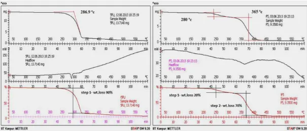
TGA-DSC thermograms of pure 5-FU and drug loaded microsponges are depicted in Figure 3. The
extrapolated onset temperature in thermograms denotes
the temperature at which the weight loss begins. The
extrapolated onset temperature of 5-FU is 286.9 oC.
The DSC thermogram of the pure 5-FU also showed a
sharp melting endotherm peak at approximately 286.9 °C followed by decomposition, which was in agreement
with those reported previously (Lee et al., 2003). The red curve is the % weight loss curve and it indicated that % Weight loss occurs in one step with 90% decomposition
of the drug between 280 °C to 350 °C. Drug-loaded Optimized microsponges showed TGA extrapolated onset temperature at 280 °C and 365 °C indicating the
presence of small drug in crystalline form which might be due to small amount of drug adsorbed onto the surface of the microsponges whereas a shifted small broad
endothermic peak at 351 oC suggested that the drug
was either totally or partially converted into amorphous form and further no characteristic peak of 5-FU was observed. The reduction of height and sharpness of the endotherm peak may be due to the presence of polymers in the microsponges. TGA thermograms of microsponges also showed a downward shift which indicated loss of mass (due to solvent evaporation, loss of moisture and degradation) upon heating. The red curve, which is the %
FIGURE 1 - SEM of Microsponges F4 before drug release (A and B), surface view of F4 before drug release (C and D), F4 after drug release (E and F).
weight loss curve showed % weight loss in two steps. In
the irst step, 20% weight loss between 260 oC to 290 oC
indicated decomposition of surface adsorbed drug present in crystalline form and in the second step, 70% weight loss in a temperature range of 340 oC to 410 oC suggested
the decomposition of amorphous form of drug entrapped within the microsponges. Therefore, physical status of drug present in microsponges was in correlation with PXRD data.
Particle size
The particle size of the microsponges ranged between
41.48 µm (F2) and 75.02 µm (F9) (Table II). On applying one-way ANOVA, it was observed that the experimental design had signiicant inluence on the particle size. As the
amount of the drug to be incorporated was kept constant,
any change in particle size was inluenced by the variation in the levels of ERS100 (polymer) and the volume of
organic solvent. At the lowest level of organic solvent, the
microsponges F2, F5 and F7 with mean diameter 61.22 µm, 71.48 µm and 75.02 µm respectively were obtained.
However, on increasing the volume of organic solvent, microsponges with smaller diameter were formulated.
Thus F4 and F9 yielded smaller microsponges of 42.15 and 58.31 respectively. Also at low levels of polymer content, microsponges F2, F4 and F6 with a mean diameter of 61.22 µm, 42.15 µm, and 55.32 µm, were
obtained. However, on increasing the polymer content, microsponges with larger diameter were formulated, i.e.
F1, F5 and F8 with a mean diameter of 69.40 µm, 71.48 µm and 65.31 µm respectively, were obtained. Therefore,
it was interpreted that a higher concentration of polymer or a lower level of organic solvents produced a more viscous
dispersion, which formed larger droplets and consequently
larger microsponges obtained.
Entrapment efficiency
The level of organic solvent and level of polymer
also had signiicant inluence on the entrapment eiciency.
The entrapment efficiency was observed to increase with an increase in the level of organic solvent that can be attributed to better solubilisation of the drug in the organic medium. The Higher volume of the organic
solvent resulted in the more uniform mixing of the drug and polymer resulting in a more uniform matrix with high drug entrapment eiciency. Microsponges F1, F4 and F 9
with high level of organic solvent had a high percentage
of EE 58.02%, 59.05%, and 60.82% respectively. Whereas
Microsponges F2 and F7 had the lowest percentage of
entrapment efficiency, i.e. 35.56% and 46.14% due to
low level of organic solvent. Similarly, on increasing
polymer content entrapment eiciency, increased, i.e. (F1) 58.02% and (F8) 54.87% had a high entrapment eiciency,
whereas microsponges with low level of polymer content
had less entrapment eiciency i.e. (F2) 35.56% and (F6)
47.40 % (Table II).
Along with above two factors a third factor, i.e. particle size also found to have an impact on the
entrapment eiciency. The observations suggested that entrapment eiciency of the microsponges increased with the decrease in the particle size. Consequently F4 and F9 with particle size 42.15 µm and 58.31 µm had high EE %, i.e. 59.05% and 60.82%, whereas F2 and F7 with particle size 61.22 µm and 75.02 µm had low EE i.e. % 35.56 %and 46.14% respectively. Therefore, Particle size
also had an impact on the entrapment efficiency, but it
was not statistically signiicant. Drug Content and Drug Loading of microsponge’s ranged from 52.2 % (F2) to 85.2% (F9) and 15.98 % (F6) to 39.75 % (F4) respectively
(Table II).
In vitro drug release study
The percent cumulative drug release in the SCF
was observed between 90.52% (F6) to 97.58% (F9)
suggesting the ability of themicrosponges to release the drug completely (Table II). All the formulationsfollowed
zero order release kinetics (Figure 4) except F2 and F8 that
followed Higuchi and Peppas release kinetics. Zero order release of drug from microsponges, has been reported by
researchers (Devrim, Canefe, 2006). The mechanism of drug release from microsponges as explained by various
researchers is co-relatable to its porous surface. The porous surface of the carrier particle enables easy penetration of the release media and its accessibility to the entrapped drug molecule. In case of F2 and F4, a high initial rate of
the drug release in 2–3 h was observed due to the surface adsorbed drug molecule that underwent quick release to attain the Equilibrium with the dissolved drug molecule,
Additionally, as suggested by various research reports, control on the drug release can also be achieved by
uniform mixing of the drug and polymer in organic media
to form homogenous phase.F2 and F7 formulated with low levels of polymer and organic phase might have resulted
in the improper encapsulation of the 5-FU in the ERS 100 matrix. Due to this, majority of the drug molecules resided
on the particulate surface as adsorbed molecules and had
the probability of undergoing quick solubilisation and hence quick drug release. But after achieving equilibrium,
the rate of drug release decreased. This effect was not predominant in F9 as it is constituted of intermediate level of ERS and high level of organic phase. While in F5, the
release itted irst order model that may be due to the fact
that the drug release from the polymeric microsponge occurs on transition of the polymer, and as the amount of
polymer in microsponges increases the time required for
phase conversion also increases. Thus, in F8, the initial drug release was due to the adsorbed molecules but with the lapse of time, ERS layer underwent transition resulting in the abrupt or burst release of 5-FU.
Statistical analysis
Statistical analysis was done by Design expert software version 8.0.7.1 (Stat-Ease, Inc., Minneapolis, USA) and the second order polynomial equations were
derived. The statistical model incorporating interactive and polynomial terms was utilized to evaluate the responses. Response surface plot (Figure 5) and 3-D Bar
Graph (Figure 6) clearly depicts the efects of independent
variables, i.e. varying the levels of volume of organic solvent and polymer content over dependent variables (i.e. Particle size, % EE and % CDR).
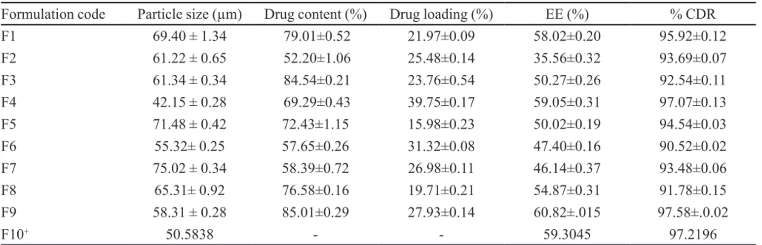
TABLE II - Compilation of evaluation parameters of 5-FU loaded microsponges
Formulation code Particle size (µm) Drug content (%) Drug loading (%) EE (%) % CDR
F1 69.40 ± 1.34 79.01±0.52 21.97±0.09 58.02±0.20 95.92±0.12
F2 61.22 ± 0.65 52.20±1.06 25.48±0.14 35.56±0.32 93.69±0.07
F3 61.34 ± 0.34 84.54±0.21 23.76±0.54 50.27±0.26 92.54±0.11
F4 42.15 ± 0.28 69.29±0.43 39.75±0.17 59.05±0.31 97.07±0.13
F5 71.48 ± 0.42 72.43±1.15 15.98±0.23 50.02±0.19 94.54±0.03
F6 55.32± 0.25 57.65±0.26 31.32±0.08 47.40±0.16 90.52±0.02
F7 75.02 ± 0.34 58.39±0.72 26.98±0.11 46.14±0.37 93.48±0.06
F8 65.31± 0.92 76.58±0.16 19.71±0.21 54.87±0.31 91.78±0.15
F9 58.31 ± 0.28 85.01±0.29 27.93±0.14 60.82±.015 97.58±.0.02
F10+ 50.5838 - - 59.3045 97.2196
+ Predicted value, % EE: Percent Entrapment Eiciency, % CDR: Percent Cumulative Drug Release. *All the values are expressed
in Mean ± SD
FIGURE 4 - In vitro drug release profile of 5-FU loaded
Factorial equation for particle size
TheResponse Surface linear model generated for
particle size was found to be signiicant with an F-value
of 11.30 (p< 0.0500) and correlation coeicient of 0.9824.
Particle Size (Y1) = 62.17 - 6.31 X1+ 7.92 X2 -1.52 X12 +
2.72 X22 + 3.94 X
12X2 + 3.06 X1 X22 – 2.03 X12X22
The co-eicient of X1 is negative, indicating that when the volume of organic solvent increased, the particle size decreased, whereas the positive coefficient of X2 indicates that particle size increased on increasing polymer
content. The P value for variable X1 and X2 were 0.0252
and 0.0099 respectively (p< 0.0500) indicated that both the
independent variables show signiicant efect on dependent
variable i.e. particle size.
Factorial equation for % EE
TheResponse Surface linear model generated for
particle size was found to be signiicant with an F-value
of 63.59 (p< 0.0500) and correlation coeicient of 0.9745.
% EE (Y2) = 51.350 + 7.695 X1 + 3.483 X2 – 3.872 X1X2
The positive co-eicient of X1 and X2 indicates that
% EE increases on increasing volumes of organic content as well as polymer content. Whereas the combination of independent variables X1X2 had a negative inluence on % EE. The P value for variable X1, X2 and X1X2 were
0.0001, 0.0031 and 0.0047 respectively (p< 0.0500) show that both the independent variables as well as combination of independent variable had a significant effect on dependent variable i.e. % EE.
Factorial equation for % CDR
The Response Surface linear model generated for %
CDR was found to be signiicant with an F-value of 11.55
(p< 0.0500) and correlation coeicient of 0.9506.
% CDR (Y3) = 92.022 + 1.476X1 + 3.766 X1 2
+ 0.160 X2 – 0.613X22 - 0.500X1X2
According to above factorial equation Independent
variables X1, X12 and X
2 had a positive influence on %
CDR. Whereas the combination of independent variables X1X2 and X22 had a negative inluence on % CDR. The X1
and X12 with p value 0.0250 and 0.0087 respectively, were signiicant model terms (p< 0.0500) whereas X2, X1X2 and
X22 were insigniicant model terms.
FIGURE 5 - Response surface plot depicting the inluence of independent variables over dependent variables.
Validation of the experimental design
The results of experimentally observed responses
and those predicted by mathematical models along with the percentage prediction errors were compared. The prediction error in the response parameters ranged
between 0.51 and 1.15% with the value of absolute error of 1.28 ± 0.70%. Lower values of error indicate the high prognostic ability of factorial equation and counter plot
methodology. “Adeq Precision” measures the signal to noise ratio. A ratio greater than 4 is desirable. The ratio of
8.505 indicates an adequate signal. Thus, this model can
be used to navigate the design space.
Selection of optimized formulation
M i c r o s p o n g e f o r m u l a t i o n F 4 w i t h h i g h e s t desirability factor of 0.944 having smallest particle size of
42.15 ± 0.28 µm, maximum % entrapment eiciency and % CDR of 59.05±0.31 and 97.07±0.13 respectively was
selected as optimized formulation. Ramps reports clearly
depict criteria’s for selection of optimized formulation
(Figure 7). F4 was further used for preparation of colon target capsules.
Modification of pectin to calcium pectinate
Use of Calcium pectinate beads as a plug in colon target capsules was a novel step and the rationale behind this was that calcium pectinate (the insoluble salt of pectin) is not degraded by gastric or intestinal enzymes, but will be degraded by colonic pectinolytic enzymes and hence now our formulation would be microbial triggered. Calcium pectinate is a natural polymer which can be used as a
major component or iller in pharmaceutical composition that could be degraded by only colonic enzymes (Jain, Gupta, Jain, 2007). Pectin is one of the most widely investigated polysaccharides in the colon-speciic drug
delivery. It can be broken down by pectinase enzymes
produced by anaerobic bacteria in the colon and control the drug release by this principle. It can also act via the pH- and time controlled mechanisms. However, due to high water solubility and swelling, pectin is not capable of
shielding the drug efectively during the passage through
the stomach and small intestine. Therefore Pectin with low
degrees of methoxylation (i.e., low methoxypectins or LM
pectin) was cross-linked with calcium ions (Ca2+, divalent cations) to produce Ca- pectinate networks that were less water soluble (Bourgeois et al., 2006). Indeed, Ca2+ ions
form “bridges” between the free carboxylated groups of
galacturonic acid moieties. The network that is formed has
been described by Grant et al. under the name of «egg-box
model». Pectin with DM < 50% (i.e., LM pectin) was used
because it contains more free carboxylic group than high methoxy pectin (HM pectin). Therefore, cross-linking between divalent cations (e.g., Ca2+) and free carboxylic
groups of the pectin are more evident in LM pectin than HM pectin (Badve et al., 2007). The spherical beads were easily prepared without any sophisticated instrument due to ionic interaction between the negatively charged
carboxylic groups of pectin molecules and the positively
charged divalent calcium ions which led to intermolecular cross-linking and instantaneously produced gelled sphere (Sriamornsak, Thirawong, Puttipipatkhachorn, 2005).
Characterization of calcium pectinate beads
Size, surface morphology and rheological characterization
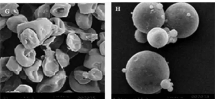
Particle size of calcium pectinate beads was of approx 105.22 ± 3.36 µm. SEM of pectin demonstrated smooth
doughnut shaped particles more or less, of uniform size (Figure 8G) and calcium pectinate beads were spherical with smooth surfaced (Figure 8H). Analysis of the
rheological characteristics of calcium pectinate conirmed
good rheological properties with bulk and tapped density of 0.504 g/cm3 and 0.558 g/cm3, Carr’s compressibility index of 9.67% and Angle of repose of 20.56 ºindicated good low
properties of calcium pectinate beads.
FIGURE 7 - Numerical optimization ramps report.
• Swelling characteristics
Pectin and calcium pectinate were compared for
their swelling characteristics. No signiicant swelling was observed for calcium pectinate (swelling ratio of 0.11±
0.04) as compared to pectin, which swelled considerably with a swelling ratio of 0.98 ± 0.02. The reduction in swelling of the calcium pectinate in SIF (pH 7.4) presents its feasibility to be used as a carrier for the colon-targeted drug delivery.
Preparation of enteric coated HPMC capsules
EUDRAGIT® L and S grades are suitable for enteric
coatings and are speciically used for controlled release in
the colon (Ramasamy et al., 2011). Coatings on the gelatin
capsules often sufer from insuicient adhesion between
the shell and the coating. Thus, previous workers in the area of enteric coating have found it necessary to pre-coat gelatin capsules with, for instance, a cellulose derivative, either to promote adhesion of polymers to the capsule shell or to improve gastric-resistance (Cole et al., 2002). When the capsule itself is made of a cellulose derivative it would
be expected, based on the experience with enteric coating
of tablets with a pre-coating of HPMC, that a pre-coating step could be eliminated. Gelatin capsules have a very glossy surface due to the fact that the amount of regular reflection from the surface is high and the amount of
difuse relection is low. In contrast, HPMC capsules have a visually matt surface with a greater amount of difuse relection, suggesting a more irregular surface. SEM’s
of the surface of HPMC and gelatin capsules are shown
in Figure 9 (I) and Figure 9 (J) where this diference is
clearly visible. During the coating process the temperature
of the capsule bed reaches 25–27 °C. At this temperature
HPMC is soluble and will start to dissolve in the eudragit
ilm providing a strongly adhesive surface. Gelatin, on the
other hand, is only slightly soluble at this temperature and its surface characteristics will remain virtually unchanged.
Figure 9 (K) shows SEM of the cross-section of a cleaved
surface through an HPMC capsule coated with EdS100
solution. The contours of the coating material are seen to follow the irregular surface of the HPMC capsule. It is suggested that the high strength of the bond between HPMC and the film is a combination of the irregular surface and the tackiness of the partially dissolved surface. No pores or cracks can be observed, due to the well controlled coating process. In addition, the critical area of overlap between the cap and the body of the capsule is also
covered with EdS100 polymers to ensure gastric integrity.
Evaluation of colon target enteric coated HPMC capsules
Integrity test of enteric coated HPMC capsules
Results of integrity test are shown in Table III.
ECHC-3, ECHC14 and ECHC15 which did not dissolve in pH 5.5 bufers were selected for further in vitro drug release study in simulated colonic luid.
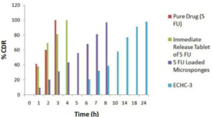
In vitro drug release study
In vitro drug release study was conducted in pH change method as per USP protocol (2 h in SGF, 2-5 h in SIF and 5-24 h in the SCF) and the results are shown in
Table IV. The immediate release marketed formulation
showed almost total release in 4 h and the pure drug in 3 h. Nearly complete drug release of microsponges in
TABLE III - Integrity test of enteric coated HPMC capsules
Parameters Formulation Code
ECHC1 ECHC2 ECHC3 ECHC4 ECHC5 ECHC6 ECHC7 ECHC8 ECHC9ECHC10 ECHC11 ECHC12 ECHC13 ECHC14 ECHC15
Coating ratio S100 L 100 S100: L100 (1:1 ) S100: L100 (2:1 ) S100: L100 (4:1)
Calcium pectinate
plug level 5 10 15 5 10 15 5 10 15 5 10 15 5 10 15
Disintegration test
in 0.1 N HCL F P P F P P F P P F P P P P P
Solubility in pH 5.5 - S NS - S S - S S - S S S NS NS
*F = Fail, P = Pass, S = Soluble, NS = Not Soluble
FIGURE 9 - SEM of a cross section of HPMC capsule surface
HPMC capsules (97.07%) in 8 h indicated the need for enteric coating of capsules for colon delivery (Figure
10). Enteric coated HPMC capsules 3, ECHC-14 and ECHC-15 showed no drug release in simulated gastric luid (pH 1.2) up to 2 h indicating the intactness of
the applied coating. On exposure to simulated intestinal
media, capsules coated with blend of EdS-100: EdL-100
showed the earlier drug release as compared to capsules
coated with only EdS-100. As the pH of solubilisation of EdL-100 is 6 and that of EdS-100 is 7.0, EdL-100 gets dissolved irst and form pores. Calcium pectinate beads
also become hydrated and forms a viscous gel layer that
slows down further seeping -in of dissolution luids and therefore release of 5-FU takes place by difusion along
with the erosion of eudragit layers (Sharma, Philip, Pathak,
2008). The formulations ECHC-14 and ECHC-15 released 15.01% and 14.56 % of 5-FU respectively, at the end of 5h
dissolution study in intestinal media. This indicated that in spite of the high water solubility of 5-FU, there was tight control of drug release in the physiological environment of the stomach and small intestine. However the integrity of ECHC -3, was maintained after 5 h studies with no drug release upto 5 h this indicated that drug released was
highly retarded on coating with EdS-100 (ECHC -3) in comparison to blend of EdS-100: EdL-100 coating. Visual observation revealed small lakes of coatings occurred about 3.5 h from the beginning of the release experiment.
The release of drug from the enteric-coated capsules can
be explained by the pore formations and bursting/ lake
formation of the coat due to the presence of high alkaline pH of dissolution media.
The ECHC-3 containing 15 % calcium pectinate plug in ES 100 coated HPMC capsules appears to be promising as it did not release the cytotoxic 5-FU in the
physiological environment of the stomach and small intestine . The relative advantage of ECHC-3 over the other
formulations ECHC-14 and ECHC-15 also depend on its
ability to release the drug in the physiological environment of the colon. Hence, after completing the dissolution study
in 1.2 pH and 6.8 pH, the dissolution study was carried out in simulated colonic luids (with pectinase enzyme and
without pectinase enzyme i.e. control study) for another
19 h. The percentage of 5-FU released from ECHC-3 at
the end of 24 h with pectinase enzyme was found to be
97.83 ± 0.12%, whereas in the control study (without
pectinase enzyme in the dissolution medium) only 40.08
± 0.02% of 5-FU was released. This diference was found to be statistically signiicant (p<0.001). This study shows
that the release of 5-FU in a physiological environment of the colon is due to the microbial degradation of calcium pectinate plug in the presence of pectinase enzyme. The dissolution study was conducted without pectinase enzyme
TABLE IV - In vitro drug release proile of ECHC
Time Cumulative % Drug Release
ECHC 3 ECHC 14 ECHC 15
0-2 h at Gastric pH (1.2) No release No release No release
2-5 h in intestinal pH (6.8) No release 12.01±0.02 10.56±0.07
Simulated Colonic pH 7.4 Pectinase Control Study Pectinase Control Study Pectinase Control Study
6 21.03±0.01 8.17±0.02 38.02±0.04 18.03±0.02 35.12±0.05 14.06±0.01
8 39.04±0.12 15.02±0.14 50.04±0.06 26.04±0.11 49.07±0.13 22.15±0.08
10 58.22±0.03 22.08±0.13 63.21±0.08 31.21±0.07 66.15±0.04 28.07±0.13
14 77.01±0.06 29.01±0.05 76.14±0.01 35.08±0.06 79.22±0.11 34.12±0.06
18 91.02±0.04 33.09±0.06 88.11±0.03 39.01±0.11 90.23±0.02 40.01±0.02
24 97.83±0.12 40.08±0.01 96.05±0.12 44.84±0.03 97.01±0.08 46.43±0.09
*All the values are expressed in Mean ±SD
(control study) to ensure that the drug was not released due to the mechanical erosion, which is likely to occur because of bowel movements in humans. Similarly formulations
ECHC-14 and ECHC-15 released 96.05 ± 0.05% and 97.01± 0.08% of 5-FU respectively in the presence of
pectinase enzyme, whereas in control study, they released
only 44.84±0.03% and 46.34±0.09% respectively. A significant difference was observed (p<0.001) in the amount of 5-FU released from formulations ECHC-14 and ECHC-15 at the end of 24 h with a pectinase enzyme
as compared to in vitro release without pectinase enzyme.
All the three formulations ECHC -3, ECHC-14 and ECHC-15 released almost complete drug by the end of
24 h in vitro release study in the presence of pectinase
enzyme. The release proiles of all the three formulations
were similar at the end of 24 h with no significant
diference (p ≤ 0.05). HPMC capsule provided a system of low permeability and a good barrier to drug difusion at the pH, where protection is required (Moawia, Tabakha,
2010). Variation in coating ratios had little inluence on the dissolution proiles of microsponges conirming the
robustness of the formulation and the better compatibility
between HPMC and the polymethacrylate ilms. Thus, enteric coated HPMC capsules plugged with a matrix of
5-FU loaded microsponges and calcium pectinate proved to be potential candidate for targeted colonic delivery to treat colorectal cancer.
ANIMAL STUDIES
In vivo roentgenography study
In vivo X-ray imaging allows the visualization of in vivo functioning of a colon speciic drug delivery system, thereby ascertaining the location of drug release. The results of X-ray imaging studies are shown in Figure
11. Figure 11 (a) shows position of capsule just after
the administration followed by its passage to stomach
after 1.5 h in Figure 11 (b) thereby indicating that the
capsule remains intact in the stomach, establishing in vivo
eiciency of the coating of Eudragit L100 and Eudragit S 100 in preventing drug release in the gastric milieu. Figure 11 (c) depicts no signiicant diference in the integrity of capsules in comparison to Figure 11 (b), thereby indicating
intactness of capsule in the small intestine at 3.5 h. The capsule could be traced in the small intestine at 3.5 h
(Figure11c), intestinal colon junction at 5 h (Figure 11 d) and inally in colon at 7.5 h (Figure 11 e). At 10 h (Figure 11 f) the intact capsule could not be traced demonstrating
its erosion in the colon. These images clearly demonstrate
the eiciency of selected formulation (ECHC-3) for its
capacity to traverse the intestine intact and deliver the
drug for site speciic targeting of 5-FU microsponges to
the colon.
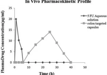
In vivo pharmacokinetics study
Figure 12 shows the plasma concentration vs. time
proiles of 5-FU after administration of ECHC and the 5-FU aqueous solution to New Zealand white rabbits at a dose of 10 mg/kg respectively. Colon speciic absorption of releasing 5-FU afected its pharmacokinetic parameters.
After oral administration of the 5-FU solution, the drug was
detected rapidly in plasma. The maximum concentration of 5-FU was 20 µg/mL after about 1 h. Thereafter, the plasma concentration decreased quickly and the drug
was not detectable as soon as 8 h. The elimination half
life was 0.12 ± 0.03 h. While in case of colon targeted
enteric coated capsules (ECHC), 5-FU appeared in plasma
after 6 h (lag time) of administration with a Cmax of
14 µg/mL at 25 h. A lag time of 6 h for the developed colon
targeted enteric coated capsules indicated the ability of the colon-targeted formulation to prevent the release of 5-FU in the stomach and small intestine. The Cmax, Tmax, t 1/2 and AUC 0-∞ were signiicantly diferent from those of the
aqueous solution. The Cmax of colon-targeted formulation
was signiicantly (p < 0.001) less than the Cmax of 5FU solution, suggested reduced systemic absorption of the drug from ECHC-3. This means that a larger fraction of the drug was available on the colonic surface for local action.AUC0–∝ for aqueous solution and
colon-targeted formulation was found to be 62.5543 µg h/mL and 49.3412 µg h/mL, respectively. Consequently the Fr
(relative bioavailability) of the colon-targeted tablet was
determined as 70.18%. Thus, in vivo pharmacokinetic studies of enteric coated HPMC capsules plugged with calcium pectinate beads and 5-FU loaded microsponges
exhibited increased lag time, delayed t max, decreased Cmax and reduced bioavailability. It can thus be concluded FIGURE 11 - X-ray images of New Zealand rabbit administered with colon targeted formulation (a) immediately after
administration, (b) 1.5 h (stomach), (c) 3.5 h (small intestine), (d) 5 h (intestinal colon junction), (e) 7.5 h (colon) and (f) 10 h
FIGURE 12 - In vivo pharmacokinetic proile of 5-FU in New
Zealand white rabbits following administration of 5-FU aqueous
solution and developed colon targeted capsules.
that the developed colon targeted formulation has the ability to avoid the drug release in the upper GIT, but can
release the active agent speciically in the colon exert local action with reduced systemic exposure.
CONCLUSION
A novel colon targeted drug delivery system that combined two approaches, i.e. pH-sensitive delivery as well as degradation by bacterial enzymes in the colon environment was successfully developed. This study clearly indicated that enteric coated HPMC capsules plugged with 5-FU loaded microsponges along with
15% calcium pectinate beads provided a tight control
over the drug release in the physiological environment of the stomach and small intestine in spite its high water solubility. Matt surface of HPMC capsule provided a good substrate for adhesion of enteric coating polymers and calcium pectinate beads also proved to be a suitable carrier for colon targeted system. Thus, the developed colon targeted drug delivery system proved to be more patient compliant by providing a better mode of treatment over present intermittent chemotherapy by injection or infusion.
ACKNOWLEDGEMENTS
The authors are thankful to IIT Kanpur and Diya Labs for providing PXRD, TGA and SEM facility. We would also like to acknowledge Shalaks Pharmaceuticals, Evonik Labs and ACG Associated Capsules Pvt. Ltd, SciTech Centre for generous gift samples of 5-FU, Eudragit polymers and HPMC capsules.
REFERENCES
BADVE, S. S.; SHER, P.; KORDE, A.; PAWAR, A. P.
Development of hollow/porous calcium pectinate beads for
loating-pulsatile drug delivery. Eur. J. Pharm. Biopharm.,
v.65, n.1, p.85-93, 2007.
BAE, S. E.; SON, J. S.; PARK, K.; HAN, D. K. Fabrication
of covered porous PLGA microspheres using hydrogen
peroxide for controlled drug delivery and regenerative
medicine. J. Control. Release, v.133, n.1, p.37-43, 2009.
B O U R G E O I S , S . ; G E R N E T, M . ; P R A D E A U , D . ; ANDREMONT, A.; FATTAL, E. Evaluation of critical
formulation parameters inluencing the bioactivity of
beta-lactamases entrapped in pectin beads. Int. J. Pharm., v.324,
n.1, p.2-9, 2006.
CHICKPETTY, S. M.; RAGA, B. V. Formulation, in vitro
drug release and in vivo human X-ray investigation of polysaccharide based drug delivery systems for targeting
5-luorouracil to the colon. Braz. J. Pharm. Sci., v. 49, n.2,
p.263-273, 2013.
COLE, E. T.; SCOTT, R. A.; CONNOR, A. L.; WILDING, I. R.; PETEREIT, H. U.; SCHMINKE, C. Enteric coated HPMC capsules designed to achieve intestinal targeting. Int. J. Pharm., v.231, n.1, p.83-95, 2002.
COSTA, P.; LOBO, J. M. S. Modeling and comparison of dissolution proiles. Eur. J. Pharm. Sci., v.13, n.2,
p.123-133, 2001.
DEVRIM, B.; CANEFE, K. Preparation and evaluation of
modified release ibuprofen microspheres with acrylic
polymers (Eudragit) by quasi emulsion solvent difusion method: efect of variables. Acta Pol. Pharm., v.63, n.6,
p.521-534, 2006.
GRAVES, R.; MOISEYEV, R.; PAMUJULA, S.; PRAETORIUS, N.; HAVEN, K.; BOSTANIAN, L. Spherical biodegradable
microsponge particles for drug delivery. Am. Assoc. Pharm. Sci. J., v.7, n.1, p.52, 2005.
HUYGHEBAERT, N.; VERMEIRE, A.; REMON, J. Alternative
method for enteric coating of HPMC capsules resulting in ready-to-use enteric-coated capsules. Eur. J. Pharm. Sci.,
JAIN, A.; GUPTA, Y.; JAIN, S.K. Potential of calcium pectinate
beads for target specific drug release to colon. J. Drug Target., v.15, n.4, p.285-294, 2007.
LEE, J. S.; CHAE, G. S.; KUN, A. T.; KHANG, G. Preparation
of 5-fluorouracil-loaded poly (l-lactide-co-glycolide) wafer and evaluation of in vitro release behaviour. Macromolecules, v.11, n.3, p.183-188, 2003.
MOAWIA, M.; TABAKHA, A. L. HPMC capsules: current status and future prospects. J. Pharm. Pharm. Sci.,v.13,
n.3, p.428-442, 2010.
ORLU, M.; CEVHER, E.; ARAMAN, A. Design and evaluation of colon speciic drug delivery system containing lurbiprofen microsponges. Int. J. Pharm., v.318, n.1-2,
p.103-117, 2006.
RAMASAMY, T. G.; KANDHASAMI, U. S.; RUTTALA, H.;
SHANMUGAM, S. Formulation and evaluation of xanthan
gum based aceclofenac tablets for colon targeted drug delivery. Braz. J. Pharm. Sci., v.47, n.2, p.299-311, 2011.
SHARMA, V.; PHILIP, A.K.; PATHAK, K. Modified polysaccharides as fast disintegrating excipients for orodispersible tablets of roxithromycin. AAPS Pharm. Sci. Tech., v.9, n.1, p.87-94, 2008.
S R I A M O R N S A K , P . ; T H I R A W O N G , N . ; PUTTIPIPATKHACHORN, S. Emulsion gel beads of
calcium pectinate capable of loating on the gastric luid:
effect of some additives, hardening agent or coating on release behavior of metronidazole. Eur. J. Pharm. Sci., v.24,
n.4, p.363-373, 2005.
SRIVASTAVA, R.; KUMAR, D.; PATHAK, K. Colonic luminal surface retention of meloxicam microsponges delivered by erosion based colon targeted matrix tablet. Int. J. Pharm.,
v.427, n.2, p.153-162, 2010.
WOLPIN, B. M.; MAYER, R. J. Systemic treatment of
colorectal cancer. Gastroenterology, v.134, n.5,
p.1296-1230, 2008.
ZHANG, Y.; LANG, M.; TANG, X.; LI, L.; SHEN, X.; Folate-functionalized nanoparticles for controlled 5-Fluorouracil delivery. J. Colloid Interface Sci., v.354, n.1, p.202-209,
2011.