MECHANISM OF BACTERICIDAL ACTIVITY OF SILVER NITRATE – A CONCENTRATION DEPENDENT
BI-FUNCTIONAL MOLECULE
Sureshbabu Ram Kumar Pandian 1, Venkataraman Deepak 1, Kalimuthu Kalishwaralal 1, Pushpa Viswanathan 2, Sangiliyandi Gurunathan 1*
1
Department of Biotechnology, Division of Molecular and Cellular Biology, Kalasalingam Academy of Research and Education,
Anand Nagar, Krishnankoil-626190, Tamilnadu, India; 2 Department of Electron microscopy, Cancer Institute (Women India
Association), Chennai – 600020, Tamilnadu, India.
Submitted: June 04, 2009; Returned to authors for corrections: September 08, 2009; Approved: February 18, 2010.
ABSTRACT
Silver nitrate imparts different functions on bacteria depending upon its concentration. At lower
concentration it induced synthesis of nanoparticles, whereas at higher concentrations it induced cell death.
Bacilluslicheniformis was used as model system. The MIC was 5 mM, and it induced catalase production,
apoptotic body formation and DNA fragmentation.
Key words: Silver nitrate, Bacillus licheniformis, nanoparticle synthesis, apoptosis, DNA fragmentation.
Gram positive bacteria are an important cause of serious
infections particularly form hospitals and are getting resistance
to many antibiotics (16). For quite a long time, silver has been
known to impart antimicrobial activity to bacteria. Prior to the
introduction of the Sulphadiazine cream, dilute solutions of
silver nitrate were used to treat infections in the 19th century
(10). Silver-based antimicrobials can be effective in the
treatment of infections on account non-toxicity of active Ag+ to
human cells (4). Interestingly, silver nitrate imparts different
functions to bacteria depending upon its concentration. At
higher concentrations, it kills bacteria, whereas, at lower
concentrations it induces them to synthesize silver
nanoparticles where Bacillus licheniformis (MTCC1483) is
used for analysis. The biosynthesis of silver nanoparticles, as
stated in our previous report (7) occurred at a concentration of
1mM of silver nitrate. This was visualized by Scanning
Electron Microscopy and XRD. The size of the nanoparticles
was ~50nm. Moreover, during the synthesis of silver
nanoparticles the bacteria remained alive, and resumed their
growth when the silver is removed from their environment (7).
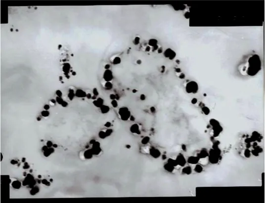
Also, the nanoparticles accumulated inside the cells during
incubation (Fig. 1). But, when the concentration of AgNO3 was
increased, it induced cell death in bacteria. To analyze the
antimicrobial function of AgNO3, bacteria was treated with
various concentrations of silver nitrate and incubated for 2
mins. Subsequently, 0.1 ml of the mixture was spread on
nutrient agar for the formation of colonies, where soluble
components in agar neutralized the action of silver. The result
showed that MIC of AgNO3 was 5 mM i.e. the organism
viability was completely inhibited when the concentration of
silver is 5 mM. Many mechanisms have been suggested to
explain the mode of bactericidal action of silver ions
previously. They are known to inhibit proteins by binding to
their thiol groups and denaturing them (9, 12) and prevent
replication of DNA by its condensation (12). One mechanism
under consideration in this report is Programmed Cell Death
(Apoptosis). Usually, Programmed Cell Death (PCD) is
associated with eukaryotic multicellular organisms. Recently,
PCD systems have been observed in bacteria also (3).
Therefore to elucidate the mechanism, primarily the
Figure 1. Transmission electron microscopic image of section of bacteria treated with 1 mM AgNO3 containing accumulated
silver nanoparticles inside the cell (at 50,000x).
morphology of the AgNO3 treated cells was continuously
monitored under phase contrast microscopy. On observation it
is noted that AgNO3 treated bacterial cells started to shrink
resulting in characteristic apoptotic body formation. Apoptotic
bodies were produced when the cell wall is permeabilized by
silver ions. This shows that AgNO3 may induce apoptosis in B.
licheniformis at higher concentration of 5 mM. Apoptotic
bodies are greatly shrunken compared with normal cells and
they are completely absent in cell populations not treated with
silver nitrate. Catalase is one of the known free radical
scavengers that scavenges hydrogen peroxide (2) a known
active free radical. To check whether AgNO3 induces the
catalase, an assay was carried out according to the method
described by Lui et al., 2006 (10). The production of catalase
was maximum at the MIC and decreased thereafter. When
Reactive Oxygen Species (ROS) levels increase the cells
was found to be more sensitive to both silver zeolite and silver
nitrate than its parent. The outbreak of oxidative stress in
bacteria is attributed to heavy metals like cadmium ions (9).
Silver ions are known to particularly inhibit thiol
group-containing enzymes, such as NADH dehydrogenase II in the
respiratory system, which is implicated as a candidate for the
site of production of reactive oxygen species in vivo (12).
Therefore, inhibition of this enzyme results in an increase in
the free radical production. The increase in catalase production
in the presence of ROS could be explained by the necessity for
cells to reduce the concentration of H2O2, which is the source
of the free radicals. It is proposed that reactive oxygen species
can induce apoptotic pathways in bacteria which could
ultimately lead to their death. Moreover, at higher
concentrations of substrate i.e. H2O2, catalase gets inactivated
is also observed in B. licheniformis. The DNA was found to
undergo fragmentation in silver nitrate-treated cells. The
fragmentation could be visualized in agarose gel with ethidium
bromide. During the treatment, DNA fragmentation could be
visualized in those cells which have been treated with
concentration of 5 mM of AgNO3 and above. Whereas, no
fragmentation was observed in those cells, that was treated
with lesser concentration of AgNO3 (Data not shown).
Induction of apoptosis can be confirmed by two factors,
irregular reduction in size of cells and DNA fragmantation. The
cells reduced in size are called shrunken cells or apoptotic
bodies (14) and DNA fragmentation is the final step in
apoptosis (1). Previously there are reports available for
induction of apoptosis in microorganisms. In E. coli, cell death
is induced by a toxin – antitoxin (mazE - mazF) mechanism.
The mazF gene encodes a stable toxin, MazF, while mazE
encodes a labile antitoxin, MazE, degraded in vivo by the
ATP-dependent ClpPA serine protease. When the antitoxin degrades
the cells undergo death (3). More interesting features are
observed in Xanthomonas sp. The bacteria undergo
programmed cell death under starvation. A caspase-3-like
enzyme activity and apoptotic bodies are implicated in this
organism, but not DNA fragmentation (5). A similar
phenomenon is observed also in Streptomyces sp, where both
apoptosis and a necrosis-like activity are observed (15). But in
Plasmodium falciparum, a human malarial parasite, DNA
fragmentation is observed when it is treated with
chloroquinone (8, 13). Moreover, antibiotics also induce
apoptosis in bacteria and current research is focused on new
apoptotis-inducing antibiotics (14). Therefore, silver ions from
silver nitrate are capable of inducing apoptosis in bacteria.
Moreover, the possible mechanisms for silver nanoparticle
synthesis and the bactericidal action of silver ions are shown in
Fig.2. At lower concentrations of silver nitrate, respiratory
nitrate reductase may be involved in the production of
nanoparticles (7). But at higher concentrations silver nitrate
inhibits the action of proteins by reacting with their thiol
groups (5) and also binds with the DNA, thus arresting its
replication (12). Our results show that catalase production
increases up to MIC and decreases thereafter. Catalase is a
known scavenger of ROS; however its protective action seems
to be restricted to low concentrations of silver nitrate (below
the MIC). Catalase may also be inhibited by high
concentrations of its substrate, hydrogen peroxide. Therefore,
silver nitrate may induce apoptosis in bacteria through many
ways.
As many organisms are developing resistance to drugs like
methycillin resistant Staphylococcus aureus, penicillin resistant
Streptococcus pneumonia which are highly associated with
infections at hospitals (11), other molecules with antimicrobial
activity are under research. The molecule can be exploited
completely if the mechanism of action is known. It can be
concluded that formation of shrunken cells and DNA
fragmentation are induced by high concentrations of silver
nitrate in B. licheniformis. However, the complete mechanism
behind cell death is not clear. But it can be concluded that
apoptosis is the major effect of the bactericidal action of silver
Figure 2. Possible mechanisms of the duality in functions of silver nitrate in bacteria. A - The left side of the figure shows the
possible mechanism of silver nanoparticle synthesis at lower concentration which may involve nitrate reductase enzyme. B - The
right side of the figure shows the possible mechanism for the induction of apoptosis by silver nitrate which may involve
ACKNOWLEDGEMENTS
The authors gratefully acknowledge the Tamilnadu State
Council for Science and Technology (TNSCST), Council of
Scientific and Industrial Research (CSIR) (Grant No:
37(1384)/09/EMR-II) and Dr H. Nellaiah for critically reading
of the manuscript.
REFERENCES
1. Bortner, C.D.; Cidlowski, J.A. (2002). Apoptotic volume decrease and the incredible shrinking cell. Cell. Death. Differ., 9, 1307 – 1310. 2. Cabiscol, E.; Tamarit, J.; Ros, J. (2000). Oxidative stress in bacteria and
protein damage by reactive oxygen species. Internatl. Microbiol., 3, 3–8. 3. Engelberg-Kulka, H.; Amitai, S.; Kolodkin-Gal, I.; Hazan, R. (2006).
Bacterial Programmed Cell Death and Multicellular Behavior in Bacteria. PLOS. Genetics., 10, 135.
4. Ewald, A.; Susanne, K.; Thull, R.G.; Gbureck, U. (2006). Antimicrobial titanium/silver PVD coatings on titanium. BioMed. Eng. OnLine., 5, 22. 5. Gautam, A.S.; Sharma, A. (2002). Involvement of caspase-3 like protein
in rapid cell death of Xanthomonas. Mol. Microbiol., 44, 393–401. 6. Hua, Z.J.; Xu, M. (2000). DNA fragmentation in apoptosis. Cell Res., 10,
205-211.
7. Kalimuthu, K.; Sureshbabu, R.; Venkatraman, D.; Bilal, M.; Gurunathan, S. (2008). Biosynthesis of silver nanocrystals by Bacillus licheniformis. Colloids. Surf. B. Biointerfaces., 65,150-153.
8. Lewis, K. (2000). Programmed Death in Bacteria. Microbiol. Mol. Biol. Rev., 64, 503–514.
9. Liau, S.Y.; Read, D.C.; Pugh, W.J.; Furr, J.R.; Russell, A.D. (1997). Interaction of silver nitrate with readily identifiable groups: relationship to the antibacterial action of silver ions. Lett. Appl. Microbiol., 25, 279– 283.
10. Lui, S.L.; Vincent, K.M.P.; Lung, I.; Burd, A. (2006). Antimicrobial activities of silver dressings: an in vitro comparison Margaret Ip. J. Med. Microbiol., 55, 59–63.
11. Maeno, E.; Ishizaki, Y.; Kanaseki, T.; Hazama, A.; Okada, Y. (2000). Normotonic cell shrinkage because of disordered volume regulation is an early prerequisite to apoptosis. PNAS. Early. Edition., 1-6.
12. Matsumura, Y.; Yoshikata, K.; Kunisak, S.; Tsuchido, T. (2003). Mode of Bactericidal Action of Silver Zeolite and Its Comparison with That of Silver Nitrate. Appl. Environ. Microbiol., 69, 4278–4281.
13. Picot, S.; Burnod, J.; Bracchi, V.; Chumpitazi, B.F.; Ambroise, T.P. (1997). Apoptosis related to chloroquine sensitivity of the human malaria parasite. Plasmodium falciparum. Trans. R. Soc. Trop. Med. Hyg., 91, 590–591.
14. Sat, B.; Hazan, R.; Fisher, T.; Khaner, H.; Glaser, G.; Engelberg-Kulka, H. (2001). Programmed Cell Death in Escherichia coli: Some Antibiotics Can Trigger mazEF Lethality. J. Bacterial., 183, 2041–2045.
15. Shirkhanzadeh, M.; Azadegan, M.; Liu, G. Q. (1995). Bioactive delivery systems for the slow release of antibiotics: incorporation of Ag+ ions into micro-porous hydroxyapatite coatings. Mater. Lett., 24, 7–12.
16. Tally, F.P.; DeBruin, M.F. (2000). Development of daptomycin for gram positive infections. J. Antimicrob. Chemother., 6, 523- 526.