https://doi.org/10.1590/0004-282X20170059 ARTICLE
The direct first pass aspiration technique
in the treatment of acute ischemic stroke
resulting from large vessel occlusions
Técnica de aspiração primária no tratamento do acidente vascular cerebral isquêmico por
oclusões de grandes vasos
Luís Henrique de Castro-Afonso1, Guilherme Seizem Nakiri1, Lucas Moretti Monsignore1, Pedro Telles Cougo-Pinto2,
Francisco Antunes Dias2, Frederico Aléssio-Alves2, Octávio Marques Pontes-Neto2, Daniel Giansante Abud1
Mechanical thrombectomy, using stent retrievers as an adjunct to intravenous thrombolysis, is the standard treatment for acute ischemic stroke (AIS) that results from carotid or proximal middle cerebral artery occlusions1,2,3.
This treatment strategy improves functional outcomes for patients if started within the first six hours of symptom onset. More complete and faster recanalization and more pronounced brain collaterals are among the most impor-tant variables directly associated with better neurologic outcomes after endovascular treatment of AIS. Although
these concepts were already known, the introduction of stent retrievers in the endovascular armamentarium rep-resented a cornerstone for achieving more complete and faster recanalizations, which consistently improves neu-rologic outcomes across results of recent trials2,3. Despite
emerging endovascular stent retriever technology, AIS resulting from large vessel occlusions (LVOs) remains a serious condition. In general, even among patients receiv-ing the best stroke management, AIS from LVOs leads to poor functional outcomes 50% to 60% of the time and
1Universidade de São Paulo, Faculdade de Medicina de Ribeirão Preto, Divisão de Neurorradiologia Intervencionista, Ribeirão Preto SP, Brasil; 2Universidade de São Paulo, Faculdade de Medicina de Ribeirão Preto, Divisão de Neurologia, Ribeirão Preto SP, Brasil.
Correspondence: Daniel Giansante Abud; Divisão de Neurorradiologia Intervencionista, Faculdade de Medicina de Ribeirão Preto da USP; Avenida Bandeirantes, 3900; 4048-090 Ribeirão Preto SP, Brasil; E-mail: [email protected]
Conflict of interest: There is no conlict of interest to declare.
Received 12 July 2016; Received in inal form 17 October 2016; Accepted 20 December 2016. ABSTRACT
Mechanical thrombectomy using stent retrievers is the standard treatment for acute ischemic stroke that results from large vessel occlusions. The direct aspiration irst pass technique (ADAPT) has been proposed as an eficient, fast, and cost-effective thrombectomy strategy. The aim of this study was to assess the safety and eficacy of ADAPT. Methods:Recanalization was assessed using the modiied thrombolysis in cerebral infarction (mTICI) score. Neurological outcomes were assessed using the National Institutes of Health Stroke Scale and modiied Rankin Scale. Results: Fifteen patients were evaluated. The mTICI score was 2b-3 in 80%, and it was 3 in 60% of patients. No intracranial hemorrhage was seen. At three months, modiied Rankin Scale scores ≤ 2 were observed in 60% of patients and the mortality rate was 13.3%. Conclusions: The ADAPT appears to be a safe, effective, and fast recanalization strategy for treatment of acute ischemic stroke resulting from large vessel occlusions.
Key words: stroke; stents; catheters.
RESUMO
A trombectomia mecânica com stent retrievers é o tratamento padrão ouro do acidente vascular cerebral isquêmico agudo (AVCi) por oclusão de grandes artérias. A técnica de aspiração primária (ADAPT) tem sido proposta como uma estratégia de trombectomia rápida e com boa custo-efetividade. O objetivo deste estudo foi avaliar a segurança e eicácia da técnica ADAPT. Métodos: A recanalização foi avaliada utilizando a escala mTICI. Os desfechos neurológicos foram avaliados utilizando as escalas do NIHSS e mRS. Resultados: Quinze pacientes foram avaliados. Foram obtidas taxas de mTICI = 2b-3 em 80% e TICI = 3 em 60% dos pacientes. Não ocorreram hemorragias intracranianas. Em 3 meses as taxas de mRS≤2 e mortalidade foram respectivamente 60% e 13.3%. Conclusão: A técnica ADAPT parece ser uma estratégia de recanalização rápida, segura e efetiva para o tratamento do AVC por oclusão de grandes artérias.
mortality 10% to 20% of the time2,3. Therefore,
ameliora-tions in stroke care are still needed and, in this context, strategies to improve complete recanalization rates and reduce procedure times should be continuously pursued. The direct aspiration first pass technique (ADAPT), using large bore aspiration catheters, has been proposed as an efficient, fast, and cost-effective thrombectomy strategy and may improve rates of complete recanalization while reducing procedure times4,5,6,7,8,9,10,11,12.
he aim of this study was to assess the safety and ei -cacy of ADAPT using the 5MAX-ACE catheter (Penumbra, Oakland, California, USA) for treatment of acute stroke resulting from LVOs.
METHODS
Patients, clinical and imaging assessments, and follow-up
We prospectively evaluated data from 15 consecutive patients who underwent mechanical thrombectomy for AIS secondary to LVO between November 2015 and January
2016. he study was approved by the ethics committee at
our institution. Patients, or their legal representatives, signed consent forms, which were previously approved by the institutional review board. All patients underwent a brain CT scan and a supra-aortic vessel CT angiography at admission to assess the arterial occlusion site. On the preprocedure brain CT, the Alberta Stroke Program Early Computed Tomography Score (ASPECTS) was assessed
for all patients. he National Institutes of Health Stroke
Scale was determined by stroke neurologists upon patient admission to hospital and again 24 hours after admission.
he modiied Rankin Scale (mRS) was assessed on admis -sion and at the three-month follow-up by a stroke neurolo-gist. Of 15 patients treated at our institution, nine (60.0%) were men, and the mean age was 66.1 years (SD = 13).
Baseline National Institutes of Health Stroke Scales ranged from 6 to 30 (mean = 21.2, SD = 7.4), and baseline mRS
ranged from 0 to 3 (median = 0, mean = 0.7, SD = 1).
In general, patients were assessed for eligibility for intravenous thrombolysis using the National Institutes of Neurological Disorders and the European Cooperative Stroke Study 3 trial criteria13,14. If indicated, patients
pre-senting with LVOs received intravenous thrombolysis and were immediately referred for thrombectomy. We included patients whose ASPECTS scores were at least 6 within the
irst six hours of symptom onset. We did not deine a lim -ited time window for performing endovascular treatment of
posterior circulation stroke. We also did not deine speciic cut-ofs in patient age, previous clinical conditions, base
-line National Institutes of Health Stroke Scale, or base-line mRS for indicating thrombectomy. Patients presenting with
LVOs who were ineligible for intravenous ibrinolysis were
treated with direct thrombectomy.
Endovascular procedure
Each patient was transferred to our angiography suite, where thrombectomy was performed by our interventional neuroradiology team using ADAPT with a 5MAX-ACE cath-eter (Penumbra, Oakland, CA, USA). Local anesthesia and conscious sedation were used. General anesthesia via intuba-tion was performed if necessary at the discreintuba-tion of the
neu-rointerventional staf and the anesthesiologist.
All procedures were performed using the femoral artery approach. An intravenous bolus (5,000 IU) of standard heparin was administered after puncture if intravenous thrombolysis was not previously indicated. If intravenous thrombolysis was indicated prior to the endovascular procedure, no heparin was administered after the femo-ral puncture. An 8-Fr guiding catheter (Guider Softip; Boston Scientific, Natick, MA) or a NeuronMax 088 sheath (Penumbra, Alameda, CA) or a 7-Fr Destination (Pinacle-Terumo) was introduced through a femoral sheath into the internal carotid artery or the most navi-gable vertebral artery. The guiding catheter was continu-ously perfused with 10 mg milrinone diluted in 1,000 mL of physiological saline (0.9%). Frontal, oblique, and lateral angiographies were completed to determine the cervical vessel related to the ischemic territory of the brain and to define the occluded intracranial vessels.
If a cervical carotid occlusion was identified, a Wallstent (Boston Scientific Target, Fremont, CA, USA) was used to perform an angioplasty stenting procedure. After this step, or if no occlusion was identified in the proximal cervical artery, a coaxial system was used to navigate to the arterial occlusion. The system consisted of a 5MAX-ACE (Penumbra), a microcatheter (3MAX [Penumbra], or Velocity (Penumbra), or a 0.027-inch
Rebar 27 microcatheter [Medtronic, Irvine, CA, USA]
thrombectomy using a stent retriever (Solitaire-FR
[Microvention-Covidien, Irvine, CA] or Trevo [Stryker, Fremont, CA]) if recanalization could not be achieved after five attempts to aspirate the thrombus, or if the angiogram revealed a distal occlusion after aspiration. Figures 1 and 2 show examples of the procedure.
Successful recanalization was deined as a modiied
thrombolysis in cerebral infarction (mTICI) score of 2b or 3 in all treatable vessels. No intra-arterial thrombolysis was administered, even if the recanalization was unsuccess-ful. Groin punctures were closed with Angio-Seal (St. Jude Medical, St. Paul, MN).
Statistical analysis
Continuous variables are presented as mean (range,
± standard deviation [SD]) or median (interquartile [IQR]).
Categorical data are presented as numbers and percentages.
he IBM SPSS Statistics software version 20.0 (Chicago,
IL, USA) was used for statistical analysis.
RESULTS
he ASPECTS scores on CT scans at admission ranged from 6 to10 (median = 10, mean = 9, SD = 1.4). he mTICI
score was 2b-3 for 80% (12/15) of patients, whereas it was 3 for
60% (9/15) of patients. he median number of device passes
was 2. Intravenous thrombolysis was performed in 46.7% of patients. Stent retrievers were used in two patients (13.3%). Embolization to other vascular territories was observed in one patient (6.6%) during traction of an entrapped 5MAX-ACE catheter from the basilar artery; the right post-inferior cerebel-lar artery remained occluded after catheter retrieval.
Procedure times ranged from 15 to 120 minutes (mean = 60.6, SD = 31.3). We had no intracranial hemor-rhage, nor any kind of hemorrhagic transformation. At three
months, the mRS ranged from 2 to 6 (median = 2, mean = 3.2,
SD = 1.6), and it was 2 or less for 60% (9/15) of the patients.
he mortality rate was 13.3% (2/15). Individual patient data
are summarized in Table 1. Baseline patient data results are
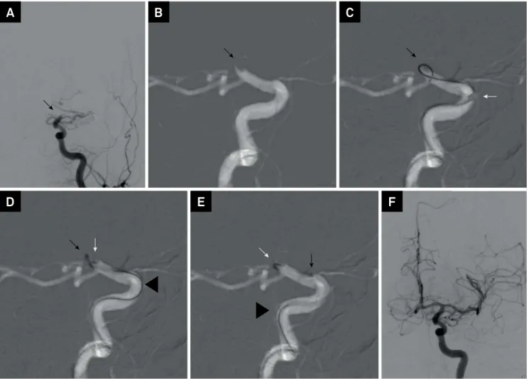
Figure 1. (A) Digital subtraction angiography (DSA), frontal view, shows an occlusion of the left internal carotid artery (LICA) (arrow); (B) road map of the LICA, lateral view, shows the occlusion at the distal carotid artery (arrow); (C) figure shows a micro-wire in the thrombus (black arrow) and the distal tip of the 5MAX-ACE catheter (white arrow); (D) a microcatheter is inserted into the thrombus (black arrow) and the distal tip of the 5MAX-ACE is engaged in the thrombus (white arrow) while micro-wire is removed (arrowhead); (E) both micro-wire (arrowhead) and microcatheter (black arrow) been removed while the 5MAX-ACE catheter remains into the thrombus ready for aspiration; (F) DSA frontal view, shows a complete recanalization of the LICA territory.
A
B
C
aggregated and summarized in Tables 2 and 3. Table 4 sum-marizes all studies published on thrombus aspiration using large bore catheters for treatment of acute stroke.
DISCUSSION
Recent trials have consistently proven the clinical ben
-eits of mechanical thrombectomy using stent retrievers for
AIS secondary to distal carotid or proximal middle cerebral artery occlusions1,2,3. A recent meta-analysis showed that
endovascular treatment resulted in good functional
neu-rologic outcomes (mRS = 0-2) for 54% of the patients and excellent functional neurologic outcomes (mRS = 0–1) for 36% of patients. he rates of symptomatic intracranial hem
-orrhage and mortality were 2.5% and 12%, respectively. he recanalization rate (mTICI = 2b–3) was 71% with a mean
procedure time (groin puncture to maximum mTICI score) of 38 minutes (range from 24 to 60 minutes). A complete recanalization (mTICI = 3) was achieved in 33% of patients2,3.
When performing an endovascular treatment of AIS, procedure times and recanalization rates are two important
variables directly associated with better outcomes;
there-fore, eforts have been made to reduce procedure times and
improve recanalization rates. Large bore aspiration devices are emerging thrombectomy devices, and they have been investigated to enhance recanalization and allow faster
procedures. he 5MAX-ACE is a large bore, highly lexible,
and atraumatic catheter capable of navigating through intracranial arteries, allowing for thrombus aspiration. Few studies have proposed a thrombectomy technique using simultaneous stent retrieval and aspiration with a distal access catheter15,16. However, despite these new
thrombectomy strategies and devices, they were not com-pared head-to-head with stent retrieval alone or while using ADAPT. In this setting, ADAPT appears to be a more
ratio-nal and cost-efective strategy12,17.
In our experience, the 5MAX-ACE catheter could be safely navigated to a variety of distal M1 segments of the middle cerebral arteries or to a selection of proxi-mal P1 segments of non-hypoplasic cerebral posterior arteries that present with favorable angles. We also noted an advantage of the 5MAX-ACE: it can be used to assess difficult cervical arteries before proceeding with
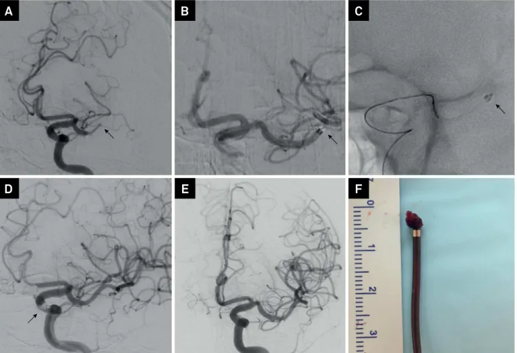
Figure 2. (A) Digital subtraction angiography (DSA) of LICA, oblique view, shows an occlusion of the distal M1 segment of the left middle cerebral artery (MCA) (arrow); (B) distal tip of the 5MAX-ACE is engaged in the thrombus (arrow); (C) distal tip of the 5MAX-ACE and a micro-catheter engaged in the thrombus (arrow); (D) DSA of LICA, oblique view, shows the exact moment of thrombus is removed from circulation entrapped in the 5MAX-ACE catheter (arrow); (E) DSA frontal view, shows a complete recanalization of the LICA territory; (F) picture of thrombus entrapped in the 5MAX-ACE catheter.
A
B
C
intracranial thrombectomy. The coaxial system allows an atraumatic distal cervical artery position of the guide catheter, even passing though critical carotid or verte-bral loops or kinks. Moreover, if aspiration alone fails, a stent retriever can be used through the large bore cath-eter, and retrieval can be performed under distal aspira-tion with the large bore catheter.
When compared to recent thrombectomy trials2,3,
we obtained unexpectedly higher rates of good clinical outcomes, considering that 40% of our patients had basilar occlusions, 20% had carotid tandem occlusions, 60% had mean recanalization times greater than six hours, and 13% had unknown times of symptom onset18,19. We obtained
a relatively high rate of mTICI = 3 (60%), a low rate of adjunctive use of a stent retriever (13%), and a low rate of emboli after thrombectomy (6.6%). Another interest-ing findinterest-ing was our zero incidence of intracranial hemor-rhage, especially considering that 80% of the patients had mTICI scores of 2b-3, and 73% were treated outside the level A evidence, which is less than six hours. Our mean
Table 1. Individual data of patients.
Patient Gender / Age
NIHSS at admission / 24 hours after
treatment
Aspects
Symptomatic intracranial hemorrhage
Artery site occluded
IV rTPA / Intubation
Recanalization time (minutes)
Procedure time (minutes)
Device passes(n)
/ Carotid stenting
mTICI
Baseline mRS / 3-months
mRS
1 M / 74 12/ago 10 N Basilar
artery N / N - 120 5 / N 0 / 3 0 / 2
2 F / 65 30/abr 10 N Basilar
artery Y / Y 810 50 2 / N 3 0 / 2
3 M / 60 19/abr 10 N Basilar
artery N / N 355 35 1 / N 3 0 / 2
4 M / 62 24 / 20 10 N Distal LICA N / N 480 60 3 / N 3 0 / 3
5 F / 76 26 / 26 9 N
Tandem proximal and distal
RICA
Y / Y - 120 7 / Y 0 0 / 6
6 M / 61 22/jul 10 N Proximal
left M1 N / N 255 15 1 / N 3 1/2
7 M / 34 20 / 18 9 N Distal LICA N / N 350 60 2 / N 3 0 / 2
8 M / 66 28 / 30 10 N Basilar
artery Y / Y 480 45 2 / N 2b 0 / 5
9 F / 73 30 / 30 6 N Basilar
artery Y / Y 475 30 2 / N 3 0 / 5
10 M / 52 14/dez 10 N
Tandem proximal RICA + right M1
N / N 450 80 1 / Y 3 2/2
11 M / 62 12/dez 8 N
Tandem proximal and distal
RICA
N / N 600 60 3 / Y 2b 2/2
12 F / 68 06/jun 6 N Basilar
artery N / N 450 40 1 / N 3 2/2
13 F / 79 30 / 30 7 N Distal LICA Y / Y 320 45 3 / N 2b 0 / 6
14 M / 68 25/dez 10 N Distal LICA Y / N 375 50 3 / N 3 0 / 2
15 F / 92 25 / 23 7 N Proximal
left M1 N / Y - 100 8 / N
2a /
2a 3/5
M: male; F: female; Y: yes; N: no; NIHSS: National Institutes of Health Stroke Scale; mRS: modiied Rankin Scale; mTICI: modiied thrombolysis in cerebral infarction score; ASPECTS: Alberta Stroke Program Early Computed Tomography Score; RICA: right internal carotid artery; LICA: left internal carotid artery.
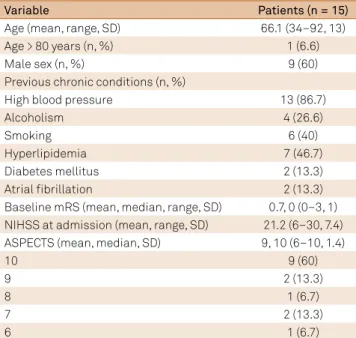
Table 2. Aggregated baseline clinical and radiologic data of patients. Variable Patients (n = 15)
Age (mean, range, SD) 66.1 (34–92, 13)
Age > 80 years (n, %) 1 (6.6)
Male sex (n, %) 9 (60)
Previous chronic conditions (n, %)
High blood pressure 13 (86.7)
Alcoholism 4 (26.6)
Smoking 6 (40)
Hyperlipidemia 7 (46.7)
Diabetes mellitus 2 (13.3)
Atrial ibrillation 2 (13.3)
Baseline mRS (mean, median, range, SD) 0.7, 0 (0–3, 1) NIHSS at admission (mean, range, SD) 21.2 (6–30, 7.4)
ASPECTS (mean, median, SD) 9, 10 (6–10, 1.4)
10 9 (60)
9 2 (13.3)
8 1 (6.7)
7 2 (13.3)
6 1 (6.7)
procedure time (one hour) was relatively long when com-pared to results of previous studies2-12. We believe that
this finding may be explained by the use of an adjunctive stent retriever in two patients, which was demonstrated to extend procedure time by 21 minutes20. Where
recana-lization could not be achieved, we considered a maximum procedure time of 120 minutes. In addition, before prepar-ing thrombectomy devices and systems, we routinely per-formed a diagnostic angiography, which may have length-ened overall procedure time ( from puncture to maximum mTICI). Although this strategy may lengthen proce-dure times, intravenous thrombolysis eventually opened
the vessel, and we were able to save devices. However,
if thrombectomy devices were prepared and ready for use before femoral puncture, overall procedure time would certainly have been shortened, but with a probability of finding an opened intracranial vessel at the first angio-graphic run.
More data must be collected to conirm whether the
ADAPT used in conjunction with stent retriever devices, compared to use of stent retrievers alone, would improve outcomes for patients presenting with AIS due to LVOs.
Moreover, studies are needed to deine the best recanaliza -tion strategy, aspira-tion or stentrievers, for distal occlusions aiming to achieve mTICI 3.
Despite encouraging results using ADAPT in our series,
these indings may have an inherent statistical bias because
of the small sample, and our data is exposed to random
efects. Another limitation of this study is the lack of a
control group.
We ind that ADAPT appears to be a safe, efective, and
fast recanalization strategy for treatment of acute ischemic stroke resulting from large vessel occlusions.
Table 3. Aggregated results.
Variable Patients (n= 15)
Site of vessel occlusion (n, %)
Tandem carotid artery 3 (20)
Distal carotid artery 4 (26.6)
Middle cerebral artery M1 2 (13.3)
Basilar artery 6 (40)
General anesthesia (n, %) 6 (40)
Device passes (median, mean, range, SD) 2, 2.6 (1–7, 1.6)
Recanalization mTICI = 2b-3 (n, %) 12 (80)
Recanalization mTICI = 3 (n, %) 9 (60)
Received intravenous r-tPA (n, %) 7 (46.7)
Use of adjunctive stentriever (n, %) 2 (13.3)
Embolization (n, %) 1 (6.6)
Procedure time (minutes, mean, range, SD) 60.6 (15–120, 31.3)
Time from symptoms onset to
recanalization (min) (mean, range, SD) 475.3 (255–810, 167)
Time from symptoms onset to
recanalization > 6 h (n, %) 9 (60)
Unknown time of symptoms onset (n, %) 2 (13.3)
Any intracranial hemorrhage (n, %) 0 (0.0)
Symptomatic intracranial hemorrhage
(n, %) 0 (0.0)
NIHSS at 24 hours (mean, range, SD) 16.1 (4–30, 9.7)
mRS at 3 months (mean, median,
range, SD) 3.2, 2 (2–6, 1.6)
mRS ≤ 2 at 3 months (n, %) 9 (60)
mRS = 6 (Mortality, n, %) 2 (13.3)
mTICI: modiied thrombolysis in cerebral infartcion score; r-TPA: recombinant tissue plasminogen activator; NIHSS: National Institutes of Health Stroke Scale; mRS: modiied Rankin Scale; SD: standard deviation.
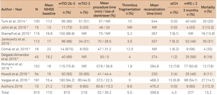
Table 4. An overview of the published studies on mechanical thrombectomy using new large bore aspiration devices.
Author - Year N
Mean NIHSS baseline
mTICI 2b-3 mTICI 3 Mean procedure time
(min) / Use of stentriever (%)
Thrombus fragmentation
n (%)
Mean recanalization
time (min)
sICH mRS ≤ 2
Mortality n (%)
n (%) n (%) n (%) 3 months
n (%)
Turk et al. 2014 4 100 17.2 95 (95) 51 (51) 37 / NR 10 544 0 (0) 40 (40) 20 (20)
John et al. 2015 5 15 13 11 (73) 5 (33) 46 / 33 NR NR 0 (0) 4 (33) 2 (13.3)
Stampl et al. 2015 6 115 16.8 100 (86.9) NR 73 / NR 5.2 267 7 (6.1) NR 16 (13.9)
Jankowitz et al.
2015 7 112 17 96 (86) 34 (31) 70 / 28.5 3.5 337 7 (6.2) 52 (46) 35 (31)
Comai et al. 2015 8 16 22 14 (87.5) 8 (50) 47 / 31.2 12.5 NR 1 (6.2) 9 (56) 4 (25)
Delgado Almandoz
et al. 2015 9 45 19.2 40 (89) NR 50 / 0 4 274 1 (2) 25 (56) 8 (18)
Romano et al.
2016 10 152 19 115 (75.6) NR 57.8 / 36.8 1.9 284.8 12 (7.8) 77 (50.6) 12 (7.8)
Kowoll et al. 2016 11 54 15 50 (93) 35 (65) 41 / 44.4 6 220 2 (4) 25 (46) 6 (11)
Vargas et al. 2016 12 191 15.4 180 (94.2) 85 (44.5) 37.3 / 22.5 0 468.3 13 (6.8) 98 (54.1) 27 (14.1)
Authors 2016 15 21.2 12 (80) 9 (60) 60.6 / 13.3 6.6 475.3 0 (0) 9 (60) 2 (13.3)
Total 815 17.5 87.5 27.8 52 / 26.2 5.5 358.8 4,3 37,7 13,2
References
1. Powers WJ, Derdeyn CP, Biller J, Coffey CS, Hoh BL, Jauch et al. Guidelines for the Early Management of Patients With Acute Ischemic Stroke Regarding Endovascular Treatment: A Guideline for Healthcare Professionals From the American Heart Association/American Stroke Association. Stroke.
2015;46(10):3020-35. https://doi.org/10.1161/STR.0000000000000074
2. Goyal M, Menon BK, Zwam WH, Dippel DW, Mitchell PJ,
Demchuk AM et al. Endovascular thrombectomy after large-vessel ischaemic stroke: a meta-analysis of individual patient data from ive randomised trials. Lancet. 2016;387(10029):1723-31. https://doi.org/10.1016/S0140-6736(16)00163-X
3. Campbell BC, Hill MD, Rubiera M, Menon BK, Demchuk A, Donnan GA et al. Safety and eficacy of solitaire stent thrombectomy: individual patient data meta-analysis of randomized trials. Stroke. 2016;47(3):798-806. https://doi.org/10.1161/STROKEAHA.115.012360
4. Turk AS, Spiotta A, Frei D, Mocco J, Baxter B, Fiorella D, et al. Initial clinical experience with the ADAPT technique: a direct aspiration irst pass technique for stroke thrombectomy. J Neurointerv Surg. 2014;6(3):231-7. https://doi.org/10.1136/neurintsurg-2013-010713
5. John S, Hussain MS, Toth G, Bain M, Uchino K, Hui FK. Initial experience using the 5MAX™ ACE reperfusion catheter in intra-arterial therapy for acute ischemic stroke. J Cerebrovasc Endovasc Neurosurg. 2014;16(4):350-7. https://doi.org/10.7461/jcen.2014.16.4.350
6. Stampl S, Kabbasch C, Muller M, Mpotsaris A, Brockmann M,
Liebig T et al. Initial experience with a new distal intermediate and aspiration catheter in the treatment of acute ischemic stroke: clinical safety and eficacy. J Neurointerv Surg. 2016;8(7):714-8. https://doi.org/10.1136/neurintsurg-2015-011801.
7. Jankowitz B, Grandhi R, Horev A, Aghaebrahim A, Jadhav A, Linares G et al. Primary manual aspiration thrombectomy (MAT) for acute ischemic stroke: safety, feasibility and outcomes in 112 consecutive patients. J Neurointerv Surg. 2015;7(1):27-31. https://doi.org/10.1136/neurintsurg-2013-011024
8. Comai A, Haglmüller T, Ferro F, Dall’Ora E, Currò Dossi R, Bonatti G. Sequential endovascular thrombectomy approach (SETA) to acute ischemic stroke: preliminary single-centre results and cost analysis. Radiol Med (Torino). 2015;120(7):655-61. https://doi.org/10.1007/s11547-015-0501-9
9. Delgado Almandoz JE, Kayan Y, Young ML, Fease JL, Scholz JM, Milner AM et al. Comparison of clinical outcomes in patients with acute ischemic strokes treated with mechanical thrombectomy using either Solumbra or ADAPT techniques. J Neurointerv Surg. 2015;8(11):1123-8. http://dx.doi.org/10.1136/neurintsurg-2015-012122
10. Romano DG, Cioni S, Vinci SL, Pero G, Comelli C, Comai A et al.. Thromboaspiration technique as irst approach for approach for endovascular treatment of acute ischemic stroke: initial experience at nine Italian stroke centers. J Neurointerv Surg. 2017;9(1):6-10. https://doi.org/10.1136/neurintsurg-2016-012298
11. Kowoll A, Weber A, Mpotsaris A, Behme D, Weber W.
Direct aspiration first pass technique for the treatment of acute ischemic stroke: initial experience at a European stroke center. J Neurointerv Surg. 2016;8(3):230-4. https://doi.org/10.1136/neurintsurg-2014-011520
12. Vargas J, Spiotta A, Fargen K, Turner R, Chaudry I, Turk A. Long term experience using the ADAPT technique for the treatment of acute ischemic stroke. NeuroIntervent Surg. 2016;pii: neurintsurg-2015-012211. https://doi.org/10.1136/neurintsurg-2015-012211
13. Hacke W, Kaste M, Bluhmki E, Brozman M, Dávalos A, Guidetti D et al. Thrombolysis with alteplase 3 to 4.5 hours after acute ischemic stroke. N Engl J Med. 2008;359(13):1317-29. https://doi.org/10.1056/NEJMoa0804656
14. The National Institute of Neurological Disorders and Stroke, rt-PA Stroke Study Group. Tissue plasminogen activator for acute ischemic stroke. N Engl J Med. 1995;333(24):1581-7. https://doi.org/10.1056/NEJM199512143332401
15. Humphries W, Hoit D, Doss VT, Elijovich L, Frei D, Loy D et al. Distal aspiration with retrievable stent assisted thrombectomy for the treatment of acute ischemic stroke. J Neurointerv Surg. 2015;7(2):90-4. https://doi.org/10.1136/neurintsurg-2013-010986
16. Massari F, Henninger N, Lozano JD, Patel A, Kuhn AL,
Howk M et al. ARTS (Aspiration-Retriever Technique for Stroke): initial clinical experience. Interv Neuroradiol. 2016;22(3):325-32. https://doi.org/10.1177/1591019916632369
17. Turk AS, Turner R, Spiotta A, Vargas J, Holmstedt C, Ozark S et al. Comparison of endovascular treatment approaches for acute ischemic stroke: cost effectiveness, technical success, and clinical outcomes. J Neurointerv Surg. 2015;7(9):666-70. https://doi.org/10.1136/neurintsurg-2014-011282
18. Son S, Choi DS, Oh MK, Hong J, Kim SK, Kang H et al. Comparison of Solitaire thrombectomy and Penumbra suction thrombectomy in patients with acute ischemic stroke caused by basilar artery occlusion. J Neurointerv Surg. 2016;8(1):13-8. https://doi.org/10.1136/neurintsurg-2014-011472
19. Mokin M, Sonig A, Sivakanthan S, Ren Z, Elijovich L, Arthur A et al. Clinical and procedural predictors of outcomes from the endovascular treatment of posterior circulation strokes. Stroke. 2016;47(3):782-8. https://doi.org/10.1161/STROKEAHA.115.011598