https://doi.org/10.1590/0004-282X20170074
REVIEW
Effect of intra-hippocampal injection of
human recombinant growth hormone on
synaptic plasticity in the nucleus basalis
magnocellularis-lesioned aged rats
Efeito da injeção intrahipocampal de hormônio do crescimento humano recombinante sobre
a plasticidade sináptica em ratos envelhecidos lesados do núcleo basalis magnocellularis
Maryam Malek1, Alireza Sarkaki2,4, Saleh Zahedi- Asl3, Yaghuob Farbood2,4, Ziba Rajaei1
Many common age-related problems are due to neuro-endocrine phenomena including memory impairment and
Alzheimer’s disease (AD)1. Alzheimer’s disease is a senile
neu-rodegenerative disorder with speciic pathological changes
in the brain that can cause progressive dementia2 and is
characterized by serious memory problems3. he causes
and mechanisms of AD are still under intensive
investiga-tion4. According to the cholinergic hypothesis, high
destruc-tion of basal forebrain cholinergic neurons, particularly the nucleus basalis magnocellularis (NBM), have been seen in
1Isfahan University of Medical Sciences, School of Medicine, Department of Physiology, Isfahan, Iran; 2Ahvaz Jundishapur University of Medical Sciences, Physiology Research Center, Ahvaz, Iran;
3Shaheed Beheshti University of Medical Sciences, Research Institute for Endocrine Sciences, Endocrine Research Center, Tehran, Iran. 4Ahvaz Jundishapur University of Medical Sciences, School of Medicine, Department of Physiology, Ahvaz, Iran.
Correspondence: Sarkaki Alireza; Department of Physiology, School of Medicine, Isfahan University of Medical Sciences, Isfahan, Iran. Email: [email protected]
Conflict of interest: There is no conlict of interest to declare.
Support: Physiology Research Center, Ahvaz Jundishapur University of Medical Sciences and Endocrine Research Center, Institute of Endocrine and Metabolism, Shaheed Beheshti University of Medical Sciences, Tehran, Iran. (This study is part of a Ph.D. thesis of M.M with Grant No. PRC-22). Received 20 October 2016; Received in inal form 15 March 2017; Accepted 30 March 2017.
ABSTRACT
In this study, we proposed that administration of hippocampal growth hormone in ageing animals with growth hormone deiciency can compensate long-term potentiation and synaptic plasticity in nucleus basalis magnocellularis (NBM)-lesioned rats. Aged male Wistar rats were randomly divided into six groups (seven in each) of sham-operated healthy rats (Cont); NBM-lesioned rats (L); NBM-lesioned rats and intrahippocampal injection of growth hormone vehicle (L + Veh); NBM-lesioned and intrahippocampal injection of growth hormone (10, 20 and 40 µg.2 µl-1) (L + GH). In vivo electrophysiological recording techniques were used to characterize maintenance of long-term potentiation at distinct times (1, 2, 3, 24 and 48 hours) after high-frequency stimulation. The population spike was enhanced signiicantly for about 48 hours following tetanic stimulation in rats treated with a dose-dependent growth hormone compared to the vehicle group (p < 0.05), possibly through neuronal plasticity and neurogenesis in affected areas.
Keywords: growth hormone; hippocampus; basal nucleus of Meynert; long-term potentiation; Alzheimer disease; cognition disorders.
RESUMO
Neste estudo, propusemos que a administração de hormônio hipocampal do crescimento em animais envelhecidos com deiciência de hormônio do crescimento pode compensar a potencialização em longo prazo e a plasticidade sináptica em ratos lesados do núcleo basalis magnocellularis (NBM). Ratos machos Wistar foram divididos aleatoriamente em seis grupos (sete ratos em cada grupo) de ratos falso-operados saudáveis (Cont); ratos lesados do NBM (L); ratos lesados do NBM e injeção intrahipocampal de veículo de hormônio do crescimento (L + Veh); ratos lesados do NBM e injeção de hormônio do crescimento (10, 20 e 40 μg.2 μl-1) (L + GH). Técnicas de registro eletroisiológico in vivo foram utilizadas para caracterizar a manutenção da potencialização em longo prazo em momentos distintos (1, 2, 3, 24 e 48 horas) após estimulação de alta frequência. O pico populacional aumentou signiicativamente cerca de 48 horas após a estimulação tetânica em ratos tratados com um hormônio do crescimento dose-dependente, em comparação com o grupo veículo (p <0,05), possivelmente através da plasticidade neuronal e da neogênese nas áreas afetadas.
the progression of AD. his nucleus has extensive choliner -gic projections containing acetylcholine and choline acetyl
transferase to the neocortex and hippocampal areas5. here
is a relationship between the severity of destruction of
cho-linergic neurons and memory impairment in AD6. he evi
-dencesuggeststhat a functional link may exist between the
cholinergic system and growth hormone (GH) secretion. For
example, the secretion of GH from the pituitary is enhanced
by acetylcholinesterase inhibitor (pyridostigmine)7 and a
primary mediator of growth hormone/insulin-like growth factor-1 (IGF-1) or GH-releasing hormone, which can stim-ulate secretion of acetylcholine from rat cortical slices
and the hippocampus respectively8,9. It hasrecently been
shown that age-related reductions in plasma GH, known
as somatopause, is associated with increased incidence of cognitive impairment and AD, and can be compensated
through GH treatment10,11. Various central efects of GH,
such as cell genesis, neurogenesis12 and angiogenesis13,
sug-gest that GH administration may be efective in preventing
the development or progression of AD9,14,15.
Molecular mechanisms of improved cognitive functions after GH treatment are not known but may be due to the direct impact of the hormone on the brain. Observations have indicated that hormones may cross the blood-brain barrier
and have conirmed the existence of GH receptors on neural
stem and progenitor cells through immunoreactivity studies of GH and its receptors in the various brain areas (especially regions involved in postnatal neurogenesis such as
neuro-spheres derived from the hippocampus)16,17,18,19,20,21.Growth
hormone treatment has been shown to promote
prolifera-tion, diferentiation and survival of these neural stem cells21.
It seems that GH and IGF-1 afect ultrastructural synaptic
and electrophysiological properties22. However, in this regard,
more studies have been done on IGF-1, but synaptic functions
of GH are independent of IGF-1, and the separate efects of
GH still need to be evaluated22. Previous studies in our
lab-oratory have shown that intrahippocampal and peripheral
injection of GH or IGF-1 can attenuate spatial learning deicit
in a dose-dependent manner in NBM-lesioned aged rats15,23.
he purpose of this study was to assess the direct central
actions of GH on electrophysiological properties of hippo-campal neurons.
METHODS
Animals
Aged male Wistar rats (350–400g, 18-20 months) were
housed in a temperature-controlled (22 ± 2ºC) and
humid-ity-controlled room (55–60%) with a 12:12 hour light/dark
cycle. he animals had ad libitum access to food and water
throughout the experimental periods. All experimental pro -cedures were in accordance with the local ethics
commit-tee for the Care and Use of Laboratory Animals. he animals
were divided randomly into six groups (seven in each group):
1) Cont: sham-operated healthy rats (injection of the same
volume of distillate water without ibotenic acid into the NBM; 2) L: bilateral NBM-lesioned aged rats with ibotenic acid injection; 3–5) L + GH10, L + GH20, and L + GH40 groups: bilateral NBM-lesioned aged groups and intrahip-pocampal human recombinant GH treatment (10, 20 and
40 µg.2 µl-1 respectively, Novo Nordisk, Bagsvaerd, Denmark);
and 6) L + Veh: bilateral NBM-lesioned aged rats that received intrahippocampal GH related solvent (benzyl alcohol solu-tion 0.9%) (2 µl) as a vehicle.
Measurement of plasma growth hormone concentrations
Blood samples were taken from the tail vein, immediately centrifuged at 1,000 rpm for 15 minutes at 4°C, plasma was kept frozen at -80°C until GH analysis with enzyme-linked immunosorbent assay method. All blood samples were drawn on the same day and time (8:00 a.m.) to avoid the
efects of the circadian rhythm on hormonal concentrations.
Surgery
NBM lesioning: rats were anesthetized with an
intra-peritoneal injection of ketamine (100 mg.kg-1 body weight)
and xylazine (10 mg.kg-1 body weight)15. he animals were
placed in a stereotaxic frame, lesions were made by inject
-ing ibotenic acid bilaterally (0.5 μg.0.1 μl-1 distillate water on
each side for 5 min, Sigma-Aldrich Chemical Co., USA) into the NBM according to the rat brain atlas (AP; -1.3, L; ±2.3,
V; -6.6)24. he injection was made through a 2 µl Hamilton
syringe connected to a short piece of polyethylene tube and an injection needle (gage 27). All animals were allowed to recover from surgery for 7–10 days.
Electrophysiology
After recovery, rats were anesthetized with an
intraperi-toneal injection of ketamine (100 mg.kg-1 body weight) and
xylazine (10 mg.kg-1 body weight); the animals were placed in
a stereotaxic frame and small holes were drilled in the skull
at the positions for inserting the reference, stimulating and recording electrodes. In addition, a separate hole was drilled in the same side of the skull to insert a stainless steel guide cannula (0.7 mm outer diameter, 10 mm length) for intra-hippocampal injection of the drug/vehicle (AP; -2.3, L; -1.2, V; -3.4). Bipolar stimulating electrodes (stainless steel; CFW Company, USA) and bipolar recording electrodes (Tungsten; CFW Company, USA) were placed in the angular bundle of the perforant path (AP; -6.96 , ML; +5 , DV; -3.4) and dentate gyrus (AP; -3.96 , ML; +1.9 , DV; -3.9) respectively, under
elec-trophysiological guidance24. Electrodes and cannulas were
ixed to the skull by dental cement after insertion. Test stim -uli were delivered to the angular bundle of the perforant path
intensity of a single stimulus. For further observation in experi -ments, the amplitude of baseline fEPSP was chosen as 40% of
the maximal fEPSP amplitude by adjusting the pulse inten -sity. Long-term potentiation (LTA) was recorded after induc-tion of single and tetanus stimulating pulses to the perforant
path using the high-frequency stimulus protocol (six pulses at 400Hz, repeated six times at 100 msec intervals and a stimu
-lus intensity that evoked an fEPSP of approximately 80% of the maximum response).
Long-term potentiation was measured at separate times by a light anesthesia (one third of the initial dosage of
ketamine/xylazine) at 1, 2, 3, 24 and 48-hour post-high-frequency stimulation and expressed as the percentages of amplitude or
slope against baseline fEPSPs, which were recorded at the begin-ning of the high-frequency stimulation and arbitrarily set at 100%.
Intra-hippocamal injection of growth hormone
Diferent doses of human recombinant GH (10, 20,
40 µg.2 µl-1, over a period of 5 min, twice daily, 9.00 a.m. and
3:00 p.m., for seven days) or GH vehicle (benzyl alcohol solu-tion 0.9%) (2 µl) was infused into the right hippocampus by a 10 µl Hamilton syringe that was connected to an infu-sion pump (WPI, 101i, USA) through a short piece of
poly-ethylene tube. Injectionsweremadevia an internal cannula
(0.4mm outer diameter). For all injections, the needle was
left in place for a further 3 min to prevent backlow and to
allow the infusion.
Histology
At the end of the experiments, animals were sacriiced with an overdose of anesthetic. he brainswereremoved,
ixedin10% formalin for at least one week and embedded
in gelatin. Frozen sections were cut in 40 μm coronal
sec-tions for identifying injection sites. Only data from animals with correct locations of lesions and injections were used in the analyses.
Statistical analysis
Data are represented as the mean ± SEM and analyzed using two-factor repeated-measures ANOVA, followed by
the Least Signiicant Diference post hoc test (SPSS 15.0).
Statistical signiicance was deined as p < 0.05.
RESULTS
Growth hormone plasma levels
he purpose of measuring GH plasma levels was just to show
lower levels of GH and the rationale of GH replacement
ther-apy in our experimental aged animals. However, the GH values were not signiicantly diferent (Figure 1) but the younggroup
had nearlytwice the concentrations (2.18 ± 0.91 ng/ml) of the
old group(1.09 ± 0.32ng/ml). he intra-assay and inter-assay
variations were 11.4 % and 11% respectively.
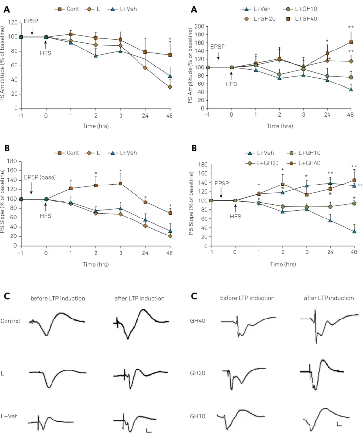
Long-term potentiation (LTP)
he percentage of amplitude or slope of LTP against base -line fEPSP, which was arbitrarily set at 100%, is shown in Figure 2. After baseline responses of fEPSP, the high-frequency stimulation was delivered at zero time to the angular bundle of perforant path; then the percentage of LTP against baseline fEPSP was recorded at separate times at 1, 2, 3, 24 and 48 hours later in bilateral NBM-lesioned aged rats with ibotenic acid injection (L); bilateral NBM-lesioned aged rats that received intrahippocampal GH related solvent (L + Veh) compared to sham-operated healthy rats (stereotaxic needle placement into
the NBM without injection of ibotenic acid) (Cont). Two-factor
repeated-measure ANOVA followed by the Least Signiicant Diference post hoc test revealed that the amplitude of LTP (A) was reduced slowly at 1 hour, 2 hours, 3 hours and 24 hours after
LTP induction in NBM-lesioned versus control rats and, at 48
hours, showed a signiicant reduction (45% vs. 74%) (p < 0.05),
which shows long-term memory impairment in NBM-lesioned
rats. he slope of the LTP (B) also showed gradual signiicant
decrements over time (at all time points after high-frequency
stimulation) compared to the control group (p < 0.05).
he efect of intrahippocampal injections of GH (10, 20,
40 µg.2 µl-1) or vehicle (benzyl alcohol solution 0.9%) (2 µl)
on the amplitude and slope of LTP showed that GH
treat-ment with 20 µg and 40 µg doses, signiicantly increased the
amplitude of LTP compared with the vehicle-treated group at 24 and 48 hours post-high-frequency stimulation (Figure 3A). Figure 3B illustrates the comparison of the LTP slope (one
way ANOVA followed by the Least Signiicant Diference post hoc test). his result showed signiicant diferences between
GH and vehicle-treated groups (*p < 0.05 and **p < 0.01).
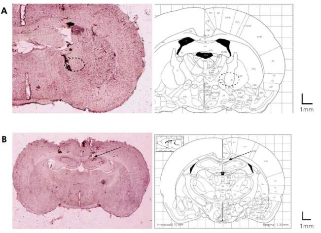
Histology
Cannula placement was veriied by histological examina
-tion of the needle tracks. Histological coronal sec-tions (40 μm)
showed the needle was inserted correctly in the NBM of the rat brain (Figure 4A) and within the right hippocampus (Figure 4B).
Figure 1. Plasma level of growth hormone in young
(3–4-month-old) and aged (18–20-month-old) healthy rats. Data are presented as means ± SEM (n = 6/group).
0 0.5 1 1.5 2 2.5 3 3.5
Aged young
GH concentration (ng/ml)
0 20 40 60 80 100 120
PS Amplitude (% of baseline)
* EPSP
HFS
0 20 40 60 80 100 120 140 160 180
-1
PS Slope (% of baseline)
Time (hrs)
* *
*
* EPSP (base)
HFS
48 24 3
2 1
0 -1
Time (hrs)
48 24 3
2 1
0
Cont L L+Veh
Cont L L+Veh
before LTP induction after LTP induction
Control
L
L+Veh
Time (hrs)
**
* *
**
Time (hrs)
** **
*
**
*
*
* * 0
20 40 60 80 100 120
PS Amplitude (% of baseline)
EPSP
HFS
-1 0 1 2 3 24 48
140 160 180 200
0 20 40 60 80 100 120
PS Slope (% of baseline)
-1 0 1 2 3 24 48
140 160 180
EPSP
HFS
L+Veh L+GH10
L+GH20 L+GH40
before LTP induction after LTP induction
GH40
GH20
GH10
L+Veh L+GH10
L+GH20 L+GH40
Figure 2. (A) Effects of bilateral nucleus basalis magnocellularis lesion with ibotenic acid injection (L), bilateral nucleus
basalis magnocellularis lesion and intrahippocampal growth hormone-related solvent (L + Veh) on amplitude and (B) slope of long-term potentiation (LTP) compared to sham-operated rats (Cont) at separate times after high-frequency stimulation (HFS) (*p < 0.05). (C) Single traces recorded before and after induction of LTP in the dentate gyrus of the hippocampus. Horizontal scale bars: 1 mV, vertical scale bars: 5 ms.
A
B
C
Figure 3. (A) The effet of bilateral nucleus basalis magnocellularis lesion and intrahippocampal human recombinant growth hormone treatment on amplitude and (B) slope of long-term potentiation (LTP) vs. vehicle-treated rats (*p < 0.05, **p < 0.01). (C) Single traces recorded before and after induction of LTP in the dentate gyrus of the hippocampus. Horizontal scale bars: 1 mV, vertical scale bars: 5 ms.
A
B
DISCUSSION
he results of the present study demonstrate, for the irst time, that central GH potentiates synaptic plasticity and memory following lesion of NBM in aged rats. he brain
cholinergic system is involved in the processing of storage
information. It originates extensively from the NBM and projects to the hippocampus and neocortex25. Lesioning of
the NBM causes degeneration of cholinergic projections,
which leads to cognitive deicits similar to Alzheimer’s dis
-ease26. In this study, an NBMlesion resulted in a decreased
amplitude and slope of the LTP at separate times after high-frequency stimulation, which represents the
sur-vival and sustainability efects of this nucleus in the LTP.
It should be noted that our control group was aged rats with
GH and memory deiciency. We expected a slight increase
in the slope or amplitude of LTP post-high-frequency stim-ulation in this group. However, there was a decline in LTP over time, especially at 24 hours and 48 hours in this group.
he age-related decline in plasma GH has been shown to be an inluencing factor in brain aging, decreased synaptic
plasticity and memory27,28. Growth hormone-deicient patients
have been shown to have short- and long-term memory loss29.
Because the purpose of our study was the administration of GH to a group that had lower plasma GH levels at the beginning of
the experiment, we measured the plasma GH level in young and
aged rats to demonstrate the reduced circulating level of GH in our aged study group. As seen in our study, the GH plasma level
in aged rats was lower than in the young group (approximately
half of the GH plasma concentration in the young). According to this result, and our previous study of impairment in spatial
learning and memory in aged compared to young rats1, we used
the aged group for GH replacement therapy and electrophysio-logical assessment. It should be noted that spot GH serum levels are very uncertain, with high variation, and serum IGF-1 levels are a better option to analyze instead, in future studies.
It has been shown that GH has an efect on the complex -ity of the neuronal dendritic tree and electrophysiological
aspects in the cerebral cortex22. It seems that the increased
response of GH on amplitude or slope of the LTP in our study (especially after 24 hours and 48 hours post-high-frequency
stimulation) demonstrated an efect of the hormone on the
synaptic structure, neurogenesis or plasticity.
he cause of instability in the LTP can be related to the
separate intervals of recording and GH-induced structural
changes. Growth hormone has positive efects on brain cell
genesis including neurogenesis, angiogenesis and synaptic
Figure 4. (A) Schematic representation of a rat brain coronal section from Paxinos and Watson and needle track in the nucleus basalis magnocellularis of the rat brain. (B) Schematic drawing of a rat brain coronal section from Paxinos and Watson, showing site of injection within the hippocampus and intrahippocampal growth hormone or vehicle needle track of the rat brain. The arrow shows the injection site of growth hormone or vehicle and circles represent the terminating site of needle.
(B)
1mm 1mm S1Tr M1 M2 RSA RSGb IG CA1 CA2 CA3 PV IMD CM IAM Rh Re XI alv InterauralBregma - 2.30 mm Interaural 6.70 mm
+5 10
5
0 +15+10 +5
0 -5 -10-150
eicacy, therefore it may be used clinically as a pharmacological
agent to enhance cell genesis in the central nervous system22,30.
he age-related decline in neurogenesis following growth fac
-tor deiciency can afect dentate gyrus functions31. It is now
clear that the adult brain contains precursor cells, including endogenous neural stem and progenitor cells that have
neu-rogenic and repair capacity in injury conditions32. Stem cell
proliferation declines with age and this may contribute to
cog-nitive impairment in the elderly33. he presence of GH recep
-tors in neural stem cells suggests the ability of GH to regulate
their activities34. Blackmore and his colleagues showed that a
seven-day intracerebroventricular infusion of GH increases neural stem cell numbers and augments neurogenesis activity
in vitro35. Furthermore, GH treatment promotes the
prolifera-tion and survival of hippocampal progenitors21. Lynch and his
colleagues determined that IGF-1 infusion in the right lateral
ventricle for 14 days ameliorates age deicits in local cerebral
glucose utilization, a function believed to be correlated with
neuronal activity36 and cerebral blood low13,37. hese activities
of GH can explain the increase of LTP magnitude after GH treat -ment, especially at 24 hours and 48 hours after high-frequency stimulation. Interestingly, reversal from long-term depression at 3 hours to LTP at 24 hours and 48 hours post-high-frequency
stimulation with a higher dose of hormone (40 µg.2 µl-1)
is another reason for this efect of GH. here is some evidence
of a negative correlation between age and hippocampal blood
low38. Administration of GH in aged rats was found to increase
the density of capillaries and blood supply of the brain cortical
surface39. In the present study, the mechanism by which GH
pro-motes memory and synaptic plasticity remains unclear. Several
researchers have proposed mechanisms based on excitatory
synaptic transmission in the hippocampus that are mediated by glutamatergic receptors (AMPA) and N-methyl-D-aspartic
acid (NMDA). hese receptors have been shown to be impor -tant for synaptic plasticity and also a common switch for many
forms of learning and memory40. Ageing is associated with a
decline of NMDA receptor subunits in the hippocampus and
NMDA receptor-mediated synaptic transmission41,42,43, whereas
GH increases the level of NMDA receptor expression in the
hippocampus44. TreatmentofGH-deicient patients with GH
replacement increases aspartate levels, the ligand of the NMDA
receptor, in the cerebrospinal luid45. Growth hormone
admin-istration reduces oxidative stress in the hippocampus of aged rats by enhancing mitochondrial eiciency, limiting the genera -tion of free radicals or increasing the activity of enzymes that
regulate oxidative stress46.
In conclusion, thisstudy showed, for the irst time, that
local GH treatment for up to 48 hours ameliorates
deterio-ration in LTP responses caused by a speciic NBM-lesion. he positive efects of GH on synaptic plasticity is possibly caused by several mechanisms. he main mechanism may be
the role of hormones in the stem cell proliferation and cen-tral nervous system neurogenesis to repair the injury areas.
his useful possibility needs further in-depth studies.
References
1. Rehman HU, Masson EA. Neuroendocrinology of ageing. Age Ageing. 2001;30(4):279-87. https://doi.org/10.1093/ageing/30.4.279 2. Fumagalli F, Racagni G, Riva MA. The expanding role of BDNF:
a therapeutic target for Alzheimer’s disease? Pharmacogenomics J. 2006;6(1):8-15. https://doi.org/10.1038/sj.tpj.6500337
3. Peng S, Wuu J, Mufson EJ, Fahnestock M. Precursor form of brain-derived neurotrophic factor and mature brain-derived neurotrophic factor are decreased in the pre-clinical stages of Alzheimer’s disease. J Neurochem. 2005;93(6):1412-21. https://doi.org/10.1111/j.1471-4159.2005.03135.x 4. Pietrzik C, Behl C. Concepts for the treatment of Alzheimer’s disease:
molecular mechanisms and clinical application. Int J Exp Pathol. 2005;86(3):173-85. https://doi.org/10.1111/j.0959-9673.2005.00435.x 5. Mesulam M. The cholinergic lesion of Alzheimer’s disease:
pivotal factor or side show? Learn Mem. 2004;11(1):43-9. https://doi.org/10.1101/lm.69204
6. Terry AV, Buccafusco JJ. The cholinergic hypothesis of age and Alzheimer’s disease-related cognitive deicits: recent challenges and their implications for novel drug development. J Pharmacol Exp Ther. 2003;306(3):821-7. https://doi.org/10.1124/jpet.102.041616 7. Arce VM, Cella SG, Locatelli V, Müller EE. Studies of growth
hormone secretion in calorically restricted dogs: effect of cholinergic agonists and antagonists, glucose and thyrotropin-releasing hormone. Neuroendocrinology. 1991;53(5):467-72. https://doi.org/10.1159/000125759
8. Nilsson-Håkansson L, Civalero I, Zhang X, Carlsson-Skwirut C, Sara VR, Nordberg A. Effects of IGF-1, truncated IGF-1 and the tripeptide Gly-Pro-Glu on acetylcholine release from parietal cortex of rat brain. Neuroreport. 1993;4(9):1111-4.
9. Shin EJ, Jhoo JH, Nabeshima T, Jhoo WK, Kwon MS, Lim YK et al. Growth hormone releaser attenuates beta-amyloid (1 - 42)-induced memory impairment in mice. J Pharmacol Sci. 2005;99(1):117-20. https://doi.org/10.1254/jphs.SC0050105
10. Sonntag WE, Brunso-Bechtold JK, Riddle DR. Age-related decreases in growth hormone and insulin-like growth factor (IGF)-1: implications for brain aging. J Anti Aging Med. 2001;4(4):311-29. https://doi.org/10.1089/10945450152850641
11. Sattler FR. Growth hormone in the aging male. Best Pract Res Clin Endocrinol Metab m. 2013;27(4):541-55. https://doi.org/10.1016/j.beem.2013.05.003
12. Åberg ND. Role of the growth hormone/insulin-like growth factor 1 axis in neurogenesis. Endocr Dev. 2010;17:63-76. https://doi.org/10.1159/000262529
13. Sonntag WE, Lynch C, Thornton P, Khan A, Bennett S,
Ingram R. The effects of growth hormone and IGF-1 deiciency on cerebrovascular and brain ageing. J Anat. 2000;197(4):575-85. https://doi.org/10.1017/S002187829900713X
14. Obermayr RP, Mayerhofer L, Knechtelsdorfer M, Tragl KH, Geyer G. The reduced release of GH by GHRH in 8 subjects aged 65-69 years is augmented considerably by rivastigmine, a drug for Alzheimer’s disease. Gerontology. 2003;49(3):191-5. https://doi.org/10.1159/000069175
16. Creyghton WM, Dam PS, Koppeschaar H, editors. The role of the somatotropic system in cognition and other cerebral functions. Semin Vasc Med. 2004;4(2):167-72. https://doi.org/10.1055/s-2004-835375 17. Donahue CP, Kosik KS, Shors TJ. Growth hormone is produced
within the hippocampus where it responds to age, sex, and stress. Proc Natl Acad Sci USA. 2006;103(15):6031-6. https://doi.org/10.1073/pnas.0507776103
18. Lobie PE, García-Aragón J, Lincoln DT, Barnard R, Wilcox JN, Waters MJ. Localization and ontogeny of growth hormone receptor gene expression in the central nervous system. Brain Res Dev Brain Res. 1993;74(2):225-33. https://doi.org/10.1016/0165-3806(93)90008-X
19. Nyberg F. Growth hormone in the brain: characteristics of specific brain targets for the hormone and their functional significance. Front Neuroendocrinol. 2000;21(4):330-48. https://doi.org/10.1006/frne.2000.0200
20. Coculescu M. Blood-brain barrier for human growth hormone and insulin-like growth factor-I. J Pediatr Endocrinol Metab. 1999;12(2):113-24. https://doi.org/10.1515/JPEM.1999.12.2.113 21. Devesa P, Agasse F, Xapelli S, Almengló C, Devesa J, Malva JO et al.
Growth hormone pathways signaling for cell proliferation and survival in hippocampal neural precursors from postnatal mice. BMC Neurosci. 2014;15(1):100. https://doi.org/10.1186/1471-2202-15-100 22. Åberg ND, Brywe KG, Isgaard J. Aspects of growth hormone and
insulin-like growth factor-I related to neuroprotection, regeneration, and functional plasticity in the adult brain. Sci World J. 2006;6:53-80. https://doi.org/10.1100/tsw.2006.22
23. Doulah AH, Rohani AH, Haddad M, Motamedi F, Farbood Y, Badavi M et al. The effect of peripheral administration of growth hormone on AD-like cognitive deiciency in NBM-lesioned rats. Neurosci Lett. 2009;466(1):47-51. https://doi.org/10.1016/j.neulet.2009.09.016 24. Paxinos G, Watson C. The rat brain in stereotaxic coordinates. 6th ed.
Amsterdam: Academic Press; 2007.
25. Zee EA, Luiten PG. Muscarinic acetylcholine receptors in the
hippocampus, neocortex and amygdala: a review of immunocytochemical localization in relation to learning and memory. Prog Neurobiol. 1999;58(5):409-71. https://doi.org/10.1016/S0301-0082(98)00092-6 26. Gaykema RP, Nyakas C, Horvath E, Hersh LB, Majtenyi C, Luiten PG.
Cholinergic iber aberrations in nucleus basalis lesioned rat and Alzheimer’s disease. Neurobiol Aging. 1992;13(3):441-8. https://doi.org/10.1016/0197-4580(92)90119-I
27. Shi L, Linville MC, Tucker EW, Sonntag WE, Brunso-Bechtold JK. Differential effects of aging and insulin-like growth factor-1 on synapses in CA1 of rat hippocampus. Cereb Cortex. 2005;15(5):571-7. https://doi.org/10.1093/cercor/bhh158
28. Ramsey MM, Weiner JL, Moore TP, Carter CS, Sonntag WE. Growth hormone treatment attenuates age-related changes in hippocampal short-term plasticity and spatial learning. Neuroscience. 2004;129(1):119-27. https://doi.org/10.1016/j.neuroscience.2004.08.001 29. Aleman A, Vries WR, Haan EH, Verhaar HJ, Samson MM,
Koppeschaar HP et al. Age-sensitive cognitive function, growth hormone and insulin-like growth factor 1 plasma levels in healthy older men. Neuropsychobiology. 2000;41(2):73-8. https://doi.org/10.1159/000026636
30. Aberg D. Role of the growth hormone/insulin-like growth factor 1 axis in neurogenesis. Endocr Dev. 2010;17:63-76. https://doi.org/10.1159/000262529
31. Kuhn HG, Dickinson-Anson H, Gage FH. Neurogenesis in the dentate gyrus of the adult rat: age-related decrease of neuronal progenitor proliferation. The J Neurosci. 1996;16(6):2027-33.
32. Gage FH. Mammalian neural stem cells. Science.
2000;287(5457):1433-8. https://doi.org/10.1126/science.287.5457.1433 33. Shetty AK, Hattiangady B, Shetty GA. Stem/progenitor cell
proliferation factors FGF-2, IGF-1, and VEGF exhibit early decline during the course of aging in the hippocampus: role of astrocytes. Glia. 2005;51(3):173-86. https://doi.org/10.1002/glia.20187 34. Pathipati P, Gorba T, Scheepens A, Gofin V, Sun Y, Fraser M. Growth hormone and prolactin regulate human neural stem cell regenerative activity. Neuroscience. 2011;190:409-27. https://doi.org/10.1016/j.neuroscience.2011.05.029 35. Blackmore DG, Reynolds BA, Golmohammadi MG, Large B,
Aguilar RM, Haro L et al. Growth hormone responsive neural precursor cells reside within the adult mammalian brain. Sci Rep. 2012;2:250. https://doi.org/10.1038/srep00250
36. Lynch CD, Lyons D, Khan A, Bennett SA, Sonntag WE. Insulin-like growth factor-1 selectively increases glucose utilization in brains of aged animals. Endocrinology. 2001;142(1):506-9. https://doi.org/10.1210/endo.142.1.8053
37. Arwert LI, Veltman DJ, Deijen JB, Lammertsma AA, Jonker C, Drent ML. Memory performance and the growth hormone/insulin-like growth factor axis in elderly: a positron emission tomography study. Neuroendocrinology. 2005;81(1):31-40. https://doi.org/10.1159/000084872
38. Heo S, Prakash RS, Voss MW, Erickson KI, Ouyang C, Sutton BP et al. Resting hippocampal blood low, spatial memory and aging. Brain Res. 2010;1315:119-27. https://doi.org/10.1016/j.brainres.2009.12.020 39. Sonntag WE, Lynch CD, Cooney PT, Hutchins PM. Decreases in
cerebral microvasculature with age are associated with the decline in growth hormone and insulin-like growth factor 1*. Endocrinology. 1997;138(8):3515-20. https://doi.org/10.1210/endo.138.8.5330 40. Tang YP, Wang H, Feng R, Kyin M, Tsien JZ. Differential effects
of enrichment on learning and memory function in NR2B transgenic mice. Neuropharmacology. 2001;41(6):779-90. https://doi.org/10.1016/S0028-3908(01)00122-8
41. Adams MM, Shi L, Linville MC, Forbes ME, Long AB, Bennett C et al. Caloric restriction and age affect synaptic proteins in hippocampal CA3 and spatial learning ability. Exp Neurol. 2008;211(1):141-9. https://doi.org/10.1016/j.expneurol.2008.01.016
42. Newton IG, Forbes ME, Linville MC, Pang H, Tucker EW, Riddle DR, et al. Effects of aging and caloric restriction on dentate gyrus synapses and glutamate receptor subunits. Neurobiol Aging. 2008;29(9):1308-18. https://doi.org/10.1016/j.neurobiolaging.2007.03.009
43. Barnes C, Rao G, Shen J. Age-related decrease in the
N-methyl-D-aspartateR-mediated excitatory postsynaptic potential in hippocampal region CA1. Neurobiol Aging. 1997;18(4):445-52. https://doi.org/10.1016/S0197-4580(97)00044-4
44. Le Grevès M, Le Grevès P, Nyberg F. Age-related effects of IGF-1 on the NMDA-, GH- and IGF-1-receptor mRNA transcripts in the rat hippocampus. Brain Res Bull. 2005;65(5):369-74. https://doi.org/10.1016/j.brainresbull.2005.01.012
45. Burman P, Hetta J, Wide L, Månsson JE, Ekman R, Karlsson FA. Growth hormone treatment affects brain neurotransmitters and thyroxine. Clin Endocrinol (Oxf). 1996;44(3):319-24. https://doi.org/10.1046/j.1365-2265.1996.617439.x 46. Donahue AN, Aschner M, Lash LH, Syversen T, Sonntag WE.
ERRATUM
http://dx.doi.org/10.1590/0004-282x20170074err
Erratum
Arq Neuropsiquiatr 2017;75(7):477-483. DOI: http://dx.doi.org/10.1590/0004-282x20170074
he order of Authors:
Maryam Malek1, Alireza Sarkaki2,4, Saleh Zahedi- Asl3 , Ziba Rajaei1 , Yaghoob Farbood2,4
Should be:
Maryam Malek1, Alireza Sarkaki2,4, Saleh Zahedi- Asl3, Yaghuob Farbood2,4, Ziba Rajaei1 ,
he Support:
Support: Physiology Research Center, Ahvaz Jundishapur University of Medical Sciences and Endocrine Research Center, Institute of Endocrine and Metabolism, Shaheed Beheshti University of Medical Sciences, Tehran, Iran.
Should be:
Physiology Research Center, Ahvaz Jundishapur University of Medical Sciences and Endocrine Research Center, Institute of
Endocrine and Metabolism, Shaheed Beheshti University of Medical Sciences, Tehran, Iran. (his study is part of a Ph.D. thesis
of M.M with Grant No. PRC-22).
he correspondence adress:
Maryam Malek; Department of Physiology, School of Medicine, Isfahan University of Medical Sciences, Isfahan, Iran
Email: [email protected]
Should be:
Sarkaki Alireza; Department of Physiology, School of Medicine, Isfahan University of Medical Sciences, Isfahan, Iran