cop
yr
ight
© ABE&M todos os direitos reser
v
ados
clinical case report
ADRIANA LOFRANO-PORTO
GUSTAVO B. BARRA
PAULA P. NASCIMENTO
PATRÍCIA G. G. COSTA
ÉRICA C. GARCIA
RODRIGO F. VAZ
ANA R. T. BATISTA
ANA C. R. DE FREITAS
BRUNO L. B. CHERULLI
FAYEZ BAHMAD JR..
LARISSA G. FIGUEIREDO
FRANCISCO A. R. NEVES
LUIZ AUGUSTO CASULARI
Section of Endocrinology, Faculty of Medicine, University Hospital of Brasília, University of Brasilia (ALP, PPN, ECG, RFV, ARTB, LAC); Sabin Institute and Laboratory of Clinical Analysis (GBB, PGGC); Radiology Center, University Hospital of Brasília (ACRF, BLBC); Department of Otolaryngology, Head & Neck Surgery, Brasilia University School of Medicine (FBJ), Brasilia, DF, Brazil; Section of Endocrinology, Faculty of Medicine, Amazon State University, Manaus, AM, Brazil (LGF); Molecular Pharmacology Laboratory, Faculty of Health Sciences, University of Brasilia (FARN), Brasilia, DF, Brazil.
Received in 26/8/2008 Accepted in 14/10/2008
ABSTRACT
Pendred Syndrome (PS) is an autossomal recessive disorder characterized by sensorineural deafness, goiter and iodide organifi cation defect. The hearing loss is associated with inner ear abnormalities, ranging from an isolated en-larged vestibular aqueduct (EVA) to a typical coclear dysplasia. Mutations in the gene that encodes pendrin (SLC26A4), a chloride/iodide transporter, have been shown to be associated with PS. We describe the clinical and molecular characteristics of a large consanguineous family harboring a mutation in the SLC26A4 gene. The proband was a 26-year-old deaf Brazilian woman who presented a bulky multinodular goiter and hypothyroidism since puberty. Five other siblings were deaf: one brother had a similar phenotype, three siblings also had goiters but normal thyroid function tests, and one brother had only a subtle thyroid enlargement. Other 4 siblings had no thyroid or hearing disorder. Parents were fi rst degree cousins and had normal hearing. The mother was healthy, except for subclinical hypothyroidism; the father was deceased. A perchlorate test in the proband showed a discharge of 21% of the incorporated iodide 2h after the administration of 1g of KClO4. Audio-logical examinations showed profound hearing loss in all deaf subjects; CT and MRI of the temporal bones showed EVA in all of them. Genomic DNA was isolated from whole blood, from the 6 affected and 4 unaffected siblings, the mother and control. The coding region of the PDS gene (exons 2-21), including exon/intron boundaries, were amplifi ed by PCR and sequenced. A single base-pair (T) deletion at position 1197 of exon 10 was detected in homozy-gous state in the 6 deaf siblings. The mother and 2 unaffected siblings were heterozygous for this mutation, which has been described by Everett et al. The 1197delT mutation is predicted to result in a frameshift and a truncated protein. The existence of PS phenocopies and intrafamilial phenotypic vari-ability are well documented. The defi nite diagnosis requires molecular analy-sis. Our study illustrates the value and challenges of mutational analysis in selected patients with PS. (Arq Bras Endocrinol Metab 2008; 52/8:1296-1303)
Keywords: Pendrin; Sensorineural deafness; Goiter
RESUMO
Síndrome de Pendred Causada por Mutação em Homozigoze no Gene SLC26A4 em uma Família Brasileira Consangüínea.
cop
yr
ight
© ABE&M todos os direitos reser
v
ados
puberdade. Outros cinco irmãos eram surdos: um irmão tinha fenotipo se-melhante, três também tinham bócio, porém com função tiroideana normal e um irmão tinha apenas um discreto aumento da tiróide. Outros quatro irmãos não apresentavam alteração tiroideana ou auditiva. Os pais eram primos de primeiro grau e tinham audição normal. A mãe era saudável, exceto por hipotir-eoidismo subclínico; o pai era falecido. O teste do perclorato no caso-índice rev-elou a liberação de 21% do iodo incorporado duas horas após a administração de 1 g de KClO4. Os exames audiológicos mostraram perda auditiva profunda em todos os indivíduos afetados; TC e RMN dos ossos temporais mostraram DAV em todos eles. O DNA genômico foi isolado do sangue total dos seis irmãos afetados e dos quatro não-afetados, da mãe e do controle. A região codifi cante do gene PDS (éxons 2-21), incluindo as junções éxon/íntron, foram amplifi cadas por PCR e seqüenciadas. Foi detectada a deleção de uma base (T) na posição 1197 do éxon 10, em homozigoze, nos seis irmãos afetados. A mãe e dois irmãos não-afetados eram heterozigotos para a mutação, que foi descrita inicialmente por Everett e cols. A mutação 1197delT provavelmente resulta em um erro de fase de leitura (frameshift) e em uma proteína truncada. A existência de
fenocó-pias da SP e a variabilidade fenotípica intrafamiliar são bem conhecidas. O diag-nóstico defi nitivo requer análise molecular. O presente estudo ilustra o valor e os desafi os da análise mutacional em pacientes selecionados com SP. (Arq Bras Endocrinol Metab 2008; 52/8:1296-1303)
Descritores: Pendrina; Surdez neurossensorial; Bócio
INTRODUCTION
P
endred syndrome (PS) is an autosomal recessive di-sorder characterized by bilateral sensorineural dea-fness, goiter and iodide organifi cation defect, with an estimated incidence of 7.5 to 10 in 100,000 individuals (1). This disorder may account to approximately 10% of the cases of hereditary deafness, making it the most common cause of syndromic deafness (1). Despite the defect in iodide organifi cation, which can be demons-trated by a positive perchlorate discharge test, thyroid function is highly variable and hypothyroidism is des-cribed in approximately half of the cases (2). The hea-ring loss is associated with inner ear abnormalities, which can be readily identifi ed with appropriate ima-ging procedures, ranima-ging from an isolated enlarged vestibular aqueduct (EVA) to a typical cochlear malfor-mation known as Mondini dysplasia. This condition is characterized by hypoplastic upper turns of the cochlea originating a common dysfunctional cavity (3,4).The gene involved in this syndrome is located at chro-mosome 7 and encodes a protein pertaining to the solute carrier family 26, member 4 (SLC26A4), a 780 amino acid protein that functions as a chloride, iodide and/or bicarbonate transporter, also known as pendrin (5). To date, more than 100 mutations spanning the whole cod-ing region of the SLC26A4 gene have been shown to be associated with PS, although the genotype-phenotype
correlations are still poor and remain unexplained. Muta-tions in the SLC26A4 cause not only classical PS but also an autosomal recessive form of isolated deafness without thyroid disease, both associated with EVA (6,7).
In this study, we report the clinical and molecular characteristics of a large consanguineous Brazilian family harboring a mutation in the SLC26A4 gene, from which six affected siblings presented with subtle phenotypic variations. Furthermore, we review recent data from studies that have contributed to the understanding of the physiopathological roles of pendrin in humans.
SUBJECTS AND METHODS
Case reports
cop
yr
ight
© ABE&M todos os direitos reser
v
ados
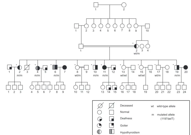
Figure 1. Pedigree of the family with Pendred syndrome caused by a 1197delT in the SLC26A4 gene. The subjects evaluated in this study are shown ((*)).
four living children were healthy and had normal hea-ring. On physical examination, her height was 160 cm and weight was 63.6 kg. Blood pressure was normal (112X70 mmHg). A large multinodular goiter with predominance of the right lobe was noted, with no other physical abnormality. Because of progressive thyroid enlargement and compressive symptoms, she underwent a total thyroidectomy at the age of 46.
Family history was remarkable for congenital deaf-ness and goiter. The proband was the third of 11 sib-lings. Five other siblings were deaf; among them, two brothers (IV-6 and IV-10) and two sisters (IV-2 and IV-11) also had a history of thyroid enlargement since early puberty that progressively evolved to large multi-nodular goiters, whereas one brother (IV-19) had only a modest diffuse thyroid enlargement.
Other four siblings had no thyroid or hearing dis-order (IV-8, IV-13, IV-15 and IV-17). Parents were fi rst degree cousins and had normal hearing. The moth-er (III-2) was healthy, except for subclinical hypothy-roidism, with negative thyroid auto-antibodies tests. The father (III-1) was deceased. Family’s pedigree is shown in Figure 1.
Clinical studies
All patients provided written informed consent to parti-cipate in this study, which was approved by the Research Ethics Committee of Faculty of Medicine, University of Brasília, Brazil.
In order to confi rm the clinical suspicion of PS, a perchlorate test was performed in the proband. Two hours after administration of 131-iodine (50 mCi), 1 g of perchlorate (KClO4) was administered, and 2 hours later, the discharge of iodide was determined. A dis-charge of less than 10% of the incorporated iodide is expected in normal individuals (2,8).
Evaluation of the thyroid function was based on serum determinations of thyroid-stimulating hormone (TSH), free thyroxine (FT4) and total triiodotyronine (T3) levels, using a chemiluminescent immunoassay (IMMULITE 2000, DPC).
cop
yr
ight
© ABE&M todos os direitos reser
v
ados
threshold (SRT), pure-tone audiometry (PTA) and transiently evoked otoacoustic emissions (OAE). Imag-ing studies from the inner ear were obtained with the use of computed tomography (CT) and magnetic reso-nance imaging (MRI) of the temporal bones. EVA was defi ned when enlargement of the vestibular aqueduct was >1.5 mm at midway between the endolymphatic sac and the vestibule (9-11).
Molecular studies
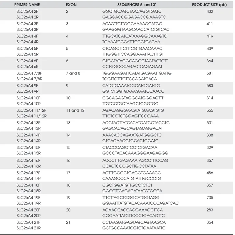
Genomic DNA samples from ten siblings (6 affected and 4 unaffected), their mother and a control were pre-pared from 50 µL whole blood by the Chelex-100 method (12). The coding region (exons 2 to 21) of the SLC26A4 gene, including exon/intron boundaries, was amplifi ed by PCR with primers designed for this study (Table 1), with the exception of exon 3 primers,
Table 1. Primers used in the SLC26A4 gene amplifi cation.
PRIMER NAME EXON SEQUENCES 5’ and 3’ PRODUCT SIZE (pb)
SLC26A4 2F SLC26A4 2R
2 GGCTGCAGCTAACAGGTGATC
GAGGACCGGAGACCGAAAGTC
432
SLC26A4 3F SLC26A4 3R
3 ACAGTTCTTGGCAAAAGCATGG
GAAGGGTAAGCAACCATCTGTCAC
411
SLC26A4 4F SLC26A4 4R
4 TTTGCATCATCATAAAGGCAAAGTC
TGAAATCCCATTTCCCTGACAA
419
SLC26A4 5F SLC26A4 5R
5 CTCAGCTTCTTTCGTGAACAAAC
TTTGGGTTCCAGGAAATTACTTTGT
439
SLC26A4 6F SLC26A4 6R
6 GTGCTATAGGCAGGCTACTAGTGTT
CCTGGCCCAGACTCAGAGAAT
364
SLC26A4 7/8F SLC26A4 7/8R
7 and 8 TGGGAAGATTCATATGAGAATTGATTG TGGTTGTTTCTTCCAGATCACA
581
SLC26A4 9F SLC26A4 9R
9 CATGTGAAATGGCATGGATGG
GGTCTGGTGAAAGAATCCAACC
583
SLC26A4 10F SLC26A4 10R
10 CGCAGAGTAGGCATGGGAGTTT
TTGTCCTGCTAAGCTCGGTGC
314
SLC26A4 11/12F SLC26A4 11/12R
11 and 12 AGACAGGGAAGTATGAAGTGTG TTTCTCCTCTGGAGTTCCCAAA
555
SLC26A4 13F SLC26A4 13R
13 AGGTAGTTATCACATGATGGTACCTG
GAGCACAGCAGTAGAGGACAT
501
SLC26A4 14F SLC26A4 14R
14 AAACACCAGAATGATGGGCTC
GTCAGAAGGTGCACTGGATC
338
SLC26A4 15F SLC26A4 15R
15 CTACCCAGCTCCTCTGACAA
GCCCTACACAAAGGGAAGAGGG
329
SLC26A4 16F SLC26A4 16R
16 ACCCTTTGAGAAATAGCCTTTCCAG
CCACTCCCGCTTGCCTATAA
357
SLC26A4 17F SLC26A4 17R
17 AGTTTGGGCTGAGGTGAAACC
CAAAGCCCATGTATTTGCCCTG
486
SLC26A4 18F SLC26A4 18R
18 CGCTGGATGTTGCCTCTCT
GGCCTTCAGACATAATGTGCCA
357
SLC26A4 19F SLC26A4 19R
19 TTTCTTAGCTGGGCATGGTAGG
GGAATTTATGTACACAAATCCCAGATCAC
705
SLC26A4 20F SLC26A4 20R
20 AGAAGCACCAGGAAAGCTTCA
GGGAATTATGTTCCCTGACAGTTC
283
SLC26A4 21F SLC26A4 21R
21 CCTAAGATGAGTAGCAGTAAGCA
GCTGCCAAATCGTCTGAATAATTC
cop
yr
ight
© ABE&M todos os direitos reser
v
ados
which were described previously (1). Amplifi cation was performed on a PTC-100 thermal cycler (MJ Research, Inc) using the following cycling conditions : initial de-naturation for 3 minutes at 94 °C; followed by 34 cy-cles of 1min at 94 °C, 1min at 55 °C, and 1 min at 72 °C; and fi nal extension for 5 min at 72 °C. The PCR products were analyzed by electrophoresis on 1.5% Tris-acetate-EDTA/ethidium bromide agarose gels under ultraviolet illumination and were later sequenced in both sense and antisense orientations, with the use of an automated sequencer ABI-377 (Perkin-Elmer Corp., Foster City, CA). The sequences were compared to the NCBI Homo sapiens SLC26A4 DNA sequence refer-ence assembly (ACCESSION NC_000007 REGION: 107088316-107145490).
RESULTS
Clinical characterization of the six siblings with PS
The perchlorate test showed a discharge of 21% of the incorporated iodide in the proband, a fi nding
consis-Table 2. Clinical and radiological aspects of six siblings with Pendred syndrome due to the 1197delT mutation in the SLC26A4 gene.
Subjetct
No. Sex Age Endolymphatic Sac
Vestibular Aqueduct
(Nl<1,5 mm) Goiter Thyroid function
(years) R L R L (volumes-cm3)
IV-2 F 51 normal enlarged 2,9 3,5 multinodular
44,2 normal
IV-4 F 47 enlarged enlarged 3,2 3,5 multinodular
186,3* hypothyroidism**
IV-6 M 45 normal enlarged 1,1 1,8 multinodular
114,2* hypothyroidism
IV-10 M 41 normal normal 1,8 2,7 multinodular
18,4* Normal
IV-11 F 38 enlarged enlarged 3,6 3,1 multinodular**
ND Normal
IV-19 M 29 enlarged enlarged 2,3 2,7
subtle enlargement
27,3
Normal
R: right; L: left; Nl: normal; F: female; M: male; ND: not determined; § thyroid volume determined by ultrasonography; # in subjest V-4, tyroid volume was determined before total thyroidectomy was perfomed; # # subject IV-4 developed hypothyroidism with positive anti-thyroglobulin antibodies, many years before thyroidectomy.; * thyroid remanscent size after parcial thyroidectomy; ** thyroid ultrasonogram could not be obtained from subject IV-11, but physical examination reveale a multinodular goiter.
tent with the defect in iodide organifi cation into thyro-globulin.
The clinical and radiologic evaluation of the affec-ted siblings and their mother are summarized in table 2. In the proband, thyroid ultrasonography showed a multinodular, heterogeneous goiter, with a lobular contour and small cystic areas with sparse calcifi cations. Histopathological examination of the proband’s thyroid specimen revealed a large colloid goiter, weighing 310 g, with small areas of follicular hyperplasia, fi brosis and calcifi cations.
de-cop
yr
ight
© ABE&M todos os direitos reser
v
ados
veloped hypothyroidism before thyroidectomy was performed, leading to the need for levothyroxine repla-cement treatment. This condition was associated with the presence of thyroid auto-antibodies in one of them (IV-4).
Audiological examination showed bilateral pro-found hearing loss (PTA > 90 dB) in all six affected si-blings. Computed tomography (CT) and magnetic resonance imaging (MRI) of the temporal bones sho-wed variably enlarged vestibular aqueducts (EVA) and uni- or bilaterally dilated endolymphatic sacs in all of them (Table 2 and Figure 2).
Figure 2. Axial volumetric hybrid MRI sequence with T2WI predominance (FIESTA). Bilateral vestibular aqueductal en-largement (a) and endolymphatic sac dilatation (b) are shown by arrows.
Figure 3. Eletropherograms of wild-type, 1197delT homozy-gote and heterozyhomozy-gote, showing the deleted T residue (square) and the resultant frameshift (underlined) in the mu-tant allele.
ATG
WT
ATG
1197deIT
FS400 STOP
780 AA
430 AA
STOP
Figura 4. Schematic representation of the predicted frameshift from codon 400, leading to a premature stop at codon 430, as a consequence of the 1197delT mutation, also named FS400.
MOLECULAR ANALYSIS
A single nucleotide (T) deletion at position 1197 of exon 10 (1197delT) was detected in homozygous state in the six deaf siblings (Figure 3). The 1197delT muta-tion, also named FS400, is predicted to result in a fra-meshift from codon 400 leading to a premature stop at codon 430 (Figure 4). The mother, one brother and one sister were heterozygous for the mutation.
DISCUSSION
co-cop
yr
ight
© ABE&M todos os direitos reser
v
ados
don 400 (Figure 4), leading to a premature stop codon at position 430, which predicts a truncated protein. The mutation 1197delT is located in the seventh trans-membrane domain of the mature protein and results in the loss of the last two and a half transmembrane do-mains with the whole carboxi-terminal portion, impai-ring its biological activity (7).
Although clinical information about the affected subjects from previous families harboring the 1197delT mutation is scanty, some variability on the thyroid phe-notype was noted. Similar to our fi ndings, the majority of patients harboring this mutation in homozygous state had goiters, with or without hypothyroidism, except two subjects in a family from Lebanon (13-16). In a previous study of other large kindred from Brazil, Kopp and cols. identifi ed a deletion of a T residue at position 249 of exon 3 of the SLC26A4 gene associated with PS. Howe-ver, differently from the present study, Kopp and cols. found intrafamilial phenotype-genotype variability, sug-gesting that the clinical diagnosis of PS may be confoun-ded by the presence of similar phenotypes caused by distinct environmental or genetic factors (17).
In the thyroid, the main function of pendrin is to re-gulate the effl ux of iodide at the apical membrane of the thyrocytes, where it acts as an iodide-chloride transporter. Specifi cally, it is presumed to play a signifi cant role on the transport of iodide into the follicular lumen, where it is coupled to thyroglobulin. If pendrin activity is impaired, as in PS, cytoplasmatic iodide accumulation occurs. Ho-wever, in the majority of the cases, the defect is incomple-te and most patients have a preserved thyroid function or only subclinical or mild hypothyroidism (18,19).
In the present study, a positive perchlorate test in the proband confi rmed the iodide organifi cation de-fect. However, although a similar iodide organifi cation status would be expected in all affected members, thyroid function and growth were variably compromi-sed. Only two affected subjects developed hypothyroi-dism prior to thyroidectomy, and the total volume of the thyroid determined by US varied from 27.3 cm3 to
186.3 cm3. These fi ndings are in accordance with
pre-vious studies, demonstrating signifi cant intrafamilial phenotypic variability (15).
The mechanisms involved in goiter formation in PS are not completely understood, but may include tran-sient increases in serum TSH in response to low thyroid hormone synthesis and increased iodide retention in thyrocytes. Environmental factors, such as nutritional iodide intake, also appear to act as important modifi ers
of the thyroid phenotype. Recent in vitro studies using
thyrocytes from PS patients and normal individuals have shown that some iodide can leave PS thyrocytes, although less effi ciently than normal cells (20). The ab-sence of goiter observed in some PS patients keeps up with these observations. Moreover, Palos and cols. de-monstrated some evidence of the existence of adaptive mechanisms associated with impaired organifi cation, that could work together in order to avoid hypothyroi-dism, such as increased deiodinase activities in goiters of PS patients and the resulting increased intrathyroidal conversion of T4 into T3.
The audiological phenotype typically seen in PS is that of a profound hearing loss, with prelingual onset and a progressively worsening course (3,6,21,22). Our fi ndings in the six homozygote patients corroborate the-se obthe-servations. The reasons for the characteristic hea-ring phenotype are probably related to the functions of pendrin in the inner ear, where it is presumed to partici-pate in the formation and resorption of the endolymph. It has been shown that defects in chloride transport may lead to toxic and osmotic injury to the neuroepithelium, resulting in the progressive sensorineural hearing loss observed in PS patients (23). The enlargement of the endolymphatic duct (ED) and sac (ES), as well as dila-tion of the surrounding structures (ventricular aqueduct and cochlea), is believed to be related to the increased osmotic pressure and volume of the endolymph, and is frequently seen in PS patients (3).
The functions of pendrin in the kidneys are less well understood. Previous studies have shown a role in mediating bicarbonate secretion and in the regulation of acid-base transport in the renal collecting duct (24). However, in PS patients, no acid-base disturbances have been noted, under physiological conditions. Re-cently, much research have focused on the potential relations of pendrin with blood pressure regulation. It has been suggested that pendrin-mediated chloride ab-sorption in the distal nephron may be an important mechanism, by which aldosterone and angiotensin II modulate the renal regulation of blood pressure. Inde-ed, many studies have pointed to the role of pendrin as an important mediator of aldosterone actions in the kidneys (25). In light of these recent fi ndings, additio-nal studies are needed to clarify the readditio-nal impact of pen-drin inactivation in humans.
cop
yr
ight
© ABE&M todos os direitos reser
v
ados
to the clinical diagnosis of PS. However, many condi-tions causing congenital hypothyroidism may mimic PS, particularly in infants. Although a positive perchlo-rate test indicates thyroid dishormonogenesis, it is not helpful to indicate the exact nature of the molecular defect. Due to the high predictive value of the detec-tion of enlarged ED and ES in PS patients, appropriate imaging studies with high resolution CT and MRI are thus mandatory to the clinical diagnosis of PS, and must be performed before genetic analysis. The defi ni-te diagnosis requires molecular analysis.
Acknowledgements: We are thankful for the Sabin Institute and Laboratory of Clinical Analysis, Brasília, Brazil, for gently provi-ding support for the molecular studies. The authors report ha-ving no confl icts of interest relevant to this article.
REFERENCES
1. Everett L, Glaser B, Beck JC, Idol JR, Buchs A, Heyman M, et al. Pendred syndrome is caused by mutations in a putative sulphate transporter gene (PDS). Nat Genet. 1997;17:411-22. 2. Reardon W, Coffey R, Chowdhury T, Grossman A, Jan H, Britton
K, et al. Prevalence, age of onset and natural history of thyroid disease in Pendred syndrome. J Med Genet. 1999;36: 595-8. 3. Cremers C, Bolder C, Admiraal RJ, Everett LA, Joosten F, Van
Hauwe P, et al. Progressive sensorineural hearing loss and a widened vestibular aqueduct in Pendred syndrome. Arch Oto-laryngol Head Neck Surg. 1998;124:501-5.
4. Propst EJ, Stockley TL, Harrison RV, Gordon KA, Papsin BC. Temporal bone imaging in GJB2 deafness. Laryngoscope. 2006;116(12):2178-86.
5. Everett LA, Green E. A family of mammalian anion transpor-ters and their involvement in human genetic diseases. Hum Mol Genet. 1999;8(10):1883-91.
6. Yang JJ, Tsai CC, Hsu HM, Shiao JY, Su CC, Li SY. Hearing loss associated with enlarged vestibular aqueduct and Mondini dysplasia is caused by splice-site mutation in the PDS gene. Hear Res. 2005;199:22-30.
7. Azaiez H, Yang T, Prasad S, Sorensen JL, Nishimura CJ, Kim-berling WJ, et al. Genotype–phenotype correlations for SL-C26A4- related deafness. Hum Genet. 2007;122(5):451-7. 8. Wolff J. Perchlorate and the thyroid gland. Pharmacol Rev.
1998;50:89-102.
9. Lowe L, Vézina L. Sensorineural hearing loss in children. Ra-diographics. 1997;17:1079-93.
10. Goldfeld M, Glaser B, Nassir E, Gomori JM, Hazani E, Bishara N. CT of the ear in Pendred syndrome. Radiology. 2005;235(2): 537-40.
11. Vijayasekaran S, Halsted MJ, Boston M. When is the vestibular aqueduct enlarged? A statistical analysis of the normative dis-tribution of vestibular aqueduct size. Am J Neuroradiol. 2007; 28:1133-8.
12. Walsh P. Chelex 100 as a medium for simple extraction of DNA for PCR-based typing from forensic material. Biotechniques. 1991;10:506-13.
13. Van Hauwe P, Everett LA, Coucke P, Scott DA, Kraft ML, Ris-Stalpers C, et al. Two frequent missense mutations in Pendred syndrome. Hum Mol Genet. 1998;7(7):1099-104.
15. Fugazzola L, Mannavola D, Cerutti N, Maghnie M, Pagella F, Bianchi P, et al. Molecular analysis of the Pendred’s syndrome gene and magnetic resonance imaging studies of the inner ear are essential for the diagnosis of true pendred’s syndrome. J Clin Endocrinol Metab. 2000;85(7):2469-75.
16. Fugazzola L, Cerutti L, Mannavola D, Crino A, Cassio A, Gaspa-roni P, et al. Differential diagnosis between pendred and pseu-do-pendred syndromes: clinical, radiologic, and molecular studies. Ped Res. 2002;51(4):479-84.
17. López-Bigas N, Melchionda S, Cid R, Grifa A, Govea N, Zelante L, et al. Identifi cation of fi ve new mutations of PDS/SLC26A4 in Mediterranean families with hearing impairment. Hum Muta-tion. 2002;18(6):548-9.
18. Kopp P, Arseven OK, Sabacan L, Kotlar T, Dupuis J, Cavaliere H, et al. Phenocopies for deafness and goiter development in a large inbred Brazilian Kindred with Pendred’s Syndrome As-sociated with a novel mutation in the PDS Gene. J Clin Endo-crinol Metab. 1999;84(1):336-41.
19. Scott DA, Wang R, Kreman TM, Andrews M, McDonald JM, Bishop JR, et al. Functional differences of the PDS gene pro-duct are associated with phenotypic variation in patients with Pendred syndrome and non-syndromic hearing loss (DFNB4). Hum Mol Genet. 2000;9(11):1709-15.
20. Yoshida A, Taniguchi S, Hisatome I, Royaux IE, Green ED, Kohn LD, et al. Pendrin Is an Iodide-Specifi c Apical Porter Re-sponsible for Iodide Effl ux from Thyroid Cells. J Clin Endocri-nol Metab. 2002;87(7):3356-61.
21. Palos F, Garcia-Rendueles MER, Araujo-Vilar D, Obregon MJ, Calvo RM, Cameselle-Teijeiro J, et al. Pendred syndrome in two Galician families: insights into clinical phenotypes through cellular, genetic, and molecular studies. J Clin Endocrinol Me-tab. 2008;93(1):267-77.
22. Kopp P, Pesce L, Solis-S JC. Pendred syndrome and iodide trans-port in the thyroid. Trends Endocrinol Metabol. 2008;19: 260-8. 23. Iwasaki S, Usami S, Abe S, Isoda H, Watanabe T, Hoshino T.
Long-term audiological feature in Pendred syndrome caused by PDS mutation. Arch Otolaryngol Head Neck Surg. 2001;127 6):705-8.
24. Everett LA, Morsli H, Wu DK, Green ED. Expression pattern of the mouse ortholog of the Pendred’s syndrome gene (Pds) su-ggests a key role for pendrin in the inner ear. Proc Natl Acad Sci USA. 1999;96(17):9727-32.
25. Royaux IE, Wall SM, Karniski LP, Everett LA, Suzuki K, Knepper MA, et al. Pendrin, encoded by the Pendred syndrome gene, resides in the apical region of renal intercalated cells and me-diates bicarbonate secretion. Proc Natl Acad Sci USA. 2001;98: 4221-6.
26. Wall SM, Pech V. The interaction of pendrin and the epithelial sodium channel in blood pressure regulation. Curr Opin Ne-phrol Hypertens. 2008;17:18-24.
Correspondence to:
Adriana Lofrano Porto
Hospital Universitário de Brasília, Laboratório de Farmacologia Molecular (UnB)