cop
yr
ight
© ABE&M todos os direitos reser
v
ados
clinical case report
MIRIAN YUMIE NISHI
RAFAELA VIEIRA CORREA
ELAINE MARIA FRADE COSTA
ANA ELISA CORREIA BILLERBECK
ANDRÉ LUIS CRUZES
SORAHIA DOMENICE
LUCIANI RENATA CARVALHO
BERENICE B. MENDONCA
Laboratório de Hormônios e Genética Molecular LIM/42, Unidade de Endocrinologia do Desenvolvimento, Hospital das Clínicas da Faculdade de Medicina da Universidade de São Paulo (HC-FMUSP) (MYN, AECB, EMFC, SD, LRC, BBM), São Paulo, SP, Brasil; Universidade de Fortaleza (RVC), Fortaleza, CE, Brazil; Associação de Diabetes Juvenil da Região Noroeste Paulista (ALC), Birigüi, SP, Brasil.
Received in 25/8/2008 Accepted 16/10/2008
with a 45,X/46,X,der(X) Karyotype with
Gene Overdosage
ABSTRACT
SHOX is exclusively expressed in the developing distal limb bones of human
embryos and in the fi rst and second pharyngeal arches. It works as a pro-moter for linear growth and as a repressor of growth plate fusion. It was re-ported, recently, that SHOX overdosage and gonadal estrogen defi ciency have led to tall stature due to continued growth. We report, in the present study, a female patient with 45,X/46,X, psu idic(X)(pter→q21::q21→pter) kary-otype, tall stature, and hypergonadotrophic hypogonadism without Turner stigmas. She did not present breast development even after long term thera-py with high estrogen doses. Fluorescence in situ hybridization depicted the presence of three copies of SHOX gene. Microsatellite studies showed pater-nal origin of der(X). Further studies in similarly affected patients will clarify if the absence of breast development, despite previous high-dose estrogen treatment, is associated to triple copy of SHOX gene. (Arq Bras Endocrinol Metab 2008; 52/8:1282-1287)
Keywords: SHOX overdosage; Tall stature; Gonadal dysgenesis
RESUMO
Estatura Alta e Hipodesenvolvimento Mamário após Reposição Estrogênica em Paciente com Hipogonadismo Hipergonadotrófi co e Cariótipo 45,X/46,X, der(X) com Superdosagem do Gene SHOX.
O gene SHOX é expresso, exclusivamente, no primeiro e no segundo arcos faríngeos, assim como nas extremidades dos ossos dos membros em em-briões humanos. SHOX normalmente atua como um promotor para o cresci-mento linear e como um repressor do fechacresci-mento da placa de crescicresci-mento. Recentemente, foi descrito que o excesso da proteína SHOX associada à de-fi ciência estrogênica gonadal leva à estatura alta devido ao contínuo cresci-mento. Neste estudo descrevemos uma paciente do sexo feminino com cariótipo 45,X/46,X,psu idic(X)(pter→q21::q21→pter), estatura alta, hipogo-nadismo hipergonadotrófi co e sem estigmas de Turner. A paciente não apre-sentou desenvolvimento de mamas, mesmo depois do tratamento prolongado com altas doses de estrógenos. FISH evidenciou a presença de três cópias do SHOX. Estudo de microssatélites demonstrou a origem paterna do der(X).
Estudos futuros em pacientes com semelhanças clínicas esclarecerão se a ausência de desenvolvimento de mamas, apesar do tratamento com altas doses de estrógenos, está associada à tripla cópia do SHOX. (Arq Bras Endo-crinol Metab 2008; 52/8:1282-1287)
cop
yr
ight
© ABE&M todos os direitos reser
v
ados
INTRODUCTION
A
t the end of short X and Y chromosome arms there is a common portion named short arm pseudoau-tosomal region (PAR-1) spaning 2.6 Mb. PAR-1 esca-pes from X inactivation process and recombines during male meiosis (1). Individuals with absence of this gion present short stature which indicates that this re-gion is related to growth (2,3). In 1997, two different groups described, simultaneously, a gene responsible for stature, SHOX (“Short Stature Homeobox-Contai-ning Gene”) (2,3), located on the PAR-1. SHOX is composed by 7 exons with 40 kb in length (4), that encodes two different proteins, one with 292 and other with 225 amino acids due to an alternative splicing (2,3). It is exclusively expressed in the fi rst and second pharyngeal arches and in the developing distal limb bo-nes of human embryos (5). SHOX normally functions as a promoter for linear growth and as a repressor for growth plate fusion and skeletal maturation (6). SHOXhaploinsuffi ciency due to deletions or mutations leads to short stature and skeletal abnormalities such as in Turner syndrome, or Madelung deformity of the fore-arm, such as in Léri-Weill dyscondrosteosis.
Recently, patients with karyotype alterations invol-ving sexual chromosomes resulting in SHOX overdosa-ge were described in the literature (7-10). This fact associated to estrogen defi ciency owing to gonadal dys-genesis seems to be the cause of excessive growth, espe-cially in the distal limb bones, in the middle to late teenage females (7,10,11).
In the present study, we report a 45,X/46,X,der(X) female patient with tall stature, low body mass index (BMI), no breast development or Turner’s stigmata and gonadal insuffi ciency in whom a SHOX gene over-dosage was suspected.
METHODS
Cytogenetic Analysis
Chromosome metaphase spreads prepared from peri-pheral blood lymphocyte cultures of the patient and her parents were analyzed by conventional staining, G and C banding techniques. The kit Vysis® Kallman
Re-gion Probe (Vysis Inc., Abbott Laboratories, Illinois, USA) and the biotinylated LLNOYCO3”M”34F5 cos-mid containing exons III to VIb of SHOX gene were used for the FISH methodology. The analysis was
per-formed using the Karyotyping Software Macktype v.5.4.1 and Mackprobe v. 4.0 (Perceptive Scientifi c Ins-truments Inc., UK) for FISH.
Molecular Analysis
Genomic DNA was obtained from peripheral blood leukocytes using the Salting-Out technique (12). The microsatellite study was carried out using the panel for the X chromosome presented in the kit Linkage Map-ping set v.2.5 MD10 (PE Applied Biosystems, The Perkin-Elmer Corporation, CA, USA). The PCR pro-ducts were submitted to electrophoresis in the ABI PRISM 310 automatic sequencer (PE Applied Biosyste-ms, The Perkin-Elmer Corporation, CA, USA) and the analysis was made by GeneScan software (PE Applied Biosystems, The Perkin-Elmer Corporation, CA, USA).
CASE REPORT
The present study was approved by the Ethics Com-mittee of the Hospital das Clinicas, The University of Sao Paulo Medical School. Written consent was obtai-ned from the patient and their parents.
A 16 year-old Brazilian girl was referred due to ab-sence of secondary sexual development. She was born at 40 weeks of gestation after an uncomplicated preg-nancy, a single daughter of non consanguineous pa-rents. At birth, she weighted 3.250 g and measured 52 cm. Her growth chart showed that she grew at the 50th
percentile until 9 months of age, at the 75th percentile
from 9-33 months, and after 3 years of age at > 95th
percentile. She underwent spontaneous pubarche at 12 years, without telarche. She presented menarche at 14 years, after estrogen replacement without adequate breast development. At 16 her height was 183 cm, 20 cm above her target height, with an eunuchoid habitus and her BMI was 15 Kg/m2. Puberty evaluation
disclo-sed Tanner II breast development, Tanner V pubic hair and normal female external genitalia (Table 1). Basal serum hormone data revealed elevated gonadotropins (LH= 40 U/L and FSH= 85 U/L), low estrogen le-vels, normal PRL, GH, IGF1 and IGFBP3 levels.
RESULTS
cop
yr
ight
© ABE&M todos os direitos reser
v
ados
Table 1. Clinical data before and after hormonal treatment.
Age (yrs)
Height (cm)
SD (height)
Weight (kg)
Breast (Tanner)
Pubic hair
(Tanner) Treatment
11.8 174.8 + 3.5 43 I IV Conjugated estrogen 0.625 mg
16.5 183 + 3.5 50 II V Valerate of estradiol 2 mg
Levonorgestrel 0.25 mg
16.8 183.5 + 3.5 51 II V Valerate of estradiol 2 mg
Levonorgestrel 0.25 mg
17 NA NA NA II NA Ethnyl estradiol 70 ug
Ciproterone acetate 4 mg
17.5 184 + 3.6 51 III V Ethnyl estradiol 70 ug
Ciproterone acetate 4 mg
18 184.2 + 3.7 52 III V Ethnyl estradiol 35 ug
Ciproterone acetate 2 mg
18.6 184.4 + 3.7 52 III V Valerate of estradiol 2 mg
Levonorgestrel 0.25 mg
NA = Not available
cop
yr
ight
© ABE&M todos os direitos reser
v
ados
Figure 4. Heredogram of the patient with the microsatellite study indicating the paternal origin of der(X). The markers DXS991 and DXS986 were not informative.
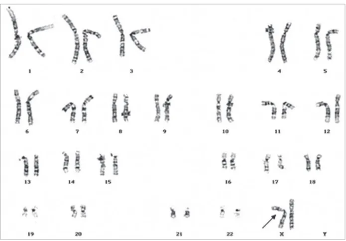
of SHOX and KAL genes and X centromere (Figures 2 and 3) in the der(X), showing that the der(X) is a psu idic(X)(pter→q21::q21→pter). Microsatellite study re-vealed the paternal origin of psu idic(X)(pter→q21::q21
→pter) (Figure 4). The markers DXS991 and DXS986 localize on Xp11.21 and Xq21.1, respectively, were not informative.
DISCUSSION
Recently, it has been reported that SHOX overdosage and gonadal estrogen defi ciency leads to tall adult hei-ght (7,9). These fi ndings suggest that SHOX overdo-sage in combination with gonadal dysgenesis leads to continuous growth since early infancy until late ado-lescence, since SHOX functions as a repressor for gro-wth plate fusion, in contrast with skeletal maturing effect of estrogens (10).
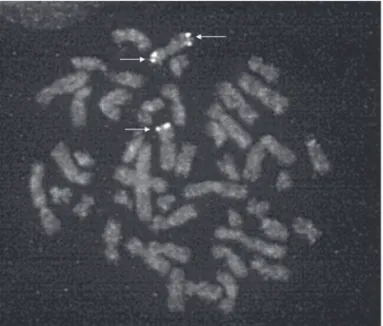
Here we reported a patient with a 45,X[4]/46,X,psu idic(X)(pter→q21::q21→pter)[46] karyotype and
go-Figure 2. FISH analysis indicating triple copies of SHOX gene in metaphase chromosome
cop
yr
ight
© ABE&M todos os direitos reser
v
ados
Table 2. Phenotype of SHOX overdosage in female patients.
Age (Yrs)
Height
(cm SD) HH
Turner Stigmas
Breast Development (after estrogen)
Cytogenetic Data Der(X)
Origin Reference
29 172 + 2.9 Present Mild webbed neck
Full development
45,X[40]/46,X,der(X)(pter→q13 or q21::p14→
or p21.2→pter[60] Paternal (10)
20 174 + 2 Present Absent Tanner II 46,X,der(X)(pter→q21::p21→pter) NR (8)
20 161.9 + 0.8 Present Absent developmentSuffi cient 45,X[28]/46,X,psu idic(X)(q28)[72] NR (9)
20 166 + 1.5 Present Absent Normal 46,X,rec(X)dupl(Xp)inv(X)(p11.22q.21.2)[30] Maternal (13)
14 172 + 2 Present
Multiple nevi, high arched
palate
NR 46,X,der(X)(pter→q21.2::p11.1→pter)[40] NR (7)
20 NR - 1.2 Present Absent NR 45,X[6]/46,X,der(X)(pter q21.1::p22.3 pter)
[12]/46,X,r(X)(p22q13)[2] Nr (14)
16 183 + 3.5 Present Absent Tanner II 45,X[4]/46,X, psu idic(X)(pter→q21::q21→pter)[46] Paternal Present Study
HH: hypergonadotropic hypogonadism; NR: not reported..
nadal dysgenesis, tall stature, no Turner stigmas and poor breast development after estrogen replacement. The altered X chromosome has a paternal origin, which contains a partial Xq deletion, two centromeres and a Xp duplication resulting in an extra SHOX gene.
To date, six female patients with an extra copy of
SHOX gene in a der(X) were described (Table 2). Three of them had tall stature as well as the present case. However, three patients present normal height despite the extra copy of SHOX associated to estro-gen deficiency indicating that an extra copy of SHOX
is not sufficient for the development of tall stature (7-10). On the other hand, Kanaka-Gantenbein, 2004 suggested that SHOX triplication per se may induce the tall stature, since their patient presented tall stature in the absence of estrogen deficiency (15). These controversial reports may be due to the fact that the mechanism of SHOX action still re-mains unclear.
Our patient showed poor breast development af-ter estrogen treatment. Normally, these patients pre-sented adequate breast development after estrogen treatment (9,10,13). However, Nakamura et al. also reported a patient with gonadal dysgenesis and an ex-tra copy of SHOX who had poor breast development after estrogen therapy (8). The reason for the poor breast development in these two patients is still unex-plained. Further studies in similarly affected patients
will clarify if the absence of breast development despi-te high-dose estrogen treatment is reladespi-ted to the extra copy of SHOX gene.
Acknowledgment: B.B.M. was supported by grant 301246/95-5 from Conselho Nacional de Pesquisa (CNPq). No potential confl ict of interest relevant to this article was reported.
REFERENCES
1. Rappold GA. The pseudoautosomal regions of the human sex chromosomes. Hum Genet. 1993;92(4):315-24.
2. Ellison JW, Wardak Z, Young MF, Gehron Robey P, Laig-Web-ster M, Chiong W. PHOG, a candidate gene for involvement in the short stature of Turner syndrome. Hum Mol Genet. 1997;6 (8):1341-7.
3. Rao E, Weiss B, Fukami M, et al. Pseudoautosomal deletions encompassing a novel homeobox gene cause growth failure in idiopathic short stature and Turner syndrome. Nat Genet. 1997;16(1):54-63.
4. Blaschke RJ, Rappold GA. SHOX: growth, Leri-Weill and Turn-er syndromes. Trends Endocrinol Metab. 2000;11(6):227-30. 5. Clement-Jones M, Schiller S, Rao E, et al. The short stature
homeobox gene SHOX is involved in skeletal abnormalities in Turner syndrome. Hum Mol Genet. 2000;22;9(5):695-702. 6. Munns CJ, Haase HR, Crowther LM, et al. Expression of SHOX
in human fetal and childhood growth plate. J Clin Endocrinol Metab. 2004;89(8):4130-5.
7. Binder G, Eggermann T, Enders H, Ranke MB, Dufke A. Tall stature, gonadal dysgenesis, and stigmata of Turner’s syn-drome caused by a structurally altered X chromosome. J Pe-diatr. 2001;138(2):285-7.
dys-cop
yr
ight
© ABE&M todos os direitos reser
v
ados
genesis, tall stature, and endometriosis. Fertil Steril. 2001;75 (6):1224-5.
9. Ogata T, Inokuchi M, Ogawa M. Growth pattern and body pro-portion in a female with short stature homeobox-containing gene overdosage and gonadal estrogen defi ciency. Eur J En-docrinol. 2002;147(2):249-54.
10. Ogata T, Kosho T, Wakui K, Fukushima Y, Yoshimoto M, Mi-haru N. Short stature homeobox-containing gene duplication on the der(X) chromosome in a female with 45,X/46,X, der(X), gonadal dysgenesis, and tall stature. J Clin Endocrinol Metab. 2000;85(8):2927-30.
11. Ogata T, Matsuo N, Nishimura G. SHOX haploinsuffi ciency and overdosage: impact of gonadal function status. J Med Genet. 2001;38(1):1-6.
12. Miller SA, Dykes DD, Polesky HF. A simple salting out proce-dure for extracting DNA from human nucleated cells. Nucleic Acids Res. 1988;16(3):1215.
13. Adamson KA, Cross I, Batch JA, Rappold GA, Glass IA, Ball SG. Trisomy of the short stature homeobox-containing gene (SHOX), resulting from a duplication-deletion of the X chro-mosome. Clin Endocrinol (Oxf). 2002;56(5):671-5.
14. Ogata T, Matsuo N, Fukushima Y, et al. FISH analysis for ap-parently simple terminal deletions of the X chromosome: identifi cation of hidden structural abnormalities. Am J Med Genet. 2001;104(4):307-11.
15. Kanaka-Gantenbein C, Kitsiou S, Mavrou A, et al. Tall stature, insulin resistance, and disturbed behavior in a girl with the triple X syndrome harboring three SHOX genes: offspring of a father with mosaic Klinefelter syndrome but with two mater-nal X chromosomes. Horm Res. 2004;61(5):205-10.
Correspondence to:
Mirian Yumie Nishi
Hospital das Clínicas, FMUSP, Laboratório de Hormônios e Genética Molecular LIM 42 - Disciplina de Endocrinologia Av. Dr Enéias de Carvalho Aguiar, 155, PAMB, 2º andar, bloco 6 05403-900 São Paulo SP