Short Report
0103 - 5053 $6.00+0.00*e-mail: [email protected]
Biotransformation of a Tetrahydrofuran Lignan by the Endophytic Fungus Phomopsis Sp.
Michelle Verza,a Nilton S. Arakawa,a Norberto P. Lopes,a Massuo J. Kato,b Mônica T. Pupo,a Suraia Saida and Ivone Carvalho*,a
aFaculdade de Ciências Farmacêuticas de Ribeirão Preto, Universidade de São Paulo,
Av. do Café, s/n, 14040-903 Ribeirão Preto-SP, Brazil
bInstituto de Química, Universidade de São Paulo, Av. Lineu Prestes, 748, 05508-900 São Paulo-SP, Brazil
A biotransformação da lignana tetraidrofurânica, (-)-grandisina, pelo fungo endofítico Phomopsis sp, obtido de Viguiera arenaria, conduziu à formação de um novo metabólito caracterizado como 3,4-dimetil-2-(4’-hidróxi-3’,5’-dimetóxifenil)-5-metóxi-tetraidrofurano. O metabólito foi analisado contra o parasita Trypanosoma cruzi, o agente causador da doença de Chagas, e mostrou uma atividade tripanocida (IC50 9,8µmol L
-1) similar ao precursor natural
(IC50 3,7 µmol L-1).
The biotrasformation of the tetrahydrofuran lignan, (-)-grandisin, by the endophitic fungus Phomopsis sp, obtained from Viguiera arenaria, led to the formation of a new compound determined as 3,4-dimethyl-2-(4’-hydroxy-3’,5’-dimethoxyphenyl)-5-methoxy-tetrahydrofuran. The metabolite was evaluated against the parasite Trypanosoma cruzi, the causative agent of Chagas’s disease, and showed a trypanocidal activity (IC50 9.8µmol L-1) similar to the natural
precursor (IC50 3.7 µmol L-1).
Keywords: biotransformation, 2,5-diaryltetrahydrofuran, endophytic fungus, (-)-grandisin, trypanocidal activity
Introduction
The lignan family is a large group of natural products with noteworthy therapeutic activity as illustrated by
podophyllotoxin derivatives (e. g. etoposide) with antitumoral
activity.1 In particular, the optically active tetrahydrofuran
lignans display a wide range of pharmacological activities,
including anti-parasite,2-4 antibacterial,5 antifungal,6
anti-tumoral,7 anti-inflammatory8,9 and platelet-activating factor
(PAF) inhibition.10-12
Several non-symmetrical 2,5-diaryltetrahydrofuran lignans, shown in Table 1, have been isolated from
Piper species (Piperaceae) containing either piperonyl, veratryl or 3,4,5-trimethoxyphenyl substituent at 2 and 5 positions and methyl groups at 3 and 4 positions attached to the tetrahydrofuran ring, as illustrated by (+)-calopiptin (1, P. schmidtii), 5-[rel-(2S,3S,4S,5S )-5’-(3’’,4’’-
dimethoxyphenyl)-3’,4’-dimethyl-2’-tetrahydro-furanyl)]-1,3-benzodioxole (2, P. wightii), (2S,3R,4S,5R
)-3,4-dimethyl-2,5-bis(3’,4’-dimethoxyphenyl)-tetrahydrofuran
(3, P. clarkii), (±)-galgravin (4, P. futokadsura, P. hancei, P. puberculum, P.wallichii), (-)-galbelgin (5, P. attenuatum,
P. futokadsura, P. thomsoni, P. wightii), (+)-grandisin (6, P. polysyphorum), (+)-machilin G (7, P. schmidtii, P. wightii), (+)-veraguensin (8, P. cuneifolium, P. futokadsura, P. puberculum) and (-)-zuonin A (9, P. schmidtii).13 Other
bis-tetrahydrofuran lignans have also been described from
Achillea lingulata (Asteraceae).14
In previous reports, Martins et al.3 described the isolation
of (-)-grandisin (6) from Piper solmsianum and Lopes and
co-workers 4,15 from Virola surinamensis (Rol.) Warb, which
belong to Piperaceae and Myristicaceae, respectively. The
tetrahydrofuran lignan (-)-grandisin (6) showed a potent
trypanocidal activity against the trypomastigote form of
Trypanosoma cruzi, responsible for Chagas´s disease in
Latin America, at 5 µg mL-1 leading to a total parasite
lysis after 24 h, without hematiae damage.4 Furthermore,
veraguensin (8) also isolated from Virola surinamensis4
preserved this anti-parasite pattern along with a potent platelet-activating factor (PAF) inhibitory activity that may be involved in the mechanisms of the parasite growing
the poor water solubility of compounds 6 and 8 for in vivo
evaluation, comprising semi-synthetic modifications such
as O-demethylation of the methoxy groups attached to the
aromatic rings, led to byproducts due to the presence of two benzylic carbons in the tetrahydrofuran ring that were
very unstable under acidic or base conditions.17
The biotransformation of the naturally occurring 2,5-diaryl-3,4-dimethyltetrahydrofurans with PAF inhibitory
activity, such as (+)-veraguensin (8), (+)-galbelgin (3)
and galgravin (4) named as 7,7’-epoxylignans, has been
investigated in order to obtain novel and bioactive derivatives. In the series, carrying a 3,4-dimethoxyphenyl substituents
pattern, both methyl groups at para-position of the aromatic
rings were metabolized and cleaved by Aspergillus niger
yielding the corresponding 4,4’-O-demethyl derivatives.18
Similar results were also achieved using furofuran lignans
with the 2,6-diaryl-3,7-dioxabicyclo[3.3.0]octane skeleton,19
such as (+)-magnolin and (+)-eudesmin. Replacing the 3,4-dimethoxyphenyl groups by 3,4,5-trimethoxyphenyl (e. g. (+)-yangabin) and 4,5-dihydroxy-3-methoxyphenyl
groups on the lignans lead to a stable para-methoxy groups
not cleaved by A. niger. On the other hand, the use of
larvae of Spodoptera litura20 or rats as a source of liver
metabolized enzymes21,22 allowed the O-demethylation
at the para-position of both aromatic rings of furofuran
lignans containing either di- or trimethoxyphenyl groups, being the pre-formed hydroxyl group frequently conjugated
to glycosides. The grandisin (6) has also been
di-O-demethylated by Lepidopterae and Coleoptera species.23
The biotransformation reactions of lignans, including podophyllotoxin, and neolignans was described in a recent
review.24
Endophytic fungi are prolific producers of natural products, since they live inside the health plant tissues
without causing any apparent disease.25 These environmental
conditions might stimulate the fungi to produce a range of metabolic enzymes as a requirement for their survival
in the presence of toxic defense compounds of plants.26
Endophytes have also been successfully used in the
biotransformation of natural products.27,28
These findings stimulated us to investigate about
20 endophytic strains from our collection of fungi29 as
biocatalyst systems and effective method for regio- and stereoselective production of novel and bioactive products. This procedure could convert unreactive methoxy groups to hydroxyl functions of a tetrahydrofuran lignan under
mild conditions which may increase the solubility of 6.
Thus, this study focuses on the biotransformation of 6
by four different endophytic fungi with the aim to obtain potential trypanocidal derivatives with improved drug-like properties.
Experimental
General procedures
GC-MS were measure at 70 eV on a Shimadzu Model: QP2010, 120 ºC (oven temp.), using a DB-5MS (30 m ×
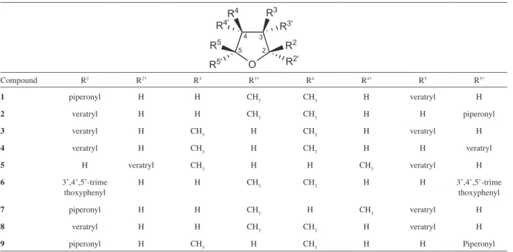
Table 1. Non-symmetrical 2,5-diaryltetrahydrofuran lignans isolated from Piper species
Compound R2 R2’ R3 R3’ R4 R4’ R5 R5’
1 piperonyl H H CH3 CH3 H veratryl H
2 veratryl H H CH3 CH3 H H piperonyl
3 veratryl H CH3 H CH3 H veratryl H
4 veratryl H CH3 H CH3 H H veratryl
5 H veratryl CH3 H H CH3 veratryl H
6 3’,4’,5’-trime
thoxyphenyl
H H CH3 CH3 H H 3’,4’,5’-trime
thoxyphenyl
7 piperonyl H H CH3 H CH3 veratryl H
8 veratryl H H CH3 CH3 H veratryl H
9 piperonyl H CH3 H CH3 H H Piperonyl
0.25 mm × 0.25 µm). ESI analysis were performed on a
ultrOTOFQ-ESI-TOF MS (End Plate: 4000 Volts, Capillary:
4500 Volts, Capillary Exit: 300 Volts, Skimmer 1: 50 Volts, Skimmer 2: 25 Volts, Transfer: 90 µs, Collision Exit Gate:
80 µs).1H NMR spectrum was measured on a Bruker
DPX-400 (DPX-400 MHz) while 2D experiments (HSQC and HMBC) were recorded in a Bruker DRX-400 (400 and 100 MHz)
using CDCl3 (Aldrich) as solvent and TMS as int. standard.
Chemical shifts were reported in units (ppm) and coupling
constants (J) in Hz. TLC: silica gel 60 F254 pre-coated (layer
thickness 0.25 mm, Merck); CC: silica gel with hexane-EtOAc 4:6, v/v. The instrumentations for HPLC analysis
consisted of a Shimadzu (SCL-10Avp,Japan) multisolvent
delivery system, Shimadzu SCL-M10vp Photodiode Array detector, and an Intel Celeron computer for analytical system control and data collection. Chromatography was carried out using the isocratic method with methanol:water 7/3, v/v within 20 minutes during the screening phase for the evaluation of grandisin biotransformation (condition A). The mobile phase used during the isolation of the metabolites was methanol:water 6:4, v/v within 60 minutes (condition B). A CLC-ODS (M)- 4.6 × 250 mm
Shimadzu column was used at a flow of 1.0 mL min-1.
The spectral data from the detector were collected over de 200-400 nm range of absorption chromatograms, and the chromatograms were plotted at 225 nm.
Grandisin was obtained from dichloromethane extracts
from twigs of Virola surinamensis, which yielded two
steroids, two lignans (compounds 6 and 8), five flavonoids
and one polyketide.4
Microorganisms and culture
The endophytic fungi have been isolated and identified
by their rDNA sequences as previously described.29 The
strains are maintained on PDA slants at 4 ºC. Initially the fungi were screened for their ability to transform grandisin. Fungi were cultivated on PDA in Petri dish for 7 days at
30 ºC and agar pieces (1 cm2) containing fungal mycelia
was used as inocullum. Strain SS42 was also identified by “Micoteca URM/ Depto. de Microbiologia/ CCB/ UFPE” (Av. Prof. Nelson Chaves, s/n, 50670-420 Recife-PE, Brazil).
A two-step culture was used for each fungus. Initially, the fungi were inoculated into 20 mL of Czapek medium, supplemented with 3% of sucrose and the cultures were incubated at 30 ºC for 3 days, with shaking at 120 ºC rpm. After cultivation the resulting mycelia were harvested by filtration and transferred to 20 mL of Czapek medium, containing 1% of sucrose and supplemented with a solution
of grandisin in DMSO, resulting 0.4 mg mL-1 as final
concentration in the media. The cultures were reincubated at the same conditions for 8 days.
Extraction of the metabolites
The culture broths were separated from mycelia by vacuum filtration, followed by 3 times partition sequentially
with ethyl ether, ethyl acetate and n-butanol (10 mL). The
solvents were evaporated and the crude extracts were analyzed by TLC and HPLC (condition A).
Isolation of the metabolites
After the preliminary screening, Phomopsis sp (VA35)
culture was scaled up to 150 mL using the same conditions
already described, including the proportion of grandisin (6)
(60 mg), but changing the fermentation time to 9 days. The ethyl ether and ethyl acetate extracts were joined because they showed similar profiles. The crude extract obtained was purified by Silica-gel column chromatography using hexane/ethyl acetate 1:1, yielding 4 fractions. Fraction 3 was further purified through HPLC (Condition B) to yield
compound 10 (1.0 mg).
In vitro trypanocidal assay
The bioassays were carried out using the blood of infected Swiss albino mice, collected by cardiac puncture
on the parasitemy peak (7th day) after infection with Y
strains of T. cruzi. The infected blood was diluted with
the blood health mice to achieve a concentration of 106
trypomastigote per mL. The assays were performed on
titration microplates (96 wells) in duplicate. The compound
10 was dissolved in dimethyl sulfoxide (DMSO) and was
added into the infected mouse to provide concentration of
0.5, 2.0, 8 and 32 µmol L-1. The plates were incubated at
4 ºC and the numbers of parasites were counted after 24 h. Negative and positive controls containing either DMSO
or crystal violet at 250 µg mL-1, respectively were run in
parallel.30
Results and Discussion
The microbial transformations of (-)-grandisin (6)
by endophytic fungi were investigated in a small scale screening test (8 mg) monitored by TLC and HPLC analyses
(condition A). Among the 20 endophytic fungi, Curvularia
lunata (SS-42), isolated from Smallanthus sonchifolius,
scale experiments because of its great ability to generate metabolites in the preliminary tests.
The metabolization of compound 6 (60 mg), with a
retention time of 40.23 min as indicated by HPLC analysis
(condition B), led to the generation of a major metabolite (10)
with a distinct retention time of 10.35 min. Compound 10
was purified by column chromatography, followed by HPLC (condition B). Condition B was established by exploring its different interaction properties with the reverse phase column (C-18), in a manner similar to that previously reported for
butyrolactones isolated from Virola surinamensis.31,32
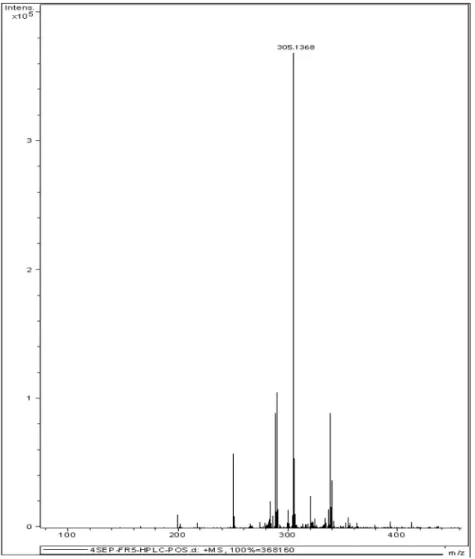
GC-MS analysis showed that compound 10 has a
molecular weight lower than grandisin with a [M]+. at
m/z 282 and fragments (m/z 68, 85, 100, 167 and 182)
suggesting the molecular formula C15H22O5. In fact, the
High Resolution Mass Spectrometry confirms the proposed
molecular formula with a detectable [M+ Na]+ 305.1368
and a corresponding theoretic mass of 305.1365.
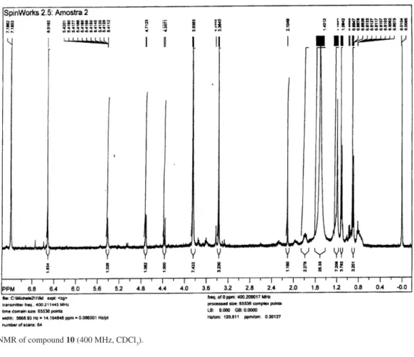
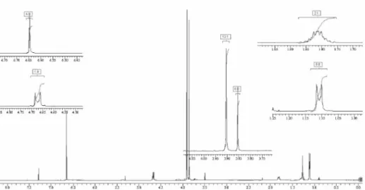
The structure of compound 10 was established by 1D
and 2D NMR data. For instance, the 1H NMR spectrum
of compound 10 showed two methoxy proton signals at d
3.37 and 3.83 with a relative integration of 3:6, which were characteristic of groups attached to aliphatic and aromatic
skeleton, respectively. Moreover, the ortho-aromatic proton
signals were assigned at d 6.52 (s, 2H), while the methyl
groups at C-13 and C-12 were observed at d 1.10 and d 0.87
as two doublets (J 6.9 Hz).
Nearly all relevant spectral details of the isolated metabolites were closely comparable to the corresponding
13C and 1H features of grandisin,4,33 but no data for the second
aromatic ring were observed by the integration of signals. In
addition, a hemiacetal proton was observed at d 4.71 (d, 1H),
attached to a carbon at d 111.1 as observed in the HMQC
experiment. Also, the signal at d 3.83 was assigned to the
methoxy groups at meta positions of the remaining aromatic
ring linked to the tetrahydrofuran nucleus, indicating the
presence of a free hydroxyl group at para position. Methoxy
aliphatic protons at d 3.37 and methyl protons at d 1.10
showed correlation in the HMBC with carbon at d 111.1,
unequivocally establishing the methoxy group at C-5. These data confirmed the presence of a single aromatic ring in
compound 10, and an extra methoxy group at C-5. The main
HMBC correlations for compound 10 are shown in Figure
1 and data described in Table 2.
Thus, from the foregoing spectral data the structure
of compound 10 was characterized as
3,4-dimethyl- 2-(4’-hydroxy-3’,5’-dimethoxyphenyl)-5-methoxy-tetrahydrofuran. To our knowledge, this compound has not been previously described in the literature.
We propose that the peculiar aliphatic methoxy
groups of compound 10 should be generated firstly by a
microbial oxidative metabolism at position C-5 followed by rearrangement to a lactone displacing the aromatic ring. Further reduction to the hemiketal function and
O-methylation of the nearly formed hydroxyl group
might explain how the fungus converted the 3,4,5
trimethoxyphenyl group of compound 6 to methoxy group
of the metabolite 10.
The HPLC separation (condition B) provided a second metabolite with a retention time of 12.50 min. GC-MS analysis showed that this compound has the same molecular
weight and fragments presented by compound 10. The
High Resolution Mass Spectrometry detected [M+ Na]+
305.1368. 1H NMR spectrum revealed the same integrals,
but insufficient data could be obtained from 2D analysis because of the small isolated quantity.
The results from 1H NMR spectrum, GC-MS and high
resolution mass spectrometry suggested that this secondary
compound might be an epimer of compound 10.
Figure 1. Selected HMBC correlations for metabolite 10 obtained in the biotrasformation process.
Table 2.1HMR (400 MHz) and 13C (100 MHz) data for compound 10 (CDCl3)
Position Conpound 10
dH dC
2 4.36 (d, J 9.6, 1H) 86.7
3, 4 1.78 (m, 2H) 45.8
5 4.71 (d, J 4.7, 1H) 111.1
6 ---- 130.8
7, 11 6.52 (s, 2H) 103.2
8, 10 ---- 143.6
9 ---- 143.2
12 0.87 (d, J 6.9, 3H) 14.0
13 1.10 (d, J 6.9, 3H) 15.7
OH 5.41 (s, 1H)
----OMe 3.37 (s, 3H) 56.1
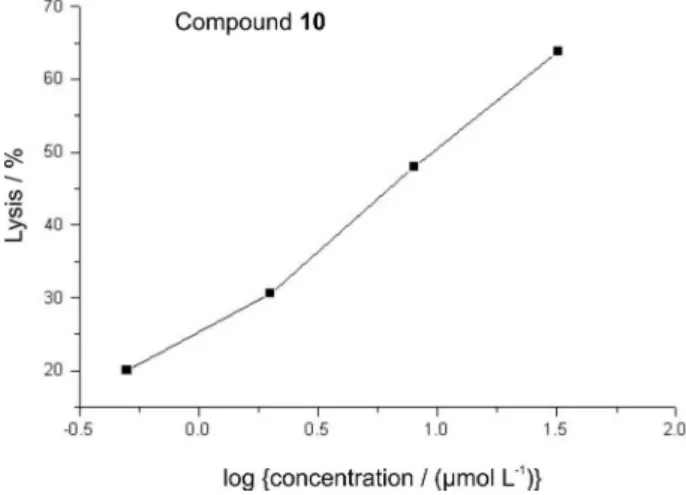
The percentage of lyses observed for the trypomastigote
form of T. cruzi treated with compound 10, as outlined in
Figure 2, was similar to the corresponding natural product
6.4 Based on experiments involving the Y strains of T. cruzi
compounds 6 and 8 were able to cause lyses of parasites
with IC50 3.7 and 2.3 µmol L-1 respectively,33,34 while the
biotransformation product 10 displayed IC50 9.8µmol L-1.
Conclusions
The product
3,4-dimethyl-2-(4’-hydroxy-3’,5’-dimethoxyphenyl)-5-methoxy-tetrahydrofuran (10) was
successfully obtained from the biotransformation of
grandisin (6) by the endophytic fungus Phomopsis sp.
The evaluation of the trypanocidal activity of compound
10 and its corresponding natural precursor 6 gave similar
results, revealing that a unique methoxylated aromatic ring bound to the central tetrahydrofuran scaffold might preserve the trypanocidal activity, this represents an interesting molecular simplification for synthetic purposes.
Acknowledgments
The authors acknowledge Fundação de Amparo a Pesquisa do Estado de São Paulo-FAPESP for financial support (Grant 04/07935-6 Bioprospecta/Biota) and fellowship, Prof. Dr. Antonio Gilberto Ferreira, Prof. Dr. Sérgio Albuquerque and Maria Angélica dos Santos Cunha Chellegatti for valuable contribution.
Supplementary Information
1H NMR, HMQC and high resolution spectra of 10 and
1H and 13C NMR spectra of 6 are available free of charge
at http://jbcs.sbq.org.br, as PDF file.
References
1. Ward, R. S.; Nat. Prod. Rep.1999, 16, 75.
2. Filho, A. A. S.; Albuquerque, S.; Silva, M. L. A.; Eberlin, M. N.; Tomazela, D. M.; Bastos, J. K.;J. Nat. Prod.2004, 67, 42. 3. Martins, R. C. C.; Lago, J. H. G.; Albuquerque, S.; Kato, M.
J.; Phytochemistry2003, 64, 667; Martins, R. C. C.; Latone, L. R.; Sartorelli, P., Kato, M. J.; Phytochemistry 2003, 55, 843. 4. Lopes, N. P.; Chicaro, P.; Kato, M. J.; Albuquerque, S.; Yoshida
M.; Planta Med.1998, 64, 667.
5. Maruyama, M., Yamauchi, S.; Akiyama K.; Sugahara, T.; Kishida, T.; Koba, Y.; Biosci. Biotechnol. Biochem.2007, 71, 677. 6. Akiyama, K.; Yamauchi, S.; Nakato, T.; Maruyama, M.;
Sugahara, T.; Kishida, T.; Biosci. Biotechnol. Biochem.2007,
71, 1028.
7. Xu, S.; Li, N.; Ning, M. M.; Zhou, C. H.; Yang, Q. R.;Wang, M. W.; J. Nat. Prod. 2006, 69, 247.
8. Schmidt, T. J.; Heilmann, J.; Planta Med.2000, 66, 749. 9. Wu, J-L.; Li, N.; Hasegawa, T.; Sakai, J-I.; Kakuta, S.; Tang,
W.; Oka, S.; Kiuchi, M.; Ogura, H.; Kataoka, T.; Tomida, A.; Tsuruo, T.; Ando, M.; J. Nat. Prod.2005, 68, 1656.
10. Jung, K. Y.; Kim, D. S.; Oh, S. R.; Park, S-H.; Lee, I. S.; Lee, J. J.; Shin, D-H.; Lee, H-K.; J. Nat. Prod.1998, 61, 808. 11. Coran, S. A.; Bambagiotti-Alberti, M.; Melani, F.; Giannellini,
V.; Vincieri, F. F.; Mulinacci, N.; Sala, R.; Moriggi, E.; Eur. J. Med. Chem. 1991, 26, 643.
12. Biftu, T.; Gamble, N. F.; Doebber, T.; Hwang, S. B.; Shen, T. Y.; Snyder, J.; Springer, J. P.; Stevenson, R.; J. Med. Chem.1986,
29, 1917.
13. Parmar, V. S.; Jain, S. C.; Bisht, K. S.; Jain, R.; Taneja, P.; Jha, A.; Tyagi, O. D.; Prasad, A. K.; Wengel, J.; Olsen, C. E.; Boll, P. M.; Phytochemistry 1997, 46 597; Ward, R. S;. Nat. Prod. Rep. 1997, 14, 43.
14. Stojanovic, G.; Hashimoto, T.; Asakawa, Y.; Palic, R.; Biochem. Syst. Ecol.2005, 33, 207.
15. Lopes, N. P.; Blumenthal, E. E. A.; Cavalheiro, A. J.; Kato, M. J.; Yoshida, M.;Phytochemistry 1996, 43, 1089.
16. Rodrigues, C. O.; Dutra, P. M. L.; Souto-Padron, T.; Cordeiro, R. S. B.; Lopes, A. H. C. S.; Biochem. Biophys. Res. Commun. 1996, 223, 735; Rodrigues, C. O.; Dutra, P. M. L.; Barros, F. S.; Souto-Padron, T.; Meyer-Fernandes, J. R.; Lopes, A. H. C. S.; Biochem. Biophys. Res. Commun.1999, 266, 36. 17. Andrade, P.; Bernardes, L. S. C.; Carvalho, I.; Abstracts of the
6th International Congress of Pharmaceutical Sciences, Ribeirão
Preto, Brazil, 2007.
18. Kasahara, H.; Miyazawa, M.; Kameoka, H.; Phytochemistry
1996, 43 111.
19. Miyazawa, M.; Kasahara, H.; Kameoka, H.; Phytochemistry
1993, 34, 1501.
20. Miyazawa, M.; Kasahara, H.; Kameoka, H.; Phytochemistry
1995, 39, 1027. Figure 2. Dose-response curve for lytic activity of compound 10 on
21. Miyazawa, M.; Kasahara, H.; Kameoka, H.; Phytochemistry
1993, 32, 1421.
22. Kasahara, H.; Miyazawa, M.; Kameoka, H.; Phytochemistry
1996, 43, 1217.
23. Ramos, C. S.: Vanin, S. A.; Kato, M. J.; Phytochemistry2008,
69, 2157.
24. Miyazawa, M.; Curr. Org. Chem.2001, 5, 975.
25. Strobel, G.; Daisy, B.; Castillo, U.; Harper, J.; J. Nat. Prod.
2004, 67, 257.
26. Yang, L.; Ning, Z. S.; Shi, C. Z.; Chang, Z. Y.; Huan, L. Y.;
J. Agric. Food Chem. 2004, 52, 1940.
27. Zikmundová, M.; Drandarov, K.; Bigler, L.; Hesse, M.; Werner, C.; Appl. Environ. Microbiol. 2002, 68, 4863.
28. Agusta, A.; Maehara S.; Ohasshi, K.; Simanjuntak, P.; Shibuta, H.; Chem. Pharm. Bull.2005, 53, 1565.
29. Guimarães, D.; Borges, W. S.; Kawano, C. Y.; Ribeiro, P. H.; Goldman, G. H.; Nomizo, A.; Thiemann, O. H.; Oliva, G.; Lopes, N. P.; Pupo,M. T.; FEMS Immunol. Med. Microbiol. 2008, 52, 134.
30. Brener Z.; Rev. Inst. Med. Trop. 1962, 4, 389.
31. Lopes, N. P., Franca, S. C.; Pereira, A. M. S.; Maia, J. G.; Kato, M. J.; Cavalheiro, A. J.; Gottlieb, O. R.; Yoshida, M.;
Phytochemistry 1994, 35, 1469.
32. Lopes, N. P.; Silva, D. H. S.; Kato, M. J.; Yoshida, M.;
Phytochemistry1998, 49, 1405.
33. Nihei, K-I. ; Konno, K.; Bernardes, L. S. C.; Lopes, N. P.; Albuquerque, S.; Carvalho, I.; Pupo, M. T.; Martins, R. C. C. ; Kato, M. J.; Arkivoc2004, vi, 112.
34. Bernardes, L. S. C.; Kato, M. J.; Albuquerque, S.; Carvalho, I.;
Bioorg. Med. Chem.2006, 14, 7075.
Received: June 10, 2008
Web Release Date: November 28, 2008
Supplementary Information
0103 - 5053 $6.00+0.00*e-mail: [email protected]
Biotransformation of a Tetrahydrofuran Lignan by the Endophytic Fungus Phomopsis Sp.
Michelle Verza,a Nilton S. Arakawa,a Norberto P. Lopes,a Massuo J. Kato,b Mônica T. Pupo,a Suraia Saida and Ivone Carvalho*,a
aFaculdade de Ciências Farmacêuticas de Ribeirão Preto, Universidade de São Paulo,
Av. do Café, s/n, 14040-903 Ribeirão Preto-SP, Brazil
bInstituto de Química, Universidade de São Paulo, Av. Lineu Prestes, 748, 05508-900 São Paulo-SP, Brazil
Figure S2.1H NMR of compound 10 (400 MHz, CDCl 3).
Figure S4.1H NMR of compound 6 (grandisin) (400 MHz, CDCl 3).