Article
0103 - 5053 $6.00+0.00*e-mail: [email protected]
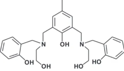
A Phenol-Based Compartmental Ligand as a Potential Chemosensor for Zinc(II) Cations
Jingwen Chen,a,b Shuhong Cao,b Dandan Wang,b Shengde Wub and Xiaoyong Wang*,a
aState Key Laboratory of Pharmaceutical Biotechnology, School of Life Sciences, Nanjing University,
Nanjing 210093, P.R. China
b School of Chemical and Biological Engineering, Yancheng Institute of Technology, Yancheng 224051, P.R. China
O ligante compartimental 2,6-bis(2-hidroxibenzil-2-hidroxietilamino) metil-4-metilfenol (L) foi sintetizado como um sensor químico em potencial para íons Zn2+. A base L coordena dois
cátions Zn2+ em metanol-água, formando um complexo dinuclear cuja formulação foi confirmada
por espectrometria de massas com ionização por “electrospray” (ESI-MS) e pelo gráfico de Job. A fluorescência de L é notavelmente aumentada por Zn2+ em comparação com os íons K+, Ca2+,
Mg2+, Cu2+, Pb2+, Mn2+, Fe3+, Fe2+, Co2+, Cd2+ e Ni2+. Isto se deve ao fato de que a complexação do
íon Zn2+ a L interrompe o processo de transferência eletrônica fotoinduzida e aumenta a rigidez do
esqueleto molecular de L. Observou-se ainda que a fluorescência de L é fortemente dependente da acidez e da polaridade dos solventes. Este composto poderá ser utilizado como uma sonda sensível a íons Zn2+ em solventes polares próticos, após uma modificação estrutural adequada.
An “end-off”-type compartmental Lewis base, 2,6-bis(2-hydroxybenzyl-2-hydroxyethylamino) methyl-4-methylphenol (L), was synthesized as a potential chemosensor for Zn2+ ions. L coordinates
two Zn2+ cations in methanol-water solution, forming a dinuclear complex whose formulation was
confirmed by ESI-MS spectroscopy and Job’s plot. The fluorescence of L is remarkably enhanced by Zn2+ as compared with K+, Ca2+, Mg2+, Cu2+, Pb2+, Mn2+, Fe3+, Fe2+, Co2+, Cd2+ and Ni2+ ions. The
fluorescence enhancement is attributed to the complexation of Zn2+ with L, which interrupts the
photoinduced electron transfer process and rigidifies the molecular skeleton of L. The fluorescence of L is greatly dependent on the acidity and polarity of the solvents. This compound may be used as a probe to sense Zn2+ ion in polar protic solvents after proper modification.
Keywords: chemosensor, fluorescence mechanism, phenol derivative, solvent effects, zinc(II)
Introduction
Zinc(II) ions play vital roles in a wide range of physiological
processes. Deficiency or imbalance of Zn2+ within the human
body can lead to a variety of diseases.1 Hence the development
of selective zinc chemosensors is of great importance for
tracking the Zn2+ status in biological systems.2 Fluorescence
chemosensors based on photoinduced electron transfer
(PET),3 intramolecular charge transfer (ICT),4 excited-state
intramolecular proton transfer (ESIPT),5 and fluorescence
resonance energy transfer (FRET) mechanisms have been developed for this purpose in the past years.2,6–10 Nevertheless,
none of them completely satisfies the criteria for a biosystem-oriented chemosensor. Therefore, efforts to design novel zinc probes are still needed.
Structural factors, such as molecular rigidity, could produce significant influence on the fluorescence efficiency of a chemosensor. An increase in planarity and a decrease in torsion may benefit the chemosensor
to enhance its fluorescence.11 As a metal ion binds to a
chemosensor, the molecular rigidity is enhanced and the above mentioned transfer processes are possibly inhibited, and thereby the fluorescence may arise. Based on such chelation enhanced fluorescence (CHEF) mechanism,
many zinc chemosensors have been designed.12–16 The
structure of a typical cation chemosensor is usually composed of two parts: an ion recognition and a
signal transduction units.8 We herein report an atypical
Experimental
Materials and general methods
All reagents and solvents, including 2-[4-(2- hydroxyethyl)-1-piperazinyl]-ethanesulfonic acid (HEPES), were of analytical grade and used without further purification. The compound
2,6-bis(2-hydroxybenzyl-2-hydroxyethylamino)methyl-4-methylphenol (L) was
synthesized by a procedure reported for an analogue, except
for minor modifications that we have described recently.17,18
The testing samples were prepared using newly double-distilled water.
The infrared spectra were recorded on a Bruker
VECTOR22 spectrometer as KBr pellets (4000-500 cm–1).
Elemental (C, H, N) analyses were performed on a Perkin-Elmer 240C analytic instrument. Electrospray mass spectra were recorded using an LCQ electron spray mass spectrometer (ESI-MS, Finnigan). The UV and fluorescence spectra were collected on a Shimadzu UV-2450 spectrophotometer and a Jasco FP-6500 spectrofluorometer equipped with a thermostated cell compartment, respectively. A quartz cuvette (1.0 cm, 3.0 mL) was used to carry out the spectroscopic titrations.
UV and fluorescence spectrophotometric titrations
The stock solution of L was prepared by dissolving L in
a methanol-water solution (90/10, v/v) containing HEPES
buffer (10 mmol L–1, pH 7.40). Aqueous solutions of Zn2+,
Mn2+, Co2+, Cu2+, Mg2+, Pb2+, Fe2+, Ca2+ and Ni2+ were
prepared from their acetate salts, and those of K+, Fe3+, and
Cd2+ were prepared from the respective chlorides and an
equimolar amount of sodium acetate. UV and fluorescence titrations were carried out by syringing aliquots (10 µL) of the Zn2+ solution (1 mmol L–1) into that of L (50 µmol L–1)
in HEPES buffer (3 mL). After each addition, the mixture
was equilibrated for ca. 5 min. Fluorescence spectra were
The UV spectra of L in HEPES-buffered
methanol-water solution are presented in Figure 2. The characteristic absorption appears in the range of 250-320 nm with a
λmax of 280 nm (ε = 4.93 × 10
3 L mol–1 cm–1), which could
be assigned to the α band of the substituted phenyls. The
absorbance of L decreases and the λmax shifts to 282 nm
(ε = 4.52 × 10³ L mol–1 cm–1) upon addition of Zn2+ to L for
the formation of −O−. The red-shift of the λ
max is indicative
of the coordination involving the phenolic oxygen donors
and Zn2+.19 The maximum absorption stops decreasing
when the ratio of Zn2+ to L approaches 2, suggesting that
a 1:2 dinuclear L−ZnII complex is formed. Two isosbestic
points at 257 and 284 nm are observed, which suggest the
existence of an equilibrium between L and L−ZnII. From
the absorption profile, the binding constant is calculated to
be 2.06 × 10⁴ L mol–1, following the reported equation.20
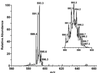
The ESI-MS spectrum obtained with the solution containing L andZn2+ in a ratio of 1:2 also supports the
formation of the dinuclear complex (Figure 3). The peak at
m/z 593.3 corresponds to one positively charged dinuclear
Figure 2. UV absorption profiles of L (50 µmol L–1) after addition of Zn2+
species [Zn2(L−3H)]+, which agrees with the result of the UV
spectra. Thus, a possible structure for the L−ZnII complex
can be proposed in Figure 4. In this structure, L provides a
NO3 donor set, i.e. an amine-N, an alcoholic-O, a phenolic-O,
and a bridging cresolic-O, for each Zn2+ ion. Considering the
coordination number of Zn2+, a bridging carboxyl group and
one or two water molecules may be involved in the metal coordination sphere, which resembles the structure we
reported previously for a similar complex.17
Fluorescence response of L to zinc(II)
Compound L exhibits moderate fluorescence with
a λem of 308 nm when excited at 276 nm in
HEPES-buffered methanol-water solution. Upon addition of Zn2+
(50 µmol L–1), the fluorescence intensity of L increases
proportionally until the molar ratio of Zn2+to L reaches 2.5
(Figure 5, inset). The result is basically in agreement with that of the UV titrations. The Stokes shift is about 30 nm and no significant change is observed for the position of
λex and λem. The Job’s plot based on the fluorescence
intensity of L−ZnII is presented in Figure 6, which confirms
that the proportion of L:Zn2+is ca. 1:2.
It is known that rigid conjugate structures tend to generate strong fluorescence,21,22 and that H+, OH– and
H2O molecules in solution are capable of forming effective
hydrogen bonds with the donor groups of a chromophore
in the ground and excited states.23 Therefore, L may form
hydrogen bonds with H+, OH– and/or H
2O in solution;
L may also form intra- or intermolecular hydrogen bonds
via its three phenolic and two alcoholic hydroxyl groups.
Such hydrogen bonds can enhance the rigidity of L and
thereby contribute to the moderate fluorescence of L in
solution.
A cooperative host-guest complexation can increase the rigidity of the host conformation and hence enhance
its fluorescence.24 Upon coordination with Zn2+, hydrogen
Figure 3. ESI-MS spectrum (positive mode) for the dinuclear L−ZnII
complex prepared in situ in methanol-water solution (90/10, v/v) at 298 K.
Figure 4. Proposed structure for the dinuclear L−ZnII complex. Charges,
counter ions, and possible water molecules are omitted for clarity.
Figure 5. Fluorescence enhancement of L (50 µmol L–1) with increasing
amount of Zn2+ in HEPES-buffered (10 mmol L–1, pH 7.40) methanol-water solution (90/10, v/v) at 298 K. λex: 276 nm; Zn2+ (from bottom to top): 0, 10, 20, 30, 40, 50, 60, 70, 80, 90, 100, 110, 120 and 130 µmol L–1; Inset: fluorescence intensity (IF) changes of L at 308 nm.
Figure 6. Job’s plot for the fluorescence of the L−ZnII system at 308 nm
in HEPES-buffered (10 mmol L–1, pH 7.40) methanol-water solution (90/10, v/v). [L + Zn2+] = 100 µmol L–1; λ
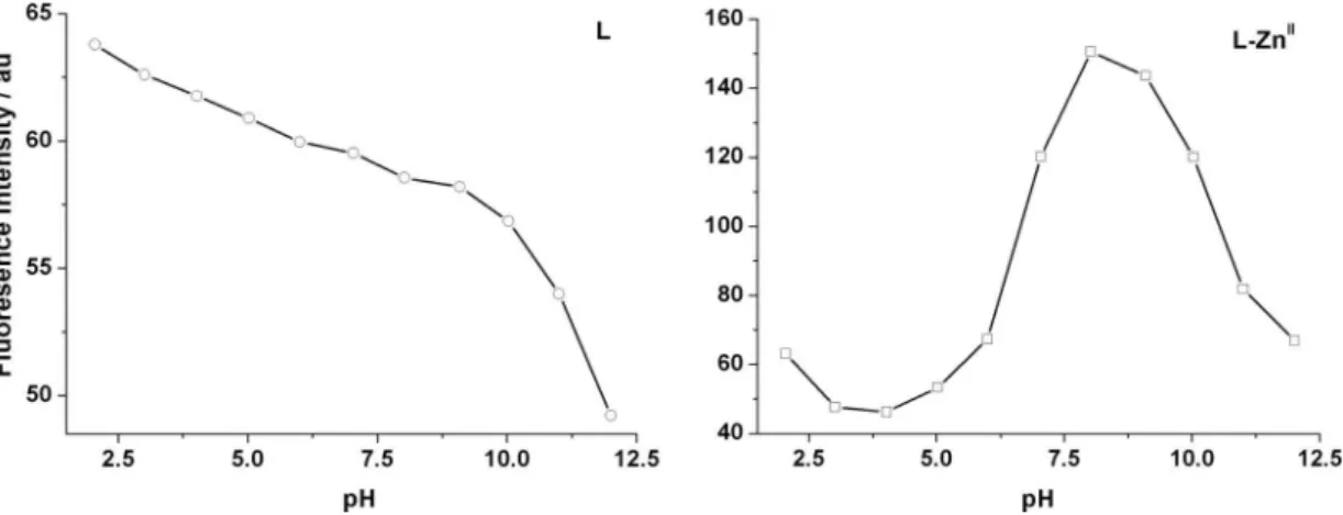
Effect of acidity on the fluorescence
The pH-dependent fluorescence of L and L−ZnII
prepared in situ was further investigated to verify the
fluorescence mechanism. The pH value of both solutions
was adjusted using HCl or KOH (10 mmol L–1) and the
alterations of the fluorescence at 308 nm were recorded
in the range of pH 2-12. As shown in Figure 7, both L
and L−ZnII show moderate fluorescence at low pH values.
The fluorescence of L decreases with the increase of pH
gradually before 10 and drastically after that. The reason for this may be the protonation of the tertiary amino N of
L at low pH and the deprotonation of the phenolic OH
and protonated tertiary amino N at high pH; the former can interrupt the PET pathway from N atoms to phenolic moieties, while the latter can promote the PET process. For
the L−ZnII system, the fluorescence behavior resembles
that of L when pH is below 4.0, suggesting the complex
is not yet formed. However, the fluorescence at 308 nm increases significantly when pH is above 4.0 and reaches
its maximum at ca. 8.0, which is about 3 times stronger
than that of L. The result suggests that the tertiary amino
N has coordinated to Zn2+ and the PET process is inhibited.
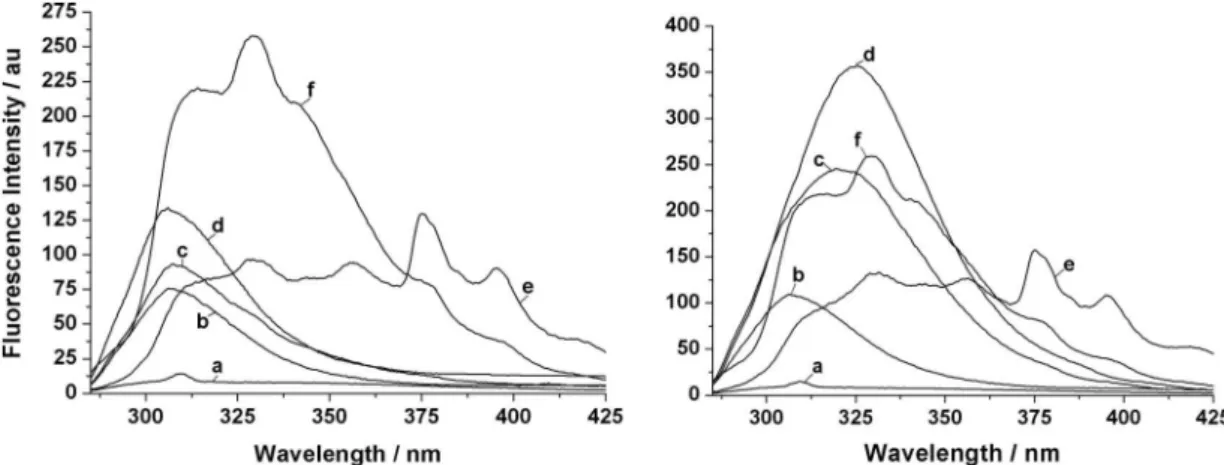
To gain a deeper insight into the fluorescence mechanism of L in the presence and absence of Zn2+, polar protic
(e.g. water, methanol, ethanol) and polar aprotic (e.g. DMSO, acetonitrile) solvents were selected to examine their effects on the fluorescence. As Figure 8 shows, the
fluorescence of L decreases remarkably with the increase
of polarity and acidity in polar protic solvents. For example, the fluorescence is almost completely quenched in water in
the absence or presence of Zn2+, which is similar to some
reported chemosensors,23,26 however, the fluorescence is
enhanced by ca. 3 times in methanol and ethanol in the
presence of Zn2+ with red shifts of 11.5 and 19 nm for the
maximum emission, respectively. By contrast with this, only subtle changes in fluorescence and curve profile of
L are observed in polar aprotic solvents, where Zn2+ has
little influence on the fluorescence. It has been proved that the solvent polarity can affect the electron transfer of a fluorescence sensor. Polar protic solvents may act as carriers to facilitate the electron transfer through the
partial-positively charged H-end.23 Thus, the electron transfer
process from the tertiary amino N to the phenolic moiety is accelerated and the fluorescence is quenched. Moreover,
polar protic solvents may facilitate L to form intra- and
intermolecular hydrogen bonds. If the intermolecular
Figure 7. Effect of pH on the fluorescence of L (50 µmol L–1, left) and L−ZnII prepared in situ (50 µmol L–1, right) at 308 nm in methanol-water solution
Figure 8. Fluorescence changes of L (50 µmol L–1) in different solvents at 298 K in the absence (left) and presence (right) of Zn2+ (50 µmol L–1). (a) H 2O; (b) MeOH/H2O (90/10, v/v); (c) MeOH; d, EtOH; (e) DMSO; (f) CH3CN.
Figure 9. Fluorescence responses of L (50 µmol L–1) to various metal
ions in HEPES-buffered (10 mmol L–1, pH 7.40) methanol-water solution (90/10, v/v) at 298 K (λex = 276 nm; λem = 308 nm). IF and IF0 represent the fluorescence intensity of L in the presence and absence of cation, respectively.
hydrogen bond is preferable, the rigidity of L would be
weakened. Anyway, the suppression of the PET process
and the rigidization of L appear to be the main reasons for
the fluorescence of L and L−ZnII.
Fluorescence responses of L to other cations
Cations such as K+, Ca2+, Mg2+, Cu2+, Pb2+, Mn2+,
Fe3+, Fe2+, Co2+, Cd2+ and Ni2+ were used instead of Zn2+
to investigate the fluorescence responses of L under the
same condition. The fluorescence of L was evaluated by
recording its changes after addition of the metal ion to the
methanol-water solution of L. To avoid possible hydrolysis
and ensure the main reaction, the metal ion solution was
added dropwise to L and the mixture was left for 5 min to
reach the equilibrium. As Figure 9shows, the alkali metal
cation K+ and the alkaline earth metal cations Ca2+ and
Mg2+ hardly interfere with the fluorescence of L at high
concentration (5 mmol L–1) due to their poor complexation
with L. However, the metal cations Pb2+, Mn2+, Co2+, Ni2+,
and especially Cu2+, Fe3+, and Fe2+,reduce the fluorescence
of L at relatively low concentration (0.5 mmol L–1),
probably due to an electron or energy transfer between metal cation and L.12,27,28 Interestingly, Cd2+, that has the
same d10 electronic configuration as Zn2+, does not enhance
the fluorescence of L. These observations indicate that L
may selectively signal the presence of Zn2+ in
methanol-water solution.
Conclusions
A phenol-based ligand (L) was synthesized as a
potential chemosensor for Zn2+. A dizinc complex is
formed by L and Zn2+ in methanol-water solution, which
greatly enhances the fluorescence of L. An inhibition of
the PET process and a ZnII-induced rigidization of L are
suggested to account for the fluorescence enhancement.
The fluorescence response of L to Zn2+ is more sensitive
in polar protic solvents than in polar aprotic solvents. In
similar conditions, L exhibits high fluorescence selectivity
for Zn2+ over other common cations. The results suggest that
L may possibly be used to sense Zn2+ after some structural
modification.
Acknowledgments
We thank the financial support from the Natural Science Program for Basic Research of Higher Education of Jiangsu
Province (Grant 06KJB150120), theChina Postdoctoral
5. Henary, M. M.; Wu, Y. G.; Fahrni, C. J.; Chem.Eur. J.2004, 10, 3015.
6. Godwin, H. A.; Berg, J. M.; J. Am. Chem. Soc.1996, 118, 6514.
7. Woodroofe, C. C.; Lippard, S. J.; J. Am. Chem. Soc.2003, 125, 11458.
8. Lim, N. C.; Freake, H. C.; Brückner, C.; Chem.-Eur. J.2005, 11, 38.
9. Hendrickson, K. M.; Geue, J. P.; Wyness, O.; Lincoln, S. F.; Ward, A. D.; J. Am. Chem. Soc.2003, 125, 3889.
10. Burdette, S. C.; Frederickson, C. J.; Bu, W.; Lippard, S. J.; J. Am. Chem. Soc.2003, 125, 1778.
11. Radke, K. R.; Ogawa, K.; Rasmussen, S. C.; Org. Lett.2005, 7, 5253.
12. De Silva, A. P.; Gunaratne, H. Q. N.; Gunnlaugsson, T.; Huxley, A. J. M.; McCoy, C. P.; Rademacher, J. T.; Rice, T. E.; Chem. Rev.1997, 97, 1515.
13. Dai, Z.; Proni, G.; Mancheno, D.; Karimi, S.; Berova, N.; Canary, J. W.; J. Am. Chem. Soc.2004, 126, 11760.
Horwood: New York, 1991.
20. Pocker, Y.; Ciula, J. C.; J. Am. Chem. Soc.1989, 111, 4728. 21. Nijegorodov, N. I.; Downey, W. S.; J. Phys. Chem.1994, 98,
5639.
22. Gao, L.; Wang, Y.; Wang, J.; Huang, L.; Shi, L.; Fan, X.; Zou, Z.; Yu, T.; Zhu, M.; Li, Z.; Inorg. Chem.2006, 45, 6844. 23. Woo, H. Y.; Liu, B.; Kohler, B.; Korystov, D.; Mikhailovsky,
A.; Bazan, G. C.; J. Am. Chem. Soc.2005, 127, 14721. 24. Nakashima, H.; Yoshida, N.; Org. Lett.2006, 8, 4997. 25. Valeur, B.; Leray, I.; Coord. Chem. Rev.2000, 205, 3. 26. Woo, H. Y.; Hong, J. W.; Liu, B.; Mikhailovsky, A.; Korystov,
D.; Bazan, G. C.; J. Am. Chem. Soc.2005, 127, 820. 27. Hirano, T.; Kikuchi, K.; Urano, Y.; Higuchi, T.; Nagano, T.;
Angew. Chem., Int. Ed. 2000, 39, 1052.
28. Mikata, Y. J.; Wakamatsu, M.; Yano, S.; Dalton Trans.2005, 545.
Received: March 4, 2008