Article
0103 - 5053 $6.00+0.00
*e-mail: [email protected]
Modification of Spherical SiO
2Particles
via
Electrolyte for High Zeta Potential
and Self-Assembly of SiO
2Photonic Crystal
Li-ying Liu,* Xiu-feng Wang, Bing Cheng and Chun-xiang Zhang
School of Materials Science & Engineering, Shaanxi University of Science & Technology, Xi’an 710021, P.R. China
Partículas esféricas de SiO2 foram preparadas através de um novo processo de co-precipitação melhorado pelo eletrólito NaCl para carregamento e posterior auto-agrupamento em cristais fotônicos pelo método de deposição vertical. Os compostos obtidos e suas estruturas foram caracterizados por microscopia eletrônica de varredura (SEM) com espectroscopia dispersiva de energia de raios-x (EDS), potencial zeta, e espectrofotometria UV-Vis. As imagens SEM indicaram que as partículas SiO2 eram esféricas e o diâmetro médio das partículas modificadas foi aproximadamente 380 nm, maior do que as partículas não modificadas (315 nm). Além disso, usando-se quantidade excessiva de NaCl (10 mg), uma distribuição bimodal do diâmetro das partículas de SiO2 foi obtida e os diâmetros
foram aproximadamente 110 nm e 420 nm, respectivamente. Comparando com as partículas não modificadas de SiO2, o espectro EDS das partículas modificadas mostrou um pico característico de Na, o qual provou a existência do elemento sódio na partícula SiO2. Os resultados de potencial Zeta revelaram que o potencial Zeta médio das partículas modificadas SiO2 em etanol foi –56,6 mV, o qual é muito maior do que para as partículas não modificadas (–48,7 mV). Portanto, os cristais fotônicos que se auto-agrupam pelas partículas modificadas de SiO2 possuem estrutura cúbica centrada na
face com menos defeitos. Além disso, a espectrofotometria de UV-Vis mostrou que o gap da banda fotônica do cristal modificado de SiO2 apresentava um deslocamento para o vermelho e o pico de absorção tornou-se mais estreito e alto.
Spherical SiO2 particles were prepared by a novel co-precipitation process improved by electrolyte NaCl for charging and further self-assembled into photonic crystal by vertical deposition method. The obtained composites and their structures were characterized by scanning electron microscope (SEM) with X-Ray energy dispersive spectroscopy (EDS), Zetasizer, and UV-Vis spectrophotometer. SEM images indicated that the SiO2 particles were spherical and the average diameter of modified SiO2
particles was about 380 nm, larger than that of the unmodified ones (315 nm). Moreover, when the amount of NaCl was excessive (10 mg), bimodal diameter distribution SiO2 particles were obtained and the diameters were about 110 nm and 420 nm respectively. Compared with the unmodified SiO2 particles, EDS spectra of the modified ones showed an obvious characteristic-peak of Na element which proved that the Na element existed in the SiO2 particle. The results of Zetasizer revealed that
the average Zeta potential of the modified SiO2 particles in ethanol was –56.6 mV, which was much higher than that of the unmodified ones (–48.7 mV). So the photonic crystal that self-assembled by the modified SiO2 particles possessed face-centred cubic structure with fewer defects. In addition, UV-Vis spectrophotometer showed that photonic band gap of the modified SiO2 photonic crystal had
a red-shift and the absorption peak became narrower and higher.
Keywords: photonic crystal, SiO2 particles, modification, electrolyte
Introduction
Photonic crystal materials, i.e. materials with periodically modulated refractive indices, are attracting more and more attention of researchers because they can control the flows of photons by means of photonic band gaps (PBG).1-5 The
investigation of these materials has ascertained their use in many applications, such as filters, sensing devices, zero-threshold lasers, high efficiency light emitting diodes, optical switch and integrated optical waveguides, etc.6-8 In order to
Liu et al. 47 Vol. 20, No. 1, 2009
Self-assembly approaches offer distinct advantages in that they are efficient, require minimal fabrication efforts, and can be easily modified for comprehensive manufacturing purposes. The references9-12 show that an
array of fine SiO2 particles made by self-assembly method is one of the photonic crystal materials. However, when the SiO2 particles are synthesized following the traditional Stöber13 method to self-assemble into photonic crystal
structure, they have a disadvantage that they are short of charge on their surface.14,15 A key factor in successfully
producing crystalline photonic crystals is the preparation of highly monodispersed and charged colloidal particles. The higher the surface charge density of the colloidal particles, the stronger the electrostatic repulsive force between the colloids, thus the photonic crystals with high quality can be fabricated more easily using self-assembly method.16 But
the surface charge on SiO2 particles is not strong enough to be suitable for assembling into an ordered pattern. In particular, in order to self-assemble into photonic crystal, the density of colloidal particles ought to be 1013 cm-3, and
in this situation, coagulation of SiO2 particles could easily take place which would prevent the formation of orderly photonic crystal,thus more attention has been paid to enhancing the surface charge density of SiO2 particles.17,18
Currently, many organic materials,19 such as oleic acid
and succinic acid,20 were chemically bonded or physically
adsorbed on the surface of SiO2 particles for charging. But preparation processes of the above methods were complicated and unstable.
Herein, a novel and simple approach to prepare; the spherical SiO2 particles was reported. The traditional stöber method was improved by adding the right amount of electrolyte NaCl to reliably increase the charge on the surface. Additionally, ultraviolet and visible absorption measurements were performed.
Experimental
All chemicals were analytical reagent (AR) grade and used as received without further purification. Monodisperse SiO2 particles were prepared by adding a premixed solution consisted of ethanol (50 mL), ammonia (4 mL, 5 mL, 6 mL, 7 mL and 8 mL respectively), DI water (5 mL) and NaCl (5 mg) to a tetraethyl orthosilicate (TEOS, 5 mL) solution in ethanol (30 mL) under stirring at 25 °C. And bimodal diameter distribution SiO2 particles were obtained when the amount of NaCl was excessive (10 mg) and the amount of ammonia was 6 mL. The mixture was stirred for 20 hours and further washed three times with ethanol. Then the obtained samples were dried in a vacuum oven at 60 °C.
To assemble the spherical SiO2 particles into photonic crystal, 110 mg SiO2 particles were dispersed in ethanol (15 mL) by ultrasonic dispersion for 2 hours. Then a pair of glass plates separated with SiO2 spacers by 2 to 10 µm were immersed in the solution. After that, the liquid went up along the plates owing to the capillary force, brought the SiO2 particles along, and deposited them on the plates as the solution deposited in an oven at 40 °C.
The morphology and structure of the samples were examined by field-emission scanning electron microscope (FESEM; JEOL JSM-6700) with X-Ray energy dispersive spectroscopy (EDS). The value of the zeta potential and diameter distribution of the samples were determined by Zetasizer Nano ZS made in Malvern Instruments Ltd. And the UV-Vis absorption spectra measurements were performed by a UV-Vis-NIR spectrophotometer (Perkin-Elmer Lambda 950).
Results and Discussion
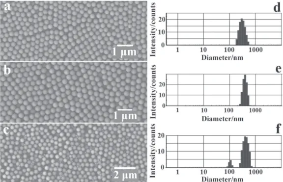
Figure 1 showed the SEM images and diameter distribution charts of the obtained SiO2 particles when the amount of ammonia was 6 mL. Figure 1a and Figure 1d indicated that the unmodified SiO2 particles were spherical, the diameter was about 315 nm and the relative deviation was about 8.6%. In comparison with the unmodified SiO2 particles, as shown in Figure 1b and Figure 1e, the SiO2 particles modified by 5 mg NaCl were also spherical, but the diameter (about 380 nm) was larger than that of the unmodified ones. Moreover, relative deviation of the modified SiO2 particles was only about 4.4%, which was much less than that of the unmodified ones. Figure 1c and Figure 1f showed that the bimodal diameter distribution SiO2 particles prepared with excessive NaCl (10 mg) were spherical and the diameters were about 110 nm and 420 nm respectively. And the relative deviation was 4.7% and 6.1% respectively.
In general, spherical SiO2 particles are prepared by the hydrolysis and condensation of TEOS in a mixture of ethanol, water, and ammonia. The hydrolysis reaction gives the singly hydrolyzed TEOS monomer [(OC2H5)3Si(OH)]
Si(OC2H5)4 + H2O → (OC2H5)3Si(OH) + C2H5OH (1)
Subsequently, this intermediate reaction product condenses eventually to form spherical SiO2 particles according to
(OC2H5)3Si(OH) + H2O → SiO2↓ + 3C2H5OH (2)
relative diameter deviation of SiO2 particles. The electric double layer is formed due to the ionization of –OH on the surface of SiO2 particles in the ethanol solution. At the beginning of the reaction, ionic concentration of the solution became higher owing to the addition of NaCl, and then the width of the electric double layer became thinner. It was beneficial to the deposition of TEOS monomers on the surface of the SiO2 particles. So the modified SiO2 particles had larger average diameter and less relative diameter deviation than that of the unmodified ones.
EDS spectra of SiO2 particles were shown in Figure 2. Both of the unmodified and modified SiO2 particles had the characteristic-peak of O and Si elements, which indicated that the main component of the particles was SiO2 as the EDS spectra showed. Additionaly, compared with the unmodified SiO2 particles (Figure 2a), the EDS spectra of modified SiO2 particles (Figure 2b) had an obvious characteristic-peak of Na element. It was probably because that Na+ ions had entered interstitial sites of the silica
tetrahedron with the increasing of the reactions. Moreover, only OH- ions and Cl− ions could exist in reaction solution,
and the EDS spectra of modified SiO2 particles did not have the characteristic-peak of Cl element shown in Figure 2b. Therefore, there were more physical absorption of OH−
ions on the surface of SiO2 particles which resulted in the fact that the electric double layer surrounding modified SiO2 particles became thicker than that of the unmodified ones. The thicker the electric double layer, the higher the
Figure 1. SEM images of unmodified (a), modified (b) and bimodal diameter distribution (c) SiO2 particles and their diameter distribution charts (d, e, and f) respectively.
Liu et al. 49 Vol. 20, No. 1, 2009
Zeta potential of SiO2 particles. Figure 3 indicated the Zeta potential of both unmodified (a) and modified (b) SiO2 particles in ethanol with different volume of ammonia. It was obvious that the average value of Zeta potential of the modified SiO2 particles (–56.6 mV) was much higher than that of the unmodified ones whose Zeta potential was about –48.7 mV.
The improvement of Zeta potential increased the electrostatic repulsive force among SiO2 particles, which was useful to self-assemble into fine photonic crystal structures by vertical deposition method. From microcosmic perspective, as the SiO2 particles deposited, the distance among SiO2 particles was shortening. When the particles were close enough, they began to assemble into ordered structures on the effect of the competition between attractive force and repulsive force. For unmodified SiO2 particles, because of the weak electrostatic repulsive force, they were prone to coagulate that resulted in the formation of photonic crystal with more defects (Figure 4a). By contrast, stronger electrostatic repulsive force of the modified SiO2 particles can adjust the self-assembly behavior, so the system can reach the thermostatic balanced state. At the moment, SiO2 particles self-assembled into fine photonic crystal with fewer defects and presented a kind of regular hexagon structure on the surface of the photonic crystal (Figure 4b).
From mathematical perspective, at least three crystalline planes must be considered to determine which structure a lattice-site is in. And an important characteristic of face-centred cubic (FCC) structure is that each lattice-site is connected with two {111} group crystalline planes and one {110} group crystalline plane as shown in the truncated octahedron (Figure 4d and 4e), which can be revealed easily in Figure 4c. Therefore, the photonic crystal assembled
by modified SiO2 particles possessed FCC structure with fewer defects.
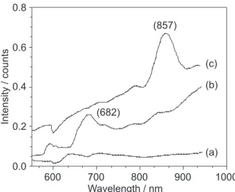
Figure 5 showed UV-Vis absorption spectra of the photonic crystal assembled by the bimodal diameter distribution SiO2 particles (a), the unmodified (b) and modified ones (c) respectively. The photonic crystal assembled by the bimodal diameter distribution SiO2 particles did not exhibit obvious absorption peaks owing to the FCC structure was not formed. But the photonic crystal assembled by unmodified and modified SiO2 particles exhibited absorption peaks at 682 nm and 857 nm respectively. Obviously, the absorption peak of the modified SiO2 photonic crystal was narrower and higher than that of the unmodified one. It was the ordered assembly of photonic crystal that led to the narrower and higher peak, doing to
Figure 4. SEM images of the photonic crystal self-assembled by unmodified (a) and modified (b and c) SiO2 particles. Diagrams of the photonic crystal structures (d and e).
Figure 5. UV-Vis absorption spectra of the photonic crystal assembled by the bimodal diameter distribution (a), unmodified (b), and modified (c) SiO2 particles.
during the higher Zeta potential. Moreover, compared with the unmodified SiO2 photonic crystal, the absorption peak of the modified one had a red-shift resulting from the increase of particle diameter. That’s because the position of the observed absorption peak agree very well with the diameter of SiO2 particles, which can be predicted by the Bragg’s law for the (111) family of planes:21
(3)
where lc is the wavelength at the peak position, D is the average diameter of SiO2 particles, f is the dielectric filling ratio (for an ideal FCC structure, f = 0.74), nsphere and nair are the refractive indexes of SiO2 particles and air respectively (in theoretical computation, nsphere = 1.46 and nair = 1).22,23
Then, the absorption peak of the modified and unmodified SiO2 photonic crystal can be easily calculated, which were 835 nm and 589 nm respectively. Thus, the difference value between calculated value and experimental value of the modified one (22 nm) was much less than that of the unmodified one (93 nm), which again proved that the photonic crystal structure assembled by modified SiO2 particles was much better.
Conclusions
Spherical SiO2 particles were synthesized by controlling the hydrolysis of Ethyl orthosilicate. It was proved that the SiO2 particles modified by the right amount of electrolyte NaCl exhibited much higher Zeta potential in ethanol, larger average diameter and less diameter deviation. Therefore, the modified SiO2 particles could self-assemble into fine photonic crystal structure with fewer defects. Additionally, UV-Vis absorption spectra indicated that the photonic crystal assembled by bimodal diameter distribution SiO2 particles showed no obvious absorption band. On the contrary, the photonic crystal assembled by modified and unmodified ones showed the absorption bands at 857 nm and 682 nm respectively. Moreover, the absorption peak of the modified SiO2 photonic crystal had a red-shift and became narrower and higher than that of the unmodified one.
Acknowledgments
This work was supported by the National Natural Science Foundation of China (Grant No. 50372038).
References
1. Yablonovitch, E.; Phys. Rev. Lett.1987, 58, 2059. 2. John, S.; Phys. Rev. Lett.1987, 58, 2486.
3. Joannopoulos, J. D.; Villeneuve, P. R.; Fan, S.; Nature1997,
386, 143.
4. Yablonovitch, E.; J. Opt. Soc. Am. B: Opt. Phys. B1993, 10, 283.
5. Toader, O.; John, S.; Science2001, 292, 1133.
6. Lee, W.; Pruzinsky, S. A.; Braun, P. V.; Adv. Mater. 2002, 14, 271.
7. Li, J.; Herman, P. R.; Valdivia, C. E.; Kitaev, V.; Ozin, G. A.;
Opt. Express2005, 13, 6454.
8. Ben-Moshe, M.; Alexeev, V. L.; Asher, S. A.; Anal. Chem.2006,
78, 5149.
9. Whitesides, G. M.; Mathias, J. P.; Seto, C. T.; Science1991,
254, 1312.
10. Jiang, P.; Bertone, J. F.; Hwang, K. S.; Chem. Mater. 1999, 11, 2132.
11. Gu, Z. Z.; Fujishima, A.; Sato, O.; Chem. Mater.2002, 14, 760.
12. Ni, P. G.; Dong, P. ; Cheng, B. Y. ; Adv. Mater.2001, 13, 437. 13. Stöber, W.; Fink, A.; J. Colloid Interface Sci.1968, 26, 62. 14. Ko, H. Y.; Lee, H. W.; Moon, J.; Thin Solid Films2004, 447,
638.
15. Holgado, M.; Blanco, A.; Ibisate, M.; Langmuir1999, 15, 4701.
16. Wang, W.; Gu, B. H.; Liang, L. Y.; J. Phys. Chem. B2003, 107, 3400.
17. Wang, W.; Gu, B. H.; Liang, L. Y.; J. Phys. Chem. B2003, 107, 12113.
18. Yoshinaga, K.; Chiyoda, C.; Ishiki, H.; Colloids Surf., A2002,
204, 285.
19. Li, Z.; Zhu, Y.; Appl. Surf. Sci.2003, 211, 315.
20. Cheng, B.; Wang, X. F.; Wang, L. S.; Wu, Y. T.; Mater. Lett.
2007, 61, 1350.
21. Goldenberg, L. M.; Wagner, J.; Stumpe, J.; Paulke, B. R.; Görnitz, E.; Langmuir2002, 18, 3319.
22. Lu, Y.; Yin, Y.; Li, Z. Y.; Xia, Y.; Langmuir2002, 18, 7722. 23. Blaaderen, A.; Vrij, A.; Langmuir1992, 8, 2921.