Isoflavonoids and Triterpenoids Isolated from
Pterodon
polygalaeflorus
Délcio D. Marquesa, Maria Iracema Lacerda Machadoa, Mário Geraldo de
Carvalhob, Luiz Augusto da C. Meleirab, and Raimundo Braz-Filhoc
a
Departamento de Química Orgânica e Inorgânica, Universidade Federal do Ceará,
C.P. 12.200, 60.455-760 Fortaleza - Ce, Brazil
b
Departamento de Química - ICE, Universidade Federal Rural do Rio de Janeiro,
23851-970 Soropédica-Itaguaí - RJ, Brazil
c
Setor de Produtos Naturais, Universidade Estadual do Norte Fluminense,
28015-620 Campos - RJ, Brazil
Received: April 30, 1997
Os isoflavonóides 6,7-dimetoxi-3’,4’-metilenodioxi-, 4’-hidroxi-3’,6,7-trimetoxi-, 3,4,6,7-tetrametoxi-, 7-hidroxi-6-metoxi-3,4-metilenodioxi-, 2’,6,7-trimetoxi-3’,4’-metilenodioxi-, 2’,3’,4’,7,7-pentametoxi- e 2’,4’,5,6,7-pentametoxiisoflavona, os triterpenóides lupeol e betulina e o ácido 4-metoxibenzóico foram isolados dos extratos acetônicos do alburno e do cerne de Pterodon polygalaeflorus. As estruturas destes produtos naturais foram caracterizadas por métodos espec-trométricos, principalmente experiências de RMN 1D e 2D de hidrogênio e carbono-13, que foram também utilizados para a atribuição inequívoca dos deslocamentos químicos dos átomos de hidrogênio e carbono-13 dos isoflavonóides.
The isoflavonoids 6,7-dimethoxy-3’,4’-methylenodioxy-, 4’-hydroxy-3’,6,7-trimethoxy-, 3,4,6,7-tetramethoxy-, 7-hydroxy-6-methoxy-3,4-methylenodioxy-, 2’,6,7-trimethoxy-3’,4’-methylenedioxy-, 2’,3’,4’,6,7-pentamethoxy- and 2’,4’,5,6,7-pentamethoxyisoflavone, together with the triterpenoids lupeol and betulin and 4-methoxybenzoic acid, were isolated from acetone extracts of the sapwood and heartwood of Pterodon polygalaeflorus. The structures of these natural products have been characterized by spectrometric methods, mainly extensive 1D and 2D NMR experiments, which were also used for complete assignments of the chemical shifts of the hydrogen and carbon-13 atoms of isoflavonoids.
Keywords: Pterodon polygalaeflorus, Leguminosae, isoflavonoids, triterpenoids,1H- and
13C-NMR data
Introduction
In a paper published recently the isolation of diterpenes from fruits of a specimen of Pterodon ploygalaeflorus
Benth, Leguminosae family, was reported1. As part of our continuing chemical investigation of this plant we have investigated the sapwood and the heartwood. The isofla-vones 1-7, p-methoxybenzoic acid (8), lupeol (9), and betulin (10), have been isolated from this plant material. The structures of these natural products were established by spectral data, mainly 1H- and 13C-NMR including
ho-monuclear and heteronuclear 2D experiments (2, 3a, 5 and 7) and NOE difference spectra (1, 2, 3a, 5 and 7), which were also used for complete assignment of the hydrogen and carbon-13 atom chemical shifts of the isoflavonoid 2, the acetyl derivatives 3a, 5 and 7.
Results and Discussion
The acetone extracts of heartwood and sapwood of
Pterodon ploygalaeflorus were submitted to chromatogra-phy on silica gel column to afford the isoflavonoids 1-7, 4-methoxybenzoic acid (8), lupeol (2) and betulin (10). The Article
isoflavones 1, 2, 5-7 were previously isolated from Milletia dura (1 and 5)2, Pterodon pubescens (2, 5 and 7)3,4 and
Condyla africana (6)5. The triterpenoids 9 and 10 are frequently found in plants6. The structural characterization of these compounds was based on spectral data, notebly the 1H- and 13C-NMR spectra, including homonuclear 1H-x 1H-COSY and heteronuclear 1H- x 13C-COSY-1J
CH and 1 H-x 13C-COSY-nJCH (n = 2 and 3)] experiments and NOE difference spectra (1H{1H}-NOE) data for the isofla-vonoids 1, 2, 3a, 5 and 7, in addition to corresponding 1H-NMR spectral data reported in the literature 2-5 (Table 2). To the best of our knowledge, the isoflavone 3 is hitherto
unreported as a natural product and 13C-NMR spectral data were only found for 2 (Table 2)7.
The homonuclear 1H- x 1H-COSY, heteronuclear 1H- x 13C-COSY-n
JCH (n = 1, spin-spin couplings between 13C and 1H- atoms via one bond; n = 2 and 3, COLOC = correlation via long-range couplings) 2D shift-correlated NMR spectra8 of 2, 3a, 5 and 7 (Tables 1-3) and NOE difference spectra8 of 1, 2, 3a, 5 and 7 (Table 4) together with the application of the shift parameters and the ob-served multiplicities of the signals of the carbon atoms deduced by comparative analysis of the PND- and DEPT-13C-NMR spectra9, were also used for the complete assign-ment of the hydrogen and carbon-13 atom chemical shifts
Table 1.13C-NMR (50.3 MHz) data for the isoflavonoids 1-3,5and 7 (CDCl3), compared values described in the literature are in parenthesis7 for 2,
TMS as internal standard and chemical shifts in δ (ppm).*
C 1 2a 3aa 5a 7a
3 124.45 123.91 (123.33) 123.86 124.27 121.54
4 175.38 175.21 (174.57) 175.14 175.76 175.24
6 147.74 147.36 (147.56) 147.68 147.29 147.30
7 154.41 154.02 (154.36) 154.35 153.33 153.98
9 152.23 151.89 (151.93) 152.12 152.27 152.05
10 117.81 117.51 (117.29) 117.72 117.08 117.55
1’ 125.85 124.51 (124.91) 130.88 112.50 118.49
2’ - - - 152.59 151.75
3’ 147.56 148.35 (148.80) 150.70 - 142.06
4’ 147.74 148.65 (148.50) 139.51 148.10 153.74
5’ - - - 140.70
-CH
2 151.96 151.77 (152.84) 152.49 153.96 153.00
5 104.90 104.35 (104.42) 104.60 104.08 104.68
8 99.50 99.18 (100.05) 99.41 99.24 99.32
2’ 109.72 112.13 (112.95) 113.55 -
-3’ - - - 94.79
-5’ 108.29 110.75 (111.52) 122.80 - 106.96
6’ 122.33 120.61 (121.21) 120.66 110.53 125.63
CH2
OCH2O 101.11 - - 100.99
-CH3
MeO-6 56.38 56.50 (55.85) 56.38 55.66 56.25
MeO-7 56.31 56.16 (56.28) 56.24 55.66 56.06
MeO-2’ - - - 55.91 60.68
MeO-3’ - 55.62 (55.64) 55.85 - 60.53
MeO-4’ - 56.02 (55.64) - - 55.76
of 2, 3a, 5 and 7, and consequently of the other isofla-vonoids isolated from Pterodon polygalaeflorus by com-parison with data now unambiguously assigned (Tables 1-4).
Thus, a series of 2D NMR experiments led to the assignment of all 1H- and 13C resonances for 2 and 3a (e. g.). In the 1H- x 13C-COSY-1JCH spectra of 2 and 3a the connectivities between the protonated carbon atoms and
Table 2.1H-NMR data for the isoflavonoids 1-7 [ 200 MHz (1, 2, 3a, 5 and 7), 100 MHz (6) and 60 MHz (4a)], in CDCl3 and TMS as internal standard,
chemical shifts in δ (ppm) and coupling constants (J, in parenthesis) in Hz.*
H 1 2 3a 4a 5 6 7
2 7.91 (s) 7.97 (s) 7.97 (s) 7.94(s) 7.88 (s) 8.05(s) 7.93(s)
5 7.61 (s) 7.63 (s) 7.60 (s) 7.74 (s) 7.59 (s) 7.67 (s) 7.63 (s)
8 6.86 (s) 6.88 (s) 6.87(s) 7.22 (s) 6.85 (s) 6.93 (s) 6.89 (s)
2’ 7.10 (d, 1.7) 7.25 (d, 1.8) 7.34 (d, 1.6) 7.1-6.7 (m) - -
-3’ - - - - 6.59 (s) 6.68 (s)
-5’ 6.85 (d, 8.0) 6.93 (d, 8.3) 7.07 (d, 8.2) 7.1-6.7 (m) - - 6.76 (d, 8.5)
6’ 6.98 (dd, 8.0, 1.7) 7.05 (dd, 8.3, 1.8) 7.00 (dd, 8.2, 1.6) 7.1-6.7 (m) 6.80 (s) 7.02 (s) 7.06 (d, 8.5)
OCH2O 5.98 (s) - - 6.00 (s) 5.93 (s) 4.00 (s)
-MeO-6 3.98 (s) 3.99 (s) 3.97 (s) 3.95 (s) 3.96 (s) 4.00 (s) 4.01 (s)
MeO-7 3.98(s) 3.99 (s) 3.98 (s) - 3.96 (s) 4.00 (s) 4.00 (s)
MeO-2’ - - - - 3.70 (s) 3.80 (s) 3.84 (s)
MeO-3’ - 3.92 (s) 3.86 (s) - - - 3.95 (s)
MeO-4’ - 3.88 (s) - - - 3.88 (s) 3.91 (s)
MeO-5’ - - - - - 3.97 (s)
-AcO-4’ - - 2.32 (s) - - -
-AcO-7’ - - - 2.40 (s) - -
-*Homonuclear 1Hx1H-COSY and heteronuclear 1Hx13C-COSY-1JCH 2D NMR spectra were also used for these assignments. Chemical shifts and coupling
constants (J) were obtained from 1D 1H-NMR spectra.
Table 3. Heteronuclear 2D shift-correlated via long-range couplings [1H-x13C - COSY- nJCH (n = 2 and 3), COLOC] NMR spectral data for the
isoflavonoids 2,3a, 5and 7.*
2 3a 5 7
C 2JCH 3JCH 2JCH 3JCH 2JCH 3JCH 2JCH 3JCH
3 H-2 H-2’; H-6’ H-2’ H-2’ H-6’ H-2 H-6’
4 H-2; H-5 H-2; H-5 H-5 H-2; H-5
6 H-5 H-8; MeO-6 H-8; MeO-6 H-8; MeO-6 H-8; MeO-6
7 H-8 H-5; MeO-7 H-8 H-5; MeO-7 H-5; MeO-7 H-5; MeO-7
9 H-8 H-2; H-5 H-8 H-5 H-5 H-8 H-2; H-5
10 H-5 H-8 H-5 H-8 H-8
1’ H-2; H-5’ H-2; H-5’ H-3’ H-5’
2’ H-6’ MeO-2’ H-6’; MeO-2’
3’ H-5’; MeO-3’ H-5’; MeO-3’ H-5’; MeO-3’
4’ H-2’; H-6’; MeO-4’ H-2’; H-6’ H-6’ H-6’; MeO-4’
5’ H-3’
6’ H-2’
the corresponding hydrogens that were observed are: CH-2 [2: δC 151.77(d) and δH 7.97 (s); 3a: δC 152.49 (d) and δH 7.97 (s)], CH-5 [2: δC 104.35 (d) and δH 7.63 (s); 3a: δC 104.60 (d) and δH 7.60 (s)], CH-8 [2: δC 99.18 (d) and δH 6.88 (s); 3a: δC 99.41(d) and δH 6.87 (s)], CH-2’ [2: δC 112.13 (d) and δH 7.25 (d, J = 1.8 Hz); 3a: δC 113.55 (d) and δH 7.34 (d, J = 1.6 Hz)], CH-5’ [2: δC 110.75 (d) and δH 6.93 (d, J = 8.3 Hz); 3a: δC 122.80 (d) and δH 7.07 (d, J = 8.2 Hz); shifted downfield by only 0.14 ppm [∆δH = 7.07 (3a)-6.93 (2)] and by 12.05 ppm [∆δC = 122.80 (3a)-110.75 (2) in the acetyl derivative (3a), as anticipated by shielding reduction of the mesomeric ortho-effect], CH-6’ [2: δC 120.61 (d) and δH 7.05 (dd, J = 8.3 and J = 1.8 Hz); 3a: δC 120.66 (d) and δH 7.00 (dd, J = 8.2 and J = 1.6 Hz)] and methoxy groups [2: δC 56.50 and δH 3.99, 56.16 and 3.99, 55.62 and 3.92, 56.02 and 3.88; 3a: δC 56.38 and δH 3.87, 56.24 and 3.98, 55.85 and 3.86] (Tables 1 and 2).
The chemical shift assignments of the quaternary carb-on atoms were established by 1H- x 13C-COSY-nJCH (n = 2 and 3, COLOC) spectra . Thus, the spectrum of 2 showed long-range correlations (Table 3): C-3 (δC 123.91) with H-2 (δH 7.97, 2JCH), H-2’ (δH 7.25, 3JCH) and H-6’ (δH 7.05, 3J
CH); C-4 with H-2 (δH 7.97, 3JCH) and H-5 (δH 7.63, 3JCH); C-6 (δC 147.36) with H-5 (δH 7.63, 2JCH), H-8 (δH 6.88, 3J
CH) and MeO-6 (δH 3.99, 3JCH); C-7 (δC 154.02) with H-8 (δH 6.88, 2JCH), H-5 (δH 7.63, 3JCH) and MeO-7 (δH 3.99, 3J
CH); C-9 (δC 151.89) with H-8 (δH 6.88, 2JCH), H-2 (δH 7.97, 3JCH) and H-5 (δH 7.63, 3JCH); C-10 (δC 177.51) with H-5 (δH 7.63, 2J
CH) and H-8 (δH 6.88, 3JCH); C-1’ (δc 124.51) with H-2 (δH 7.97, 3JCH) and H-5’ (δH 6.93, 3JCH); C-3’ (δC 148.33) with H-5’ (δH 6.93, 3JCH) and MeO-3’ (δH 3.92, 3JCH); C-4’ (δC 148.65) with H-2’ (δH 7.25, 3JCH), H-6 (δH 7.05, 3JCH) and MeO-4’ (δH 3.88, 3JCH). The spectra of 3a, 5 and 7 (δH 7.97, 3JCH) revealed analogous results as shown in Table 3.
Table 4. NOE difference spectra (1H-{1H}-NOE) data for the isoflavonoids 1, 2, 3a, 5 and 7.
{H} NOE enhancements
Compound H δH H δH %
1 (MeO)2-6,7 3.98 5 7.61 5
8 6.86 10
5 7.61 MeO-6 3.98 15
2 7.91 2’ 7.10 7
6’ 6.98 10
2 MeO-4’ 3.88 5’ 6.93 6
MeO-3’ 3.92 2’ 7.25 8
(MeO)2-6,7 3.99 5 7.63 6
8 6.88 5
3a MeO-3’ 3.86 2’ 7.34 9
(MeO)2-6,7 3.97, 3.98 5 7.60 6
8 6.87 6
5 MeO-2’ 3.70 3’ 6.59 11
2 7.88 > 1
2MeO-6,7 3.96 5 7.59 4
8 6.85 12
7 MeO-2’ 3.84 2 7.93 5
MeO-4’ 3.91 5’ 6.76 7
2 MeO-6,7 4.00, 4.01 5 7.63 7
Homonuclear NOE difference (1Hx{1H}-NOE) spectra (Table 4) of compounds 1, 2, 3a, 5 and 7 contributed to the assignments (Tables 1 and 2). The EIMS and IR spectra were also used (vide experimental).
Experimental
General experimental procedures
Mps were determined on a Mettler PF-5 melting point analyser. IR spectra were recorded in KBr using a Perkin-Elmer 720 infrared spectrometer. UV spectra were re-corded in MeOH on a Varian-UV/ VIS 634-5 spectrometer. 1H- and 13C-NMR spectra were obtained on a Bruker AC-200 spectrometer with standard pulse sequences oper-ating at 200 MHz and 50.3 MHz, respectively, except the 1H- NMR of 4a and 6 which were recorded on a Varian EM-360 (60 MHz) and Varian XL-100 spectrometers, re-spectively. The chemical shift values are reported in δ (ppm) and the coupling constants (J) are in Hz; carbon multiplicities were determined by DEPT experiments; 1 H-x 1H- - COSY, 1H- x 13C - COSY - 1JCH, 1H- x 13C - COSY - nJCH (n = 2 and 3, COLOC), NOE difference spectra NMR experiments were carried out using Bruker commercial microprograms. Low-resolution EIMS (70 eV) data were obtained on a GC/MS Finningan 3300F/ 9500 apparatus. Chromatography was performed using Merck Kieselgel 0.05-0.20 mesh and TLC with Merck Kieselgel 60 F254. TLC plates were examined under UV illumination and after exposure to iodine vapour.
Plant material
A specimen of Pterodon polygalaeflorus was collected in Monte Alegre - Bom Jesus, Piauí State, Brazil and identified by Professor Afrânio Gomes Fernandes (Univer-sidade Federal do Ceará, Fortaleza, Ceará, Brazil). A voucher specimen is deposited at the Herbarium Prisco
Bezerra of the Departamento de Biologia - Universidade Federal do Ceará.
Isolation ofpterodon polygalaeflorus constituents
Acetone extraction of the heartwood
Dried and powdered heartwood (4.4 Kg) was continu-ously extracted with hot acetone. Upon removal of the solvent a residue (189 g) remained. This residue was chro-matographed on a silica gel (756 g) column giving fractions H-1 to H-5, in this order, by elution with n-hexane-CHCl3 (1:1), CHCl3, CHCl3 - acetone (1:1), acetone and MeOH. Fraction H-2 (43 g, eluted with CHCl3) was rechromatogra-phed on a silica gel column using cyclohexane- CHCl3 (1:1) and CHCl3 -acetone (8:2, 7:3 and 1:1) as eluents to obtain fraction H-2a to H-2d, respectively. Chromatogra-phy of fraction H-2a furnished 1 (56 mg), H-2b afforded 1 (75 mg) and 7 (284 mg), H-2c yielded 1 (74 mg) and 2 (605 mg) and H-2d funished 2 (310 mg) and 5 (560 mg).
Fraction H-3 (20 g), was eluted with CHCl3-acetone (1:1) and rechromatographed on a sílica gel column fur-nishing fractions H-3a to 3c, in this order, by elution with CHCl3-EtOAc (9.5:0.05, 9:1 and 7.5:2.5). These fractions afforded 1 (61 mg), 2 (398 mg) and 5 (544 mg), 3 (200 mg), 6 (283 mg) and 4 as acetyl derivative [4a (100 mg), ob-tained by treatment with Ac2O/Py], respectively, after re-chromatographed on silica gel columns.
Acetone extraction of the sapwood
Dried and powdered sapwood (4.4 Kg) was continu-ously extracted with hot acetone. The residue (200 g) obtained was chromatographed on a silica gel column using CHCl3, CHCl3-acetone (1:1), acetone and MeOH as elu-ents to furnish fractions 5-1 to 5-4. Fraction 5-1 (10 g) was rechromatographed on a sílica gel column to give 1 (50 mg), 8 (20 mg), 9 (539 mg) and 10 (130 mg).
O
O
R3
R
OR1
OR2
MeO MeO
6' 5' 4'
3' 2'
1' 10
9 8
7
6
5 4
3 2 1
O
O
OR2
OR1
MeO RO
R R1 R2 R R1 R2 R3
1 Me ----CH2---- 5 H ----CH2---- OMe
2 Me Me Me 6 H Me Me OMe
3 Me Me H 7 OMe Me Me H
3a Me Me Ac
4 H ----CH2
----6,7-Dimethoxy-3’,4’-methylenedioxyisoflavone (1)
Colorless crystals from MeOH, m.p. 240-241 °C. Spec-tral data are in accordance with values described in the literature2. 13C-NMR: Table 1. 1H-NMR: Table 3. NOE difference spectra (1H-{1H}-NOE) data: Table 4.
3’,4’,6,7-Tetramethoxyisoflavone (2)
Colorless crystals from MeOH, m.p. 188-189 °C. Spec-tral data are in accordance with literature values3,4. 13 C-NMR: Table 1. 1H- NMR: Table 2. Heteronuclear 2D 1 H-x 13C shift-correlated via long-range coupling (1Hx13 C-COSY-nJCH, n = 2 and 3): Table 3. NOE difference spectra data: Table 4.
4-Hydroxy-3’,6,7-trimethoxyisoflavone (3)
Colorless crystals, m.p. 279-281 °C. IR νmax cm -1: 3240 (OH), 1620 (C=O), 1590, 1510 (aromatic). 1H-NMR (60 MHz, CF3COOH) δH: 8.80 (s, H-2), 7.84 (s, H-5), 7.50 (s, H-8), 7.10 (m, H-2’, H-5’ and H-6’), 4.27(s, MeO), 4.27 (s, MeO) and 4.04 (s, MeO). EIMS m/z (rel. int.): 328 (100, [M].+), 313 (6, [M-Me.]+), 285 (5, [M-Me.-CO]+), 181 (8, 3b), 180 (7, 3c), 148 (19, 3d).
4’-O-Acetyl-3’,6,7-trimethoxyisoflavone (3a)
Treatment of the isoflavone 3 (100 mg) with (Ac2O) (2 mL) and pyridine (2 mL) at room temperature for 24 h,
and usual work-up, produced 3a (98 mg), colorless crystals, m.p. 116-118 °C. IR νmax cm -1: 1760 (ester), 1625 (C=O), 1600, 1510 (aromatic). EIMS m/z(rel. int.): 370 (3, [M].+), 328 (100, [M-CH2C=O].+), 327 (20, [M-Ac.]+), 181 (3, 3b), 180 (4, 3c). Heteronuclear 2D 1H- x 12C shift-correlated via long-range compling (1Hx13C-COSY-nJCH, n = 2 and 3): Table 3. 13C-NMR: Table 1. 1H-NMR: Table 2. NOE dfference spectra (1H-{1H}-NOE) data: Table 4.
Methylation of 3
A solution of 3 (100 mg) in anhydrous acetone (40 mL) was treated with Me2SO4 (0.5 mL) in the presence of calcinated K2CO3, under reflux during 24 h. After filtra-tion, the acetone was evaporated and the residue washed with 50% NH4OH. The remaining residue was crystallized from MeOH to give 2.
6-Methoxy-7-0-acetyl-3’,4’-methylenedioxyisoflavone (4a)
Colorless crystals from MeOH, m.p. 204-205 °C. IR νmax cm-1: 1740 (ester), 1650 (C=O), 1610, 1480 (aro-matic). EIMS m/z (rel. int.) 354 (42, [M].+), 312 (100, [M-CH2C=O] .+), 311 (20, [M-Ac]+), 166 (10, 4b), 146 (18, 4c). 1H-NMR: Table 2.
2’,6,7-Trimethoxy-4’,5’-methylenedioxyisoflavone(5) Colorless crystals from CHCl3 + MeOH, m.p. 237-239 °C. Spectral data are in accordance with literature values2-413C-NMR: Table 1. 1H-NMR: Table 2. Heteronu-clear 2D 1H- x 13C shift-correlated via long-range coupling (1H x 13C-COSY-nJCH, n = 2 and 3): Table 3. NOE differ-ence spectra (1H-{1H}-NOE): Table 4.
2’,4’,5’,6,7-Pentamethoxyisoflavone(6)
Colorless crystals from MeOH, m.p. 169-172 °C. Spec-tral data are in accordance with literature values5. 1 H-NMR: Table 2.
2’,3’,4’,6,7- Pentamethoxyisoflavone (7)
Colorless crystals, m.p. 170-172oC. Spectral data are in accordance with literature values3.4. 13C-NMR: Table 1. 1H-NMR: Table 1. Heteronuclear 1H- x 13C 2D shift-cor-related via long-range coupling (1Hx13C-COSY-nJ
CH, n = 2 and 3): Table 3. NOE difference spectra (1H-{1H}-NOE) data: Table 4.
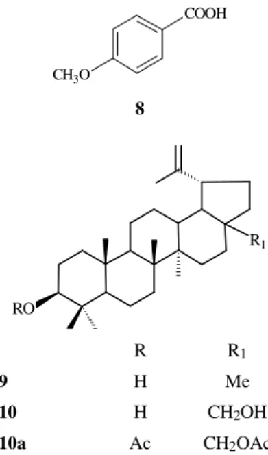
RO
R1
R R1
9 H Me
10 H CH2OH
10a Ac CH2OAc
COOH
CH3O
8
+ C MeO
MeO
OH
O O
O
C RO
CH3O
+
.
+
.
C OR1
OR2
C H
4-Methoxybenzoic acid(8, p-anisic acid)
Colorless crystals, m.p. 182-184 °C. (Lit.10 m.p. 184 °C). 1H-NMR (60 MHz, CF3COOH) δ: 8.15 (d, J = 9.0 Hz, 2H-2,6), 7.08 (d, J = 9.0 Hz, 2H-3,5), 4.02 (s, MeO-4). EIMS m/z (rel. int.): 152 (98, [M].+), 135 (100, [M-OH]+), 107 (13, [M-OH-CO and/or M-COOH]+).
Lupeol(9)
Colorless crystals from MeOH, m.p. 211-214 °C. [Lit.11 m.p. 215-216 °C (Me2CO). Spectral data, mainly the chemical shifts and multiplicities of the signals of the carbon-13 deduced by comparative analysis of the PND-and DEPT-13C-NMR, and comparison with literature val-ues12 were used in the identification of this natural product.
Betulin (10)
Colorless crystals from MeOH, m.p. 249-251 °C. [Lit.13 m.p. 251-252 °C (EtOH)]. Spectral data, mainly the chemi-cal shifts and multiplicities of the signals of the carbon-13 deduced by comparative analysis of the PND- and DEPT-13C-NMR, including the diacetyl derivative (10a) and com-parison with literature values12 were used in the identification of this compound.
Acknowledgments
This work was supported by CNPq fellowships and by grants from Conselho Nacional de Desenvolvimento Cien-tífico e Tecnológico (CNPq), Financiadora de Estudos e Projetos (FINEP), Coordenação de Aperfeiçoamento de Pessoal de Nível Superior (CAPES) and Programa de Apoio ao Desenvolvimento Científico e Tecnológico (PADCT). The authors are also grateful to Professor
Afrânio Gomes Fernandes, Universidade Federal do Ceará, for collection and identification of the plant material.
References
1. Campos, A.M.; Silveira, E.R.; Braz-Filho, R.; Teixeira, T.C. Phytochemistry1994, 36, 403. 2. Ollis, W.D.; Rhodes, C.A.; Sutherland, I.O.
Tetrahe-dron 1967, 23, 4741.
3. Braz-Filho, R.; Gottlieb, O.R.; Assumpção, R.M.V.
An. Acad. Brasil. Ci. 1970, 42 (Supl.), 111.
4. Braz-Filho, R.; Gottlieb, O.R.; Assumpção, R.M.V.
Phytochemistry 1971, 10, 2835.
5. Campbell, R.M.V.; Harper, S.H.; Kemp, A.D. J. Chem. Soc. C1969, 1787.
6. Buckingham, J., Ed.; Dictionary of Organic Com-pounds; Chapman and Hall: London, 1982, 5th Edi-tion (B-00951 and L-00498).
7. Tha, H.C.; Zilliken, F.; Breitmaier, E. Can. J. Chem. 1980, 58, 1211.
8. Sanders, J.K.M.; Hunter, B.K. In Modern NMR Spec-troscopy: A Guide for Chemists; Oxford University Press; Oxford, 1993, 2nd Edition.
9. Breitmaier, E.; Voelter, W. In Carbon-13 NMR Spec-troscopy
: High - Resolution Methods and Applications in Or-ganic Chemistry and Biochemistry; VCH: Weinheim, 1987, 3rd Edition.
10. Reference 6: M-00529. 11. Reference 6: L-00498.
12. Mahato, S.B.; Kundu, A.P. Phytochemistry1994, 37,
1517.

![Table 3. Heteronuclear 2D shift-correlated via long-range couplings [ 1 H- x 13 C - COSY- n J CH (n = 2 and 3), COLOC] NMR spectral data for the isoflavonoids 2, 3a, 5 and 7 .*](https://thumb-eu.123doks.com/thumbv2/123dok_br/18988241.459662/3.918.97.824.726.1043/table-heteronuclear-shift-correlated-couplings-coloc-spectral-isoflavonoids.webp)