This Accepted Author Manuscript is copyrighted and published by Elsevier. It is posted here by agreement between Elsevier and University of Brasilia. Changes resulting from the publishing process - such as editing, corrections, structural formatting, and other quality control
mechanisms - may not be reflected in this version of the text. The definitive version of the text was subsequently published in [Tissue and Cell, Volume 34, Issue 5, October 2002, Pages 356– 364, doi:10.1016/S0040816602000368].You may download, copy and otherwise use the AAM for non-commercial purposes provided that your license is limited by the following restrictions: (1) You may use this AAM for non-commercial purposes only under the terms of the CC-BY-NC-ND license.
(2) The integrity of the work and identification of the author, copyright owner, and publisher must be preserved in any copy.
(3) You must attribute this AAM in the following format: [agreed attribution language, including link to CC BY-NC-ND license + Digital Object Identifier link to the published journal article on Elsevier’s ScienceDirect® platform].
________________________________________________________________________
Este Manuscrito do Autor Aceito para Publicação (AAM) é protegido por direitos autorais e publicado pela Elsevier. Ele esta disponível neste Repositório, por acordo entre a Elsevier e a Universidade de Brasília. As alterações decorrentes do processo de publicação - como a edição, correção, formatação estrutural, e outros mecanismos de controle de qualidade - não estão refletidas nesta versão do texto. A versão definitiva do texto foi posteriormente publicado em [Tissue and Cell, Volume 34, Número 3, Outubro de 2002, Páginas 356–364,
doi:10.1016/S0040816602000368]. Você pode baixar, copiar e utilizar de outra forma o AAM para fins não comerciais , desde que sua licença seja limitada pelas seguintes restrições: (1) Você pode usar este AAM para fins não comerciais apenas sob os termos da licença CC- BY- NC-ND.
(2) A integridade do trabalho e identificação do autor, detentor dos direitos autorais e editor deve ser preservado em qualquer cópia.
The ultrastructure of the spermatozoa of Epipedobates flavopictus
(Amphibia, Anura, Dendrobatidae), with comments on its evolutionary
significance
Adrian A Garda Guarino R Colli Odair Aguiar-Júnior2 Shirlei M Recco-Pimentel Sônia N Báo
Abstract
We describe, for the first time, the spermatozoon ultrastructure of a dendrobatid frog, Epipedobates flavopictus. Mature spermatozoa of E. flavopictus are filiform, with a moderately curved head and a proportionally short tail. The acrosomal vesicle is a conical structure that covers the nucleus for a considerable distance. A homogeneous subacrosomal cone lies between the acrosome vesicle and the nucleus. The nucleus contains a nuclear space at its anterior end, and electron-lucent spaces and inclusions. No perforatorium is present. In the midpiece, the proximal centriole is housed inside a deep nuclear fossa. Mitochondria are scattered around the posterior end of the nucleus and inside the undulating membrane in the anterior portion of the tail. In transverse section the tail is formed by an U-shaped axial fiber connected to the axoneme through an axial sheath, which supports the undulating membrane. The juxta-axonemal fiber is absent. The spermatozoon of E. flavopictus has several characteristics not observed before in any anurans, such as a curved axial fiber, absence of a juxta-axonemal fiber, and presence of mitochondria in the typical undulating membrane. Our results endorse the view that, in anurans, the conical perforatorium and subacrosomal cone are homologous and that Dendrobatidae should be grouped within Bufonoidea rather than Ranoidea.
Keywords: sperm; ultrastructure; Anura; Dendrobatidae; Epipedobates
Introduction
Reasons for this lack of resolution range from the limited utility of external morphology (Inger, 1967) to the great paucity of data for tropical lineages. Analyses using alternative data sets, such as molecular markers (de Sá & Hillis, 1990; Hillis et al., 1993 and Hay et al., 1995), have slowly added new insights to the problem but also have refuted well-established clades. Filling the gaps on existing data sets and exploring new kinds of characters are important ways to improve phylogenetic hypotheses among anurans (Ford, 1993).
The ultrastructure of spermatozoon has been used as an alternative data set to investigate the phylogeny of many taxa such as fishes (Jamieson, 1991 and Tanaka et al., 1995), amphibians (Lee and Jamieson, 1992 and Lee and Jamieson, 1993; Jamieson et al., 1993 and Scheltinga et al., 2001), reptiles (Jamieson, 1995; Teixeira et al., 1999a and Teixeira et al., 1999b), and invertebrates (Jamieson, 1987). An advantage of sperm ultrastructure data is that they provide more conservative characters for groups with highly derived body plans, such as Amphisbaenia, which cannot be scored for some traditional morphological traits (Teixeira et al., 1999b). Spermatozoon ultrastructure data have also been useful in clarifying relationships among Polyplacophora, where traditionally used characters are either too conserved or too variable (Buckland-Nicks, 1995). Spermatozoon morphology, therefore, seems to be useful for groups where, for some reason, external morphology cannot be scored, either because of evolutionary conservativeness (as in some traits of Polyplacophora) or specialization (as for Amphisbaenia).
Some conjectures on anuran phylogeny have been made based upon spermatozoon ultrastructure and the cladistic significance of some characters has been investigated. For example, the conical perforatorium has been proposed as a tentative synapomorphy of Bufonoidea (Lee & Jamieson, 1993), whereas the presence of an undulating membrane or a rod-shaped perforatorium have been scored as symplesiomorphies of Anura. Yet, due to the paucity of data on spermatozoon morphology for several families, no comprehensive cladistic analysis of sperm ultrastructure data, such as those made for squamate reptiles (Jamieson, 1995 and Teixeira et al., 1999b) and fishes (Tanaka et al., 1995), has been conducted for anurans (but see Scheltinga et al., 2001).
the family Dendrobatidae, Epipedobates flavopictus, from the central Brazilian Cerrado. Further, we discuss the evolutionary significance of the results regarding the evolution of the subacrosomal cone in anurans and the phylogenetic position of dendrobatid frogs.
Material and methods
We collected two individuals of E. flavopictus on September 1998, at Minaçu, Goiás, Brazil (13°38′ S, 48°15′ W), and one individual on November 1995 at Serra do Cipó, Minas
Gerais, Brazil, during the reproductive season. We illed ani al r in lo aine on o e a do en in and de o i ed e a e ole o er e ol i a da niver idade de Bra lia
(CHUNB 09581, 09582) and M e de i ria Na ral ‘Profe or Ad o Jo é ardo o’, Universidade Estadual de Campinas (ZUEC 11434). We removed testes by dissection and placed them in a Petri dish with phosphate buffer (PBS) pH 7.2 and saline solution. We cut testes into small pieces and fixed them overnight at 4 °C in a solution of 2.5% glutaraldehyde, 5 mM CaCl2, and 5% sucrose in 0.1 M sodium cacodylate buffer pH 7.2. After rinsing in sodium cacodylate buffer, we postfixed testes for 1 h in 1% osmium tetroxide, 0.8% potassium ferricyanide, and 5 mM CaCl2 in 0.1 M sodium cacodylate buffer pH 7.2, and left them overni in a ol ion of 0.5% ran l a e a e for ‘in lo ’ on ra . We ro eeded wi
dehydration in a series of ascending acetone (30–100%) and embedded the material in S rr’ epoxy resin. We made ultrathin sections with glass and diamond knifes on a Leica Reichert ultramicrotome and stained the sections with uranyl acetate and lead citrate. We observed the sections in a Jeol® 100C transmission electron microscope and took micrographs at 80 kV. We also made light microscope observations of spermatozoa under interferential contrast using a Zeiss® Axiophot microscope.
Results
Acrosome complex
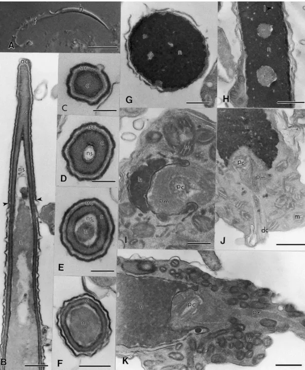
The acrosome of E. flavopictus sperm is located at the anterior portion of the head and is composed of a single and narrow vesicle, filled with a homogeneous material of low electron density ( Fig. 1B–E). Under the acrosome vesicle, the subacrosomal cone forms a conical cap that reaches the anterior portion of the nucleus. In cross-section, the acrosome and subacrosomal cone are circular ( Fig. 1C–F). The acrosome vesicle surrounds the anteriormost portion of the nucleus, below which the nucleus thickens and is enveloped only by the subacrosomal cone ( Fig. 1B–F).
Nucleus
Below the nuclear envelope, a nuclear space that probably results from the condensation of chromatin process is seen (Fig. 1B, D & E). The nucleus is circular in transverse section (Fig. 1G) and conical in longitudinal section (Fig. 1B). The anterior portion of the nucleus is enveloped by the acrosome complex and gradually tapers (nuclear shoulders absent) to a point within the subacrosomal cone (Fig. 1E & F). The chromatin is highly condensed and electron-dense. Despite the high degree of condensation, several small electron-lucent nuclear lacunae and inclusions are seen (Fig. 1G & H).
Midpiece
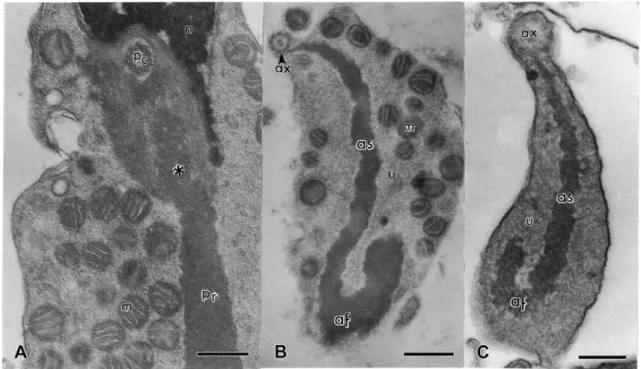
Fig. 2. Spermatozoa of E. flavopictus. Transmission electron micrographs of the midpiece and tail. (A) Longitudinal section through the midpiece showing the insertion of the paraxonemal rod in the nuclear fossa (*). ×42 000. (B) Transverse section of the tail showing the S-shaped paraxonemal rod, mitochondria inside the undulating membrane, U-shaped axial fiber in transverse section, and axial sheath. ×24 000. (C) Transverse section of the posterior portion of the tail; note the absence of mitochondria. ×60 000. Abbreviations: af, axial fibre; as, axial sheath; ax, axoneme; m, mitochondrion; n, nucleus; pc, proximal centriole; pr, paraxonemal rod; , nd la in e rane. S ale ar : (A) 0.3 μ ; (B) 0.5 μ ; ( ) 0.2 μ .
Tail
In transverse section, the tail of E. flavopictus sperm consists of the axoneme, undulating membrane and axial fiber ( Fig. 2B). In transverse section the axial fiber is curved or U-shaped and apparently of the same composition of an electron-dense structure that supports the undulating membrane, to which it is connected. Since other structures are found inside the undulating membrane of E. flavopictus, such as cytoplasm and mitochondria, it is ne e ar o na e i r re o w i e a ial fi er i onne ed, ereaf er alled ‘a ial ea ’. I i for all defined a e onne ion e ween e a ial fi er and he axoneme (or juxta-axonemal fiber, when it is present). In E. flavopictus the axial sheath is directly connected to the axoneme through the doublet 3, without a juxta-axonemal fiber ( Fig. 2B & C).
seen in the mature spermatozoon, the mitochondria are organized in a mitochondrial collar around the flagellum.
Discussion
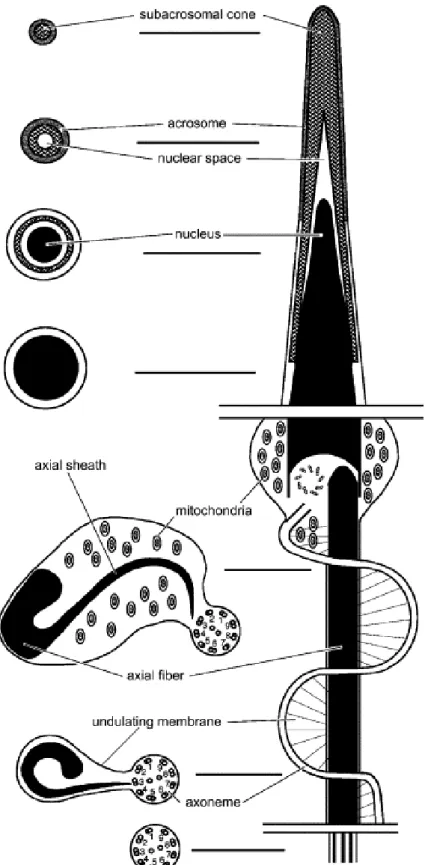
The basic structure of the spermatozoon of E. flavopictus ( Fig. 4) is similar to that of most neobatrachians ( Ford, 1993) described to date, such as Bufo arenarum ( Burgos & Fawcett, 1956), B. marinus ( Swan et al., 1980; Lee & Jamieson, 1993), Melanophryniscus cambaraensis ( Báo et al., 2001), Odontophrynus cultripes ( Báo et al., 1991), Lepidobatrachus laevis ( Waggener & Carroll, 1998), and Pachymedusa dacnicolor ( Rastogi et al., 1988). However, it possesses several traits not seen in these other species and which may possibly be shared with other dendrobatids.
Epipedobates flavopictus has a subacrosomal cone below the acrosomal vesicle. We advance that this is homologous with the subacrosomal cone of Ascaphus truei and the conical perforatorium of bufonoids, contrary to the proposition of Lee and Jamieson, 1992 and Lee and Jamieson, 1993 and Jamieson et al. (1993). These authors indicated that the subacrosomal cone seen in A. truei, urodeles, and basal amniotes is absent in all other anurans they examined and that in bufonoids a similar structure, the conical perforatorium, lies beneath the acrosome vesicle. Further, they provide four reasons why the conical perforatorium is not homologous with the subacrosomal cone of A. truei: (1) a rod-shaped endonuclear perforatorium exists in A. truei; (2) the close proximity of the subacrosomal cone, in its posterior region, to the nucleus and acrosome vesicle in A. truei, whereas in bufonoids the conical perforatorium lies free in the subacrosomal space; (3) the homogeneous nature of the subacrosomal material in A. truei, whereas loose bundles of coarse fibers running parallel to the nucleus are seen in bufonoids; and (4) the subacrosomal cone in A. truei is less electron-dense than the acrosome vesicle, while in bufonoids the conical perforatorium is more electron-dense than the acrosome vesicle. Later, Jamieson (1999) suggested that the conical perforatorium may be homologous with the subacrosomal cone, but provided no rationale to his proposition.
the tapering end of the nucleus in the spermatozoon of B. arenarum, form a perforatorium. Had Burgos and Fawcett (1956) chosen a different name (without implying a function) for the a e r re, a ‘ a ro o al one’, ar en (1) a ove wo ld vani .
Second, arguments (2)–(4) above are not independent. The more detached aspect of the presumed conical perforatorium relative to the nucleus and acrosome vesicle, and its higher electron density in bufonoids are a direct consequence of its fibrous arrangement. Moreover, in the bufonoids Myxophyes fasciolatus ( Lee & Jamieson, 1992, Fig. 3E), O. cultripes [ Báo et al., 1991, Figs 12 & 13, mislabeled as acrosome (A)], and M. cambaraensis ( Báo et al., 2001, Fig. 4) the presumed conical perforatorium is much more homogeneous, forming an almost continuous layer in transverse section around the nucleus, with no coarse fibers being observed. We regard this condition as intermediate between that found in A. truei and the state typical of most bufonoids.
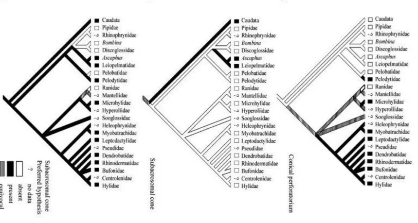
Finally, if the view of Lee and Jamieson, 1992 and Lee and Jamieson, 1993 and Jamieson et al. (1993) is correct, six steps would be required during the evolution of anurans, when mapping the characters conical perforatorium and subacrosomal cone onto a current phylogeny of the group (Hay et al., 1995) (Fig. 5). According to this view, the conical perforatorium was absent in the common ancestor of anurans and salamanders and evolved on e in e ‘ fonoid’ linea e (Fi . 5A). F rthermore, the subacrosomal cone was originally
absent in anurans and evolved independently twice in the group (Fig. 5B). Conversely, if James (1970) was correct in regarding the conical perforatorium of bufonoids as homologous with the subacrosomal cone of A. truei, then only four evolutionary steps are required and the presence of the subacrosomal cone would be plesiomorphic for bufonoids ( Fig. 5C). If inferring genealogy rests on the principle of parsimony, i.e. choosing genealogical hypothesis that mini ize req ire en for o o la ie ( Farri , 1982), en Ja e ’ view i o e referred.
The acceptance of the hypothesis proposed by James (1970) implies that the condition seen in the acrosome of Bombina ( Furieri, 1975), Discoglossus ( Sandoz, 1975), and Xenopus ( Bernardini et al., 1986), where the subacrosomal cone is absent, is not intermediate between A. truei and bufonoids as suggested by Lee and Jamieson (1993, Fig. 7). In addition, the proposition made by Lee and Jamieson (1993) and Jamieson (1999) that the conical perforatorium is a synapomorphy of bufonoids is not supported. Instead, the replacement of a perforatorial rod (as in Ascaphus and basal amniotes) with a modified fibrillar subacrosomal cone is a bufonoid synapomorphy.
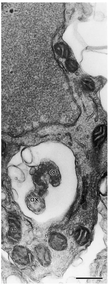
The numerous and randomly distributed mitochondria distinguish E. flavopictus from several other Neobatrachia so far examined, which usually have a mitochondrial collar, as in Bufonidae ( Burgos & Fawcett, 1956; Swan et al., 1980; Lee & Jamieson, 1993) and Leptodactylidae ( Pugin-Rios, 1980 and Bao et al., 1991). The presence of mitochondria creates a large separation between the axial sheath and the plasma membrane in the anterior tail region of E. flavopictus. In all other amphibians with an undulating membrane, the plasma membrane is closely adhered to the axial sheath. Interestingly, early spermatids of E. flavopictus have a mitochondrial collar detached from the midpiece ( Fig. 3), as in Bufonidae ( Burgos & Fawcett, 1956; Swan et al., 1980; Lee & Jamieson, 1993).
The reduction of the mitochondrial collar during the spermiogenesis, the absence of a juxta-axonemal fiber, and the somewhat degenerated structure of the axial fiber in E. flavopictus ( Fig. 2B) agrees with the proposition made by Lee and Jamieson (1993) that a reduction in complexity is a major trend in the evolution of anuran spermatozoa.
Our results suggest that the family Dendrobatidae should be placed within the
‘bufonoid’ lineage, as proposed by several authors (Hedges & Maxson, 1993; Hillis et al., 1993 and Hay et al., 1995). The acrosome structure resembles that seen in Leptodactylidae (Pugin-Rios, 1980, Amaral et al., 1999 and Bao et al., 2001), Bufonidae (Burgos & Fawcett, 1956; Rastogi et al., 1988; Lee & Jamieson, 1993; Meyer et al., 1997), and Myobatrachidae (Lee & Jamieson, 1992). All of these families share a plesiomorphic condition of the acrosome, where a subacrosomal cone lies below the conical acrosome vesicle, as in the archaeobatrachian A. truei ( Jamieson et al., 1993) and some Urodeles ( Picheral, 1967). This feature differs markedly from the condition seen in ranoids such as Ranidae ( Poirier & Spink, 1971; Pugin-Rios, 1980 and Yoshizaki, 1987) and Rhacophoridae ( Mainoya, 1981, Mizuhira et al., 1986 and Jamieson, 1999), where the acrosome vesicle sits on top of the nucleus and the subacrosomal cone is absent. Similarly, the subacrosomal cone is also absent in some archaeobatrachians, such as Pelobatidae ( James, 1970) and Pipidae ( Reed & Stanley, 1972; Bernardini et al., 1986). Furthermore, despite some peculiarities, such as the shape of the axial fiber and the absence of a juxta-axonemal fiber, the tail of E. flavopictus is similar to that generally observed in bufonoids, where an axial fiber is connected to the axoneme through an axial sheath inside an undulating membrane. All ranoids so far studied (Ranidae, Rhacophoridae, and Microhylidae) possess a tail with only the axoneme.
Hyperoliidae, and Brachycephalidae) must be investigated in order to built a consistent data set that enables parsimony analysis.
Acknowledgements
We thank Ariovaldo A. Giaretta, Cristiane G. Batista, and Osmindo R. Pires Jr. for providing specimens, an anonymous reviewer for helpful suggestions on an earlier version of e anus rip , and all e s a of e abora rio de isiologia ni al a niversidade de ras lia is or as suppor ed by apesp, by a as er’s fello s ip fro oordena o de perfei oa en o de essoal de vel uperior—CAPES to A.A.G., and by research fellowships from CNPq to G.R.C. (# 302343/88-1) and S.N.B. (# 520130/97-9).
References
Amaral, M.J.L.V., Fernandes, A.P., Báo, S.N. and Recco-Pimentel, S.M. 1999. An ultrastructural study of spermiogenesis in three species of Physalaemus (Anura, Leptodactylidae). Biocell, 23, 211–221.
Asa, C.S. and Phillips, D.M. 1988. Nuclear shaping in spermatids of the Thai leaf frog Megrophrys montanta. Anat. Rec., 220, 287–290.
Báo, S.N., Dalton, G.C. and Oliveira, S.F. 1991. Spermiogenesis in Odontophrynus cultripes (Amphibia, Anura, Leptodactylidae): ultrastructural and cytochemical studies of proteins using E-PTA. J. Morphol., 207, 303–314.
Báo, S.N., Vieira, G.H.C. and Fernandes, A.P. 2001. Spermiogenesis in Melanophryniscus cambaraensis (Amphibia, Anura, Bufonidae): ultrastructural and cytochemical studies of carbohydrates using lectins. Cytobios, 106, 203–216.
Bernardini, G., Stipani, R. and Melone, G. 1986. The ultrastructure of Xenopus spermatozoon. J. Ultrastruct. Mol. Struct. R., 94, 188–194.
Buckland-Nicks, J. 1995. Ultrastructure of sperm and sperm–egg interaction in Aculifera. In: Jamieson, B.G.M., Ausio, J. and Justine, J. (eds) Advances in Spermatozoal Phylogeny and Taxonomy. Muséu a ional d’His oire a urelle, aris, ran e, pp 129–153.
Burgos, M.H. and Fawcett, D.W. 1956. An electron microscope study of spermatid differentiation in the toad Bufo arenarum Hensel. J. Biophys. Biochem. Cytol., 2, 223–239.
de Sá, R.O. and Hillis, D.M. 1990. Phylogenetic relationships of the pipid frogs Xenopus and Silurana: an integration of ribosomal DNA and morphology. Mol. Biol. Evol., 7, 365–376.
Farris, J. 1982. The logical basis of phylogenetic analysis. In: Platnick, N. and Funk, V. (eds) Advances in Cladistics: Proc. Second Meet. Willi Hennig Soc. Columbia University Press, New York, pp 7–36.
Ford, L.S. 1993. The phylogenetic position of dart poison frogs (Dendrobatidae) among anurans: an examination of competing hypothesis and their characters. Ethol. Ecol. Evol., 5, 219–231.
Ford, L.S. and Cannatella, D.C. 1993. The major clades of frogs. Herpetol. Monogr., 7, 94–117.
Furieri, P. 1975. The peculiar morphology of the spermatozoon of Bombina variegata (L.). Monit. Zool. Ital., 9, 185–201.
Hay, J.M., Ruvinsky, I., Hedges, S.B. and Maxson, L.R. 1995. Phylogenetic relationships of amphibian families inferred from DNA sequences of mitochondrial 12S and 16S ribosomal RNA genes. Mol. Biol. Evol., 12, 928–937.
Hedges, S.B. and Maxson, L.R. 1993. A molecular perspective on lissamphibian phylogeny. Herpetol. Monogr., 7, 27–42.
Hillis, D.M., Ammerman, L.K., Dixon, M.T. and DeSá, R.O. 1993. Ribosomal DNA and the phylogeny of frogs. Herpetol. Monogr., 7, 118–131.
Inger, R.F. 1967. The development of a phylogeny of frogs. Evolution, 21, 369–384.
James, W.S. 1970. The Ultrastructure of Anuran Spermatids and Spermatozoa. Unpublished Doctor in Philosophy, University of Tennessee, Knoxville.
Jamieson, B.G.M. 1987. The Ultrastructure and Phylogeny of Insect Spermatozoa. Cambridge University Press, Cambridge, UK, pp 1–320.
Jamieson, B.G.M. 1991. Fish Evolution and Systematics: Evidence from Spermatozoa. Cambridge University Press, Cambridge, UK, pp 1–319.
Jamieson, B.G.M. 1995. The ultrastructure of spermatozoa of the Squamata (Reptilia) with phylogenetic considerations. In: Jamieson, B.G.M., Ausio, J. and Justine J. (eds) Advances in Spermatozoal ylogeny and axono y Muséu a ional d’His oire a urelle, Paris, France, pp 359–383.
Jamieson, B.G.M. 1999. Spermatozoal phylogeny of the vertebrata. In: Gagnon, C. (ed) The Male Gamete: From Basic Science to Clinical Applications. Cache River Press, Vienna, IL, pp 303–331.
Jamieson, B.G.M., Lee, M.S.Y. and Long, K. 1993. Ultrastructure of the spermatozoon of the internally fertilizing frog Ascaphus truei (Ascaphidae: Anura: Amphibia) with phylogenetic considerations. Herpetologica, 49, 52–65.
Lauder, G.V. 1994. Homology, form, and function. In: Hall, B.K. (ed) The Hierarchical Basis of Comparative Biology. Academic Press, San Diego, pp 151–196.
Lee, M.S.Y. and Jamieson, B.G.M. 1992. The ultrastructure of the spermatozoa of three species of myobatrachid frogs (Anura, Amphibia) with phylogenetic considerations. Acta Zool. (Stockholm), 73, 213–222.
Lee, M.S.Y. and Jamieson, B.G.M. 1993. The ultrastructure of the spermatozoa of bufonid and hylid frogs (Anura, Amphibia): implications for phylogeny and fertilization biology, Amphibia): implications for phylogeny and fertilization biology. Zool. Scr., 22, 309–323.
Lee, Y.H. and Kwon, A.S. 1992. Ultrastructure of spermiogenesis in Hyla japonica (Anura, Amphibia). Acta Zool. (Stockholm), 73, 49–55.
Lee, Y.H. and Kwon, A.S. 1996. Ultrastructure of spermatozoa in urodela and primitive anura (Amphibia) with phylogenetic considerations. Korean J. Syst. Zool., 12, 253–264.
Mainoya, J.R. 1981. Observations on the ultrastructure of spermatids in the testis of Chiromantis xerampelina (Anura: Rhacophoridae). Afr. J. Ecol., 19, 365–368.
Meyer, E., Jamieson, B.G.M. and Scheltinga, D.M. 1997. Sperm ultrastructure of six Australian hylid frogs from two genera (Litoria and Cyclorana): phylogenetic implications. J. Submicrosc. Cytol. Pathol., 29, 443–451.
Mizuhira, V., Futaesaku, Y., Ono, M., Ueno, M., Yokofujita, J. and Oka, T. 1986. The fine structure of the spermatozoa of two species of Rhacophorus (arboreus, schlegelii). I. Phase-contrast microscope, scanning electron microscope, and cytochemical observations of the head piece. J. Ultrastruct. Mol. Struct. R., 96, 41–53.
Picheral, B. 1967. Structure et organisation du spermatozoÏde de Pleurodeles waltlii Michah (Amphibien, Urodèle). Arch. Biol. (Liege), 78, 193–221.
Poirier, G.R. and Spink, G.C. 1971. The ultrastructure of testicular spermatozoa in two species of Rana. J. Ultrastruct. Res., 36, 455–465.
Pugin-Rios, E. 1980. Étude comparative sur la structure du permatozoÏde des amphibiens anoures. Comportament des gamètes lors de la fécondation. Unpublished Université de Rennes, France, Rennes.
Rastogi, R.K., Bagnara, J.T., Iela, L. and Krasovich, M.A. 1988. Reproduction in the Mexican leaf frog, Pachymedusa dacnicolor. IV. Spermatogenesis: a light and ultrasonic study. J. Morphol., 197, 277–302.
Reed, S.C. and Stanley, H.P. 1972. Fine structure of spermatogenesis in the South African clawed toad Xenops laevis Daudin. J. Ultrastruct. Mol. Struct. R., 41, 277–295.
Scheltinga, D.M., Jamieson, B.G.M., Eggers, K.E. and Green, D.M. 2001. Ultrastructure of the spermatozoon of Leiopelma hoschostetteri (Amphibia, Anura, Leiopelmatidae). Zoosystema, 23, 157–171.
Selmi, M.G., Brizzi, R. and Bigliardi, E. 1997. Sperm morphology of salamandrids (Amphibia, Urodela): implications for phylogeny and fertilization biology. Tissue Cell, 29, 651–664.
Swan, M.A., Linck, R.W., Ito, S. and Fawcett, D.W. 1980. Structure and function of the undulating membrane in spermatozoan propulsion in the toad Bufo marinus. J. Cell Biol., 85, 866–880.
Tanaka, S., Kurokawa, H. and Masako, H. 1995. Comparative morphology of the sperm in chondrichthyan fishes. In: Jamieson, B.G.M., Ausio, J. and Justine, J. (eds) Advances in Spermatozoal ylogeny and axono y Muséu a ional d’His oire a urelle, Paris, France, pp 313–320.
Teixeira, R.D., Colli, G.R. and Báo, S.N. 1999a. The ultrastructure of the spermatozoa of the lizard Micrablepharus maximiliani (Squamata Gymnophthalmidae) with considerations on the use of sperm ultrastructure characters in phylogenetic reconstruction. Acta Zool. (Stockholm), 80, 47–59.
Teixeira, R.D., Colli, G.R. and Báo, S.N. 1999b. The ultrastructure of the spermatozoa of the worm-lizard Amphisbaena alba (Squamata, Amphisbaenidae), and the phylogenetic relationships of amphisbaenians. Can. J. Zool., 77, 1254–1264.
Waggener, W.L. and Carroll, E.J. 1998. Spermatozoon structure and motility in the anuran Lepidobatrachus laevis. Dev. Growth Differ., 40, 27–34.