Ana Cristina Jesus Costa
Study of stability of multilayers
films based on natural polymers
Ana Cristina Jesus Costa
Study of s
tability of multila
yer
s films based on natural pol
ymer
s
Ana Cristina Jesus Costa
Study of stability of multilayers
films based on natural polymers
Trabalho efetuado sob a orientação da
Professora Doutora Natália Alves
Dissertação de Mestrado
Acknowledgements
For this work was essential the support of the following persons:
I would like to thank Professor Natália Alves, my supervisor, who guided me with her knowledge in this work, always giving me the best advice.
I also would like to thank the people who helped me in 3B’s labs specially Catarina Vale and Sofia Caridade for their patience and dedication.
A special thanks to my friend Andréa Marinho e Carolina Macedo for all the support in the most difficult moments of this process, and for her always motivating words. The last but not the least, I want to thank very much my family in special my parents, brother and sister-in-law, for all the help and understanding during this work, without them this could not have been possible.
To all of you, my sincerely,
Abstract
The use of natural polymers in areas such as tissue engineering has increased thanks to relevant properties exhibited by these as biocompatibility or biodegradability, leading to the development of new therapies in this area.
Tissue engineering has the main objective to regenerate or replace damaged tissues having as methodology the cell culture on support structures, scaffolds. These should be optimized favoring cell growth and avoiding immune responses when implanted. In the present work, multilayer films of chitosan and alginate were prepared using the layer-by-layer technique. In order to investigate the stability of these films to the action of enzymes, in situ tests were made through the microbalance quartz crystal (QCM-D) technique, at distinct pH. Moreover, the degradation for long times (until 30 days) was analysed for the first time by the QCM-D technique. The morphology of the produced films after degradation was analyzed by SEM. The experiments allowed to conclude that the constructed films (CHT/ALG)7+CHT are stable when they were immersed for 3 days in PBS as well as in enzymatic solutions, at either pH 5.5 or 7.4 and at 25ºC. It was also found that mass loss due to degradation is only evident after 7 days in the presence of lysozyme.
Resumo
A utilização de polímeros naturais em áreas como a engenharia de tecidos tem aumentado, graças às propriedades relevantes apresentadas por estes tais como biocompatibilidade ou biodegradabilidade, levando ao desenvolvimento de novas terapêuticas nesta área.
A Engenharia de tecidos tem como principal objetivo regenerar ou substituir tecidos lesados tendo como metodologia o cultivo de células sobre estruturas de suporte, os scaffolds. Estes devem ser otimizados de forma a favorecerem o crescimento celular e evitando respostas imunitárias quando implantados.
Neste trabalho, filmes multicamada de quitosano e alginato, foram preparados utilizando a técnica de layer-by-layer. De forma a investigar a estabilidade destes filmes sob a ação de enzimas, foram realizados testes in situ através da técnica de microbalança de cristais de quartzo (QCM-D), variando o pH. Além disso, a degradação para longos períodos (até 30 dias) foram analisados pela primeira vez pela técnica de QCM-D. A morfologia dos filmes produzidos após a degradação foi analisada por SEM. Através das análises efetuadas foi possível concluir que os filmes (CHT/ALG)7 + CHT são estáveis quando estes são imersos durante 3 dias em solução de PBS, assim como em soluções enzimáticas a pH 5.5 ou 7.4, a 25 °C. Verificou-se também que a perda de massa devido à degradação só é evidente após 7 dias na presença de lisozima.
Contents
Acknowledgements ... iii
Abstract ... iv
Resumo ... v
List of Abbreviations and Acronyms ... viii
List of Figures ... ix
List of tables ... x
Chapter 1 - General Introduction ... 12
1.1 Motivation... 12 1.2 Polysaccharides ... 13 1.3 Tissue engineering ... 14 1.4 Degradation ... 17 1.4.1 Chitosan Degradation ... 17 1.4.2 Alginate Degradation ... 19 1.5 References ... 20
Chapter 2 - Materials and Methods ... 25
2.1 Chitosan (CHT) ... 25 2.2 Alginate (ALG) ... 27 2.3 Lysozyme ... 28 2.4 Alginate lyase ... 28 2.5 Techniques ... 29 2.5.1 Layer-by-Layer ... 29
2.5.2 Quartz Cristal Microbalance with dissipation monitoring (QCM-D) ... 30
2.5.3 Scanning Electron Microscope (SEM) ... 32
3.1 Introduction ... 38
3.2 Materials and methods ... 40
3.2.1 Materials ... 40
3.2.2 Methods... 40
3.2.2.1 Quartz Crystal Microbalance with dissipation monitoring (QCM-D) .... 40
3.2.2.2 Construction of multilayers films ... 41
3.2.2.3 Scanning electronic microscopy ... 42
3.3 Results and Discussion ... 42
3.3.1 QCM-D experiments ... 42
3.3.2 Scanning electronic microscopy ... 48
3.4 Conclusions ... 50
3.5 References ... 51
Chapter 4- Conclusion Remarks ... 53
List of Abbreviations and Acronyms
ALG - Alginate
AFM – Atomic force microscope CHT – Chitosan
DNA – Deoxyribonucleic acid LbL – Layer-by-Layer
NaCl – Sodium chloride
PBS – Phosphate buffered saline PEC – Polyelectrolyte Complex
QCM-D – Quartz crystal microbalance with dissipation monitoring SEM – Scanning electron microscope
List of Figures
Figure 1.1 - Polysaccharides resources. ... 13
Figure 1.2 - Polyelectrolyte complex of chitosan and sodium alginate.22 ... 16
Figure 1.3 - Degradations stages25 ... 18
Figure 1.4 - Schematic illustration of chitosan hydrolysis.33 ... 18
Figure 1.5 - Alginate degradation mechanism by alginate lyase (β-elimination.)40 ... 19
Figure 2.1 - Chemical structure of chitosan. ... 25
Figure 2.2- Protonation of chitosan. ... 25
Figure 2.3 - Alginate Sodium (ALG). 12 ... 27
Figure 2.4 - Lysozyme structure. ... 28
Figure 2.5- Layer-by-Layer film deposition.17 ... 29
Figure 2.6 – Quartz Cristal equipment. 20 ... 31
Figure 2.7 - Scanning electronic microscope equipment. ... 33
Figure 2.8 - Interaction volume in SEM. ... 33
Figure 3.1 - Normalized frequency (Δf5/5) and dissipation changes (ΔD5). 1) Chitosan injection 2) washing step (NaCl) 3) Alginate injection. ... 42
Figure 3.2 - Cumulative thickness evolution of polymeric film as a function of the number of deposited layers. The line represents a linear regression with R2=0.9888. ... 43
Figure 3.3 - a) QCM monitoring with normalized frequency (Δf5/5) and dissipation change (ΔD5) during (CHT/ALG)7+CHT build up and degradation with 1mg/mL of lysozyme solution at pH=5.5, and respective B) cumulative thickness evolution estimated using Voigt viscoelastic model. ... 44
Figure 3.4 - a) QCM monitoring with normalized frequency (Δf5/5) and dissipation (ΔD5)
change during (CHT/ALG)7+CHT build up and degradation of PBS at pH=5.5, and respective b) cumulative thickness evolution estimated using a Voigt viscoelastic model. ... 45
Figure 3.5 - a) QCM monitoring with normalized frequency (Δf5/5) and dissipation (ΔD5)
change during (CHT/ALG)7+CHT build up and degradation of PBS at pH=7.4, and respective b) cumulative thickness evolution estimated using a Voigt viscoelastic model. ... 45
Figure 3.6 - a) QCM monitoring with normalized frequency (Δf7/7) and dissipation (ΔD7)
change during (CHT/ALG)7+CHT build up and degradation of 1mg/mL lysozyme solution in PBS at pH=5.5, and respective b) cumulative thickness evolution estimated using a Voigt viscoelastic model. ... 46
Figure 3.7 - a) QCM monitoring with normalized frequency (Δf5/5) and dissipation (ΔD5)
change during (CHT/ALG)7+CHT build up and degradation of 1mg/mL lysozyme solution in PBS at pH=7.4, and respective b) cumulative thickness evolution estimated using a Voigt viscoelastic model. ... 46
Figure 3.8 - a) QCM monitoring with normalized frequency (Δf7/7) and dissipation (ΔD7)
change during (CHT/ALG)7+1 build up and degradation of 0,1mg/mL alginate lyase in PBS, at pH=5.5 b) Thickness change during (CHT/ALG)7+1 build up and degradation of 0,1mg/mL alginate lyase in PBS, at pH=5.5 ... 47
Figure 3.9 - QCM offline monitoring during (CHT/ALG)7+1 degradation of 0.013mg/mL
lysozyme solution in PBS at pH=5.5. ... 48
Figure 3.10 - Comparative SEM images for 3 days degradation with lysozyme with
different concentrations (0.013mg.mL-1, 1mg.mL-1) at pH=5.5 and pH=7.4. ... 49
List of tables
Chapter 1
Chapter 1 General Introduction
1.1 Motivation
In 1993, Langer and Vacanti presented for the first time the concept of Tissue Engineering as being “an interdisciplinary field that applies the principles of engineering and life science toward the development of biological substitutes that restore, maintain, or improve tissue function or a whole organ”. Tissue engineering is based in three components: cells, scaffolds and grow factors and the objective is replacing or restoring function in diseased or injured tissues. Tissue engineering emerged as an alternative to tissue and organ reconstruction.
Scaffold is one of the most important components of tissue engineering. It has the function support to cells culture, under certain conditions, until obtaining a functional tissue. In parallel the implant material should degrades and the by-product shall be metabolized out from the body. This is a great challenge involving the design of a scaffold due to the number of variables that need to be considered.
A large variety of polymers, with different structures and properties, has been used for many different applications. Chitosan, hyaluronic acid or sodium alginate are examples of polymers that exhibit relevant properties of biomaterials such as biocompatibility and biodegradability, and where the synergy between the materials is very important to build materials which later can be used in the biomedical area, including tissue engineering.
In this work, the use of natural polymers in the biomedical area will initially be explained in chapter 1, focusing essentially on tissue engineering which is the main propose for the chitosan and alginate film studied in this thesis. It also addresses the importance of degradation in polymers being more detailed with the polymers used.
A description of all the material and techniques used in film stability studied in this work is made in chapter 2. Already in Chapter 3 presents, the description of the work performed, the results obtained and their discussion.
Finally, in chapter 4 are presented all conclusions obtained with this experimental work and some proposals for future works.
1.2 Polysaccharides
Replacement of fossil feedstocks with renewable ones is one of the main challenges of modern plastics industry. Natural polymers represent a specific class of materials among polymers based on natural resources. Typical examples are cellulose, hemicelluloses, lignin, silk and starch. Another class of materials consists of the natural-based or bio-based synthetic polymers, the monomers of which derive from renewable resources. 1,2 Polysaccharides are macromolecules formed by the union of several monosaccharides. In nature, polysaccharides have various resources: from algal origin (e.g. alginate), plant origin (e.g. pectin, guar gum), microbial origin (e.g. dextran, xanthan gum) or animal origin (chitosan, chondroitin).3
Figure 1.1 - Polysaccharides resources.
Polysaccharides have been found to participate in many biological processes, such as cell–cell communication, embryonic development, infection of bacteria and/or virus, and cellular immunity.4,5 The biological effects that polysaccharides can exert are limited to therapeutic activities for diseases of humans and animals, and toxic activity responsible for causing human and animal disease. Although polysaccharides have been used for decades in various industrial applications, e.g. pharmaceuticals, biomaterials, foodstuff and nutrition, and biofuels, growing understanding and deeper investigations
of the importance of polysaccharides in life science are driving the development of polysaccharides for novel applications.6
The use of natural polysaccharides and/or their derivatives are especially attractive because of their availability, renewability and low toxicity. Moreover, the usual biodegradability and biocompatibility of these natural polymers, coupled with their capacity for chemical modification confers them ideal properties for their use in biomedical applications such as tissue engineering scaffolds or drug release systems. 7– 10
1.3 Tissue engineering
Tissue engineering has evolved since the appearance of the idea of combining cells and scaffolds to create artificial tissues with the objective to develop biological substitutes to replace, restore and regenerate defective tissues and organ of the human body. Tissue engineering emerged as a potential alternative therapeutic process to treat severely injured patients with minimally invasive techniques. Commonly, TE strategies utilize combination of cells, biodegradable scaffolds, and bioactive molecules to recapitulate natural processes of tissue regeneration.11,12 Cooperative interplay of the components is imperative to achieve biologically functional engineered tissue and this method is contingent upon the development and implementation of advanced biomaterials.
In tissue engineering the main objective are the scaffolds because these should present adequate biological, chemical, physical and mechanical properties adapted the physiological conditions in vivo. Therefore, in their design must be taken in account several factors such as:
(I) Biocompatibility – the scaffold must be biocompatible; cells must adhere, function normally, and migrate onto the surface and eventually through the scaffold and being proliferative. After implantation, the scaffold must induce a negligible immune reaction in order to prevent it, causing such a severe inflammatory response.
(II) Biodegradability – scaffolds does not work as a permanent implant, so it’s important to allow the body’s own cells, over time, to eventually replace the implanted scaffold. Therefore, scaffolds must be biodegradable to allow cells to produce their own extracellular matrix. The by-products of this degradation should be non-toxic and able to exit the body without interference with other organs.
(III) Mechanical Properties – scaffolds should have mechanical properties consistent whit the anatomical site where they will be implanted. Producing scaffolds with adequate mechanical properties is one of the greatest challenges. In case of engineered bone and cartilage, the implanted scaffold must have sufficient mechanical integrity to function from the time of implantation to the completion of the remodelling process.
(IV) Scaffold architecture – scaffolds should have an interconnected pore structure and high porosity, to ensure cellular penetration and adequate diffusion of nutrients to the cells within the construct and to extra-cellular matrix formed by these cells. Additionally, a porous interconnected structure is required to allow diffusion of waste products out of scaffolds, and the products of scaffolds degradation should be able to exit the body without interference with other organs and surrounding tissues.
(V) Manufacturing technology – is important that scaffold to become clinically and commercially viable, therefore it should be cost effective and it should be possible to scale-up from making one at time in a research laboratory to small batch production.13
In Tissue engineering, many polymers have been investigated for the scaffolds construction, including synthetic polymers such as poly(glycolic acid) (PGA) or poly(lactic acid) (PLA).14 These turned out to be a good solution but also have some disadvantages such as the risk of rejection when implanted due to reduced biocompatibility. Thus, the natural polymers are the best option to the scaffolds construction. These present the ability to promote the cell adhesion and growth as well as the fact that they are biologically active. Examples of these materials are collagen, alginate or chitosan. However the use of scaffolds fabricated from a single-phase biomaterial caused some problems due to their poor mechanical properties, which can make them difficult to use.
One solution to this problem is the combination of materials, where the properties of materials used can complement each other. One example is the combination between chitosan and alginate, which has been extensively studied in biomedical application, mainly in tissue engineering. Iwasaki et al. reported the use of alginate based chitosan hybrid polymeric fibers for scaffolds in cartilage tissue engineering.15 Similar studies present chitosan-alginate scaffolds manufactured for cartilage tissue engineering.16,17 Alves et. al. reported the self-assembling and crosslinking of polyelectrolyte multilayer films of chitosan and alginate18 which presents the ability to this two polymers to form a novel biocompatible multilayer film based in alternative depositions of this. Baysal et. al. present chitosan/alginate crosslinked hydrogels: preparation, characterization and application for cells growth purposes.19
One of the factors that have enabled the development of tailored biomaterials using these polymers has been their potential to form a polyelectrolyte complex (PEC).20 Polyelectrolyte complex are the association between oppositely charged particles through electrostatic interactions.21
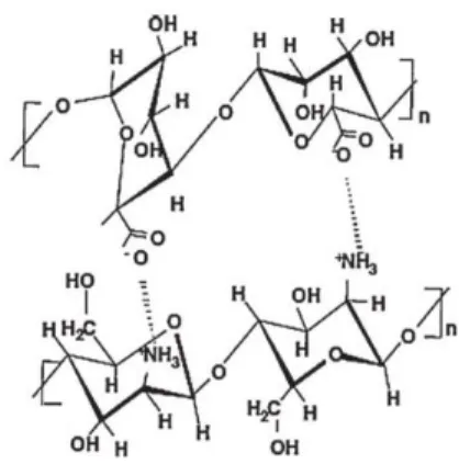
Chitosan and alginate form a polycomplex membrane via ionic cross-linking (Figure 1.2) that exhibited tensile strength and yet lower elongation break, than polymers. This membrane also showed lower methanol permeability as compared to both chitosan and alginate alone.22
The combination of biocompatible, suitable mechanical and degradation properties constitutes one of the main advantages for the use of these two polymers, showing that they have potential to be used as scaffolds in tissue engineering.
1.4 Degradation
Degradation is understood as a chemical reaction, which causes the division of the polymer chains by physical or chemical agents, consequently leading to the modification of properties.
Two types of polymer degradation could occur, depending on the variables involved in the process: (1) in absence of living organisms – abiotic degradation; (2) in presence of microorganisms – biodegradation.
The abiotic degradation depends on abiotic factors such as sunlight, heat or humidity and from there can occurs hydrolysis or oxidation reactions can occurs.
Biodegradation is the degradation that occurs in natural polymers. This happens by the action of enzymes produced by microorganisms such as fungi or bacteria, which are both present in environment or animal organisms, and causes the conversion of organic compounds in more simple organic compounds.23 So, enzymes will hydrolyze the polymer chains and there are variables that affect the biodegradation process such as temperature, light and pH.24
The degradation of polymers in biomedical applications is very important because it is necessary to ensure that no component or by-product of the degradation of polymers used can induce inflammatory or toxic reactions in the body.
1.4.1 Chitosan Degradation
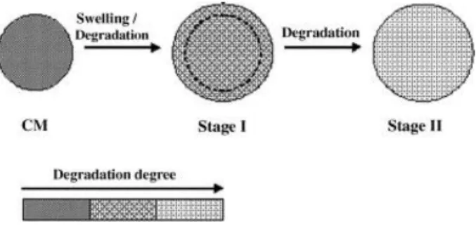
When implanted in the human body, chitosan matrices are degraded due to the presence of lysozyme, in a process that involves two steps. When the matrix is immersed in the lysozyme solution, the water permeated into the matrix will cause swelling,
phenomenon, bonds cleavage will also occur resulting in the degradation of chitosan. When the swelling is maximum continual degradation leads to weight loss (second stage), the matrices tends to be thinner, and swelling degree decrease.25 So, degradation and swelling are directly related, as illustrated in Figure 1.3.
Figure 1.3 - Degradations stages25
Several studies have been published about the degradation of chitosan by enzymatic hydrolysis, in particular by the action of lysozyme.26–30 In fact, it has been demonstrated that chitosan is degraded under the action of lysozyme, which exists in many organisms including the human body.29 In this case, the normal concentrations of this enzyme, can range from 4 to 13mg/L in serums and 450 to 1230mg/l in case of tears.25 It has been estimated that at least a normal subject produces 500 mg of lysozyme per day, but the lifetime of the protein in plasma is very short: approximately 75% is eliminated within 1h, mainly through clearance via the kidneys and only traces are detectable in the urine of healthy subjects. 30,31
The products resulting from the degradation of chitosan are biocompatible chitosan oligosaccharides of variable length, as illustrated in Figure 1.4.32
Figure 1.4 - Schematic illustration of chitosan hydrolysis.33
The activity of lysozyme increases with decreasing pH or increasing temperature in a certain range, which causes the environmentally dependent degradation behavior of
1.4.2 Alginate Degradation
Alginate is synthetized abundantly in organisms as cell walls and the intracellular matrix of browns algae or bacteria. One of the most important areas where alginate is used is in biomedical applications in the form of hydrogels35, microparticles36, films37 and fibers38. Products originated by the degradation of alginate exhibit biological functions as promotion of root growth in higher plants, enhancing penicillin production in
Penicillium chrysogenum, promotion of proliferation of cells and bringing down blood
pressure in human.39
Alginate degradation occurs via β-elimination of glycosidic bonds as illustrate in Figure 1.5. 40–42 A double bond is formed between the C4 and C5 carbons of the six-membered ring from which the 4-O-glycosidic bond is eliminated, depolymerizing alginate and simultaneously yielding a product containing 4-deoxy-L-erythro-hex-4-enopyranosyluronic acid as the non-reducing terminal moiety.43–45 Glycosidic linkages of alginate are susceptible to a variety of degradation mechanisms, including acid-, alkaline- and enzymatic catalyzed hydrolysis.
For this work, the form of degradation studied will be the enzymatic degradation by alginate lyase. This enzyme are specific for alginate degradation and the mechanism is similar to that of alkaline degradation to glycuronans.46
1.5 References
1. Imre, B. & Pukánszky, B. Compatibilization in bio-based and biodegradable polymer blends. Eur. Polym. J. 49, 1215–1233 (2013).
2. Dang, J. M. & Leong, K. W. Natural polymers for gene delivery and tissue engineering. Adv. Drug Deliv. Rev. 58, 487–499 (2006).
3. Liu, Z., Jiao, Y., Wang, Y., Zhou, C. & Zhang, Z. Polysaccharides-based nanoparticles as drug delivery systems. Adv. Drug Deliv. Rev. 60, 1650–1662 (2008).
4. Cheng, J. J., Lin, C. Y., Lur, H. S., Chen, H. P. & Lu, M. K. Properties and biological functions of polysaccharides and ethanolic extracts isolated from medicinal fungus, Fomitopsis pinicola. Process Biochem. 43, 829–834 (2008).
5. Wijesinghe, W. A. J. P. & Jeon, Y.-J. Biological activities and potential industrial applications of fucose rich sulfated polysaccharides and fucoidans isolated from brown seaweeds: A review. Carbohydr. Polym. 88, 13–20 (2012).
6. Liu, J., Willför, S. & Xu, C. A review of bioactive plant polysaccharides: Biological activities, functionalizatison, and biomedical applications. Bioact. Carbohydrates
Diet. Fibre 5, 31–61 (2014).
7. Alonso-Sande, M., Teijeiro-Osorio, D., Remuñán-López, C. & Alonso, M. J. Glucomannan, a promising polysaccharide for biopharmaceutical purposes. Eur.
J. Pharm. Biopharm. 72, 453–462 (2009).
8. García-González, C. a., Alnaief, M. & Smirnova, I. Polysaccharide-based aerogels - Promising biodegradable carriers for drug delivery systems. Carbohydr. Polym.
86, 1425–1438 (2011).
9. Suh, J. K. & Matthew, H. W. Application of chitosan-based polysaccharide biomaterials in cartilage tissue engineering: a review. Biomaterials 21, 2589–2598 (2000).
10. Lee, S.-H. & Shin, H. Matrices and scaffolds for delivery of bioactive molecules in bone and cartilage tissue engineering. Adv. Drug Deliv. Rev. 59, 339–359 (2007). 11. Lee, H., Chung, H. J. & Park, T. G. Design Principles in Biomaterials and Scaffolds.
Principles of Regenerative Medicine (2011). doi:10.1016/B978-0-12-381422-7.10031-8
12. Furth, M. E. & Atala, A. Principles of Tissue Engineering. Principles of Tissue
Engineering (2014). doi:10.1016/B978-0-12-398358-9.00006-9
13. O’Brien, F. Biomaterials & scaffolds for tissue engineering. Mater. Today 14, 88– 95 (2011).
14. Hutmacher, D. W. Scaffolds in tissue engineering bone and cartilage. Biomaterials
cartilage tissue engineering: Evaluation of chondrocyte adhesion to polyion complex fibers prepared from alginate and chitosan. Biomacromolecules 5, 828– 833 (2004).
16. Li, Z. & Zhang, M. Chitosan-alginate as scaffolding material for cartilage tissue engineering. J. Biomed. Mater. Res. Part A 75A, 485–493 (2005).
17. Sibaja, B. et al. Preparation of alginate–chitosan fibers with potential biomedical applications. Carbohydr. Polym. 134, 598–608 (2015).
18. Alves, N. M., Picart, C. & Mano, J. F. Self assembling and crosslinking of polyelectrolyte multilayer films of chitosan and alginate studied by QCM and IR spectroscopy. Macromol. Biosci. 9, 776–785 (2009).
19. Baysal, K., Aroguz, A. Z., Adiguzel, Z. & Baysal, B. M. Chitosan/alginate crosslinked hydrogels: preparation, characterization and application for cell growth purposes.
Int. J. Biol. Macromol. 59, 342–8 (2013).
20. Lawrie, G. et al. Interactions between alginate and chitosan biopolymers characterized using FTIR and XPS. Biomacromolecules 8, 2533–2541 (2007). 21. Lankalapalli, S. & Kolapalli, V. R. M. Polyelectrolyte Complexes: A Review of their
Applicability in Drug Delivery Technology. Indian J. Pharm. Sci. 71, 481–487 (2009).
22. Ma, J. & Sahai, Y. Chitosan biopolymer for fuel cell applications. Carbohydr. Polym.
92, 955–975 (2013).
23. Oliveira, Mariana B. ; Mano, J. F. Natural-based and Stimuli-respositive polymers for Tissue Engineering and Regenerative Medicine. Polym. Regen. Med. Appl.
from nano- to macro-structures 49–90 (2015).
24. Azevedo, H. S. & Reis, R. Understanding the enzymatic degradation of biodegradable polymers and strategies to control their degradation rate.
Biodegrad. Syst. tissue … 177–202 (2005). doi:10.1201/9780203491232.ch12
25. Ren, D., Yi, H., Wang, W. & Ma, X. The enzymatic degradation and swelling properties of chitosan matrices with different degrees of N-acetylation.
Carbohydr. Res. 340, 2403–2410 (2005).
26. Onishi, H. & Machida, Y. Biodegradation and distribution of water-soluble chitosan in mice. Biomaterials 20, 175–182 (1999).
27. Vårum, K. M., Myhr, M. M., Hjerde, R. J. N. & Smidsrød, O. In vitro degradation rates of partially N-acetylated chitosans in human serum. Carbohydr. Res. 299, 99–101 (1997).
28. Nordtveit, R. J., Vårum, K. M. & Smidsrød, O. Degradation of partially N-acetylated chitosans with hen egg white and human lysozyme. Carbohydr. Polym. 29, 163– 167 (1996).
29. Lee, K. Y., Ha, W. S. & Park, W. H. Blood compatibility and biodegradability of partially N-acylated chitosan derivatives. Biomaterials 16, 1211–1216 (1995).
30. Tomihata, K. & Ikada, Y. In vitro and in vivo degradation of films of chitin and its deacetylated derivatives. Biomaterials 18, 567–575 (1997).
31. Mireille Dumoulin, Russell J. K, Johnson, Vittorio Belloti, C. M. D. in Protein
Misfolding, and Conformational Diseases Volume 6, pp 285–308 (2007).
32. Kim, I.-Y. et al. Chitosan and its derivatives for tissue engineering applications.
Biotechnol. Adv. 26, 1–21 (2008).
33. Zhang, Z., Li, C., Wang, Q. & Zhao, Z. K. Efficient hydrolysis of chitosan in ionic liquids. Carbohydr. Polym. 78, 685–689 (2009).
34. Verheul, R. J. et al. Influence of the degree of acetylation on the enzymatic degradation and in vitro biological properties of trimethylated chitosans.
Biomaterials 30, 3129–35 (2009).
35. Prang, P. et al. The promotion of oriented axonal regrowth in the injured spinal cord by alginate-based anisotropic capillary hydrogels. Biomaterials 27, 3560– 3569 (2006).
36. Fundueanu, G., Nastruzzi, C., Carpov, A., Desbrieres, J. & Rinaudo, M. Physico-chemical characterization of Ca-alginate microparticles produced with different methods. Biomaterials 20, 1427–1435 (1999).
37. Wang, L., Khor, E., Wee, A. & Lim, L. Y. Chitosan-alginate PEC membrane as a wound dressing: Assessment of incisional wound healing. J. Biomed. Mater. Res.
63, 610–618 (2002).
38. Kokubo, T. et al. Apatite-forming ability of alginate fibers treated with calcium hydroxide solution. J. Mater. Sci. Mater. Med. 15, 1007–1012 (2004).
39. Inoue, A. et al. Characterization of an Alginate Lyase, FlAlyA, from Flavobacterium sp. Strain UMI-01 and Its Expression in Escherichia coli. Mar. Drugs 12, 4693–4712 (2014).
40. Breguet, V., Von Stockar, U. & Marison, I. W. Characterization of alginate lyase activity on liquid, gelled, and complexed states of alginate. Biotechnol. Prog. 23, 1223–1230 (2007).
41. Aarstad, O. A., Tøndervik, A., Sletta, H. & Skjåk-Bræk, G. Alginate sequencing: an analysis of block distribution in alginates using specific alginate degrading enzymes. Biomacromolecules 13, 106–16 (2012).
42. Holme, H. K., Lindmo, K., Kristiansen, A. & Smidsrød, O. Thermal depolymerization of alginate in the solid state. Carbohydr. Polym. 54, 431–438 (2003).
43. Kim, H. S., Lee, C. G. & Lee, E. Y. Alginate lyase: Structure, property, and application. Biotechnol. Bioprocess Eng. 16, 843–851 (2011).
44. Wong, T. Y., Preston, L. a & Schiller, N. L. ALGINATE LYASE: review of major sources and enzyme characteristics, structure-function analysis, biological roles, and applications. Annu. Rev. Microbiol. 54, 289–340 (2000).
degradation of alginate in the gelled state: Effect of gelling ions and lyase specificity. Carbohydr. Polym. 110, 100–106 (2014).
46. Gacesa, P. Alginate-modifying enzymes. A proposed unified mechanism of action for the lyases and epimerases. FEBS Lett. 212, 199–202 (1987).
Chapter 2
Chapter 2 - Materials and Methods
2.1 Chitosan (CHT)
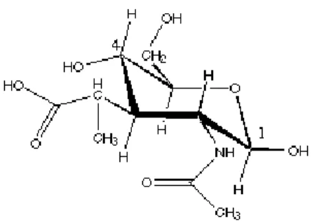
Chitosan is a linear, semi-crystalline polysaccharide, composed of (1→4)-2-acetamido-2-deoxy-b-D-glucan (N-acetyl D-glucosamine) and (1→4)-2-amino-2-deoxy- b-D-glucan (D-glucosamine) units, illustrated in Figure 2.1. This polysaccharide is not extensively present in the environment but can be easily obtained by deacetylation of a natural polymer- chitin. 1
Chitin can be found in the shells of crustaceans, cell walls of green algae and yeasts and is therefore the second most abundant biopolymer in nature. 1,2
Figure 2.1 - Chemical structure of chitosan.
The presence of amino groups in the chitosan structure differentiates chitosan from chitin, and gives to this polymer many peculiar properties.1 Chitosan, because of the semi-crystalline structure of chitin with extensive hydrogen bonds, is normally insoluble in aqueous solutions above pH 7. However when in dilute acids (pH<6), the free amino groups are protonated and the molecule becomes soluble (Figure 2.2), which opens prospects to a wide range of applications, particularly in the field of cosmetics. 3,4
Chitosan has a wide range of applications, as can be seen in Table 1. 5 This is caused by easy access to this natural resource, low cost and because it has a great advantage of being prepared in different forms such as hydrogels, microspheres and films.6
Principal application for chitosan
Agriculture
Defensive mechanism in plants Stimulation of plant growth Seed coating, Frost protection
Time release of fertilizers and nutrients into the soil
Water & waste treatment
Removal of metal ions
Ecological Polymer (eliminate synthetic polymers)
Reduce odors
Food & beverages
Not digestible human( dietary fibre) Blind lipids (reduce cholesterol) Preservative, fungistatic, antibacterial coating for fruits
Protective
Cosmetics
Treat acne
Improve suppleness of hair Reduce static electricity in hair Tone skin
Oral care ( toothpaste, chewing gun)
Biopharmaceutics
Immunological, antitumoral Haemostatic and anticoagulant Healing, bacteriostatic
Table 2.1- Principal applications of chitosan.2
Chitosan exhibits good biocompatibility and biodegradability and has been widely explored for applications in controlled drug delivery and tissue engineering.7 It has been reported that it can stimulate the activity growth factors and play an important role as a structural component of scaffolds for tissue regeneration. 8 The molecular weight and degree of N-deacetylation (DD) are thought to be the two most important determinants of the properties of chitosan.
2.2 Alginate (ALG)
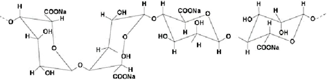
Alginate is a natural polysaccharide, consisting of linear chains of α-L-glucuronic acid (G) and β-D-mannuronic acid (M) and is found in marine brown algae.9 It is the water solubility and present negative charge attribute for the carboxylic acid groups presents on its units – Figure 2.3.
Alginate is a biomaterial that has found numerous applications in biomedical science and engineering due to its favorable properties, including biocompatibility, biodegradability and non-toxicity. The fact that it is very abundant and provide a low cost are also very important factors.10,11
Alginate can be easily modified in any form such as hydrogels, microspheres, microcapsules, sponges, films and fibers. This property can increase the applications of alginate in various fields such as tissue engineering and drug delivery.
Figure 2.3 - Alginate Sodium (ALG).12
Alginate is also a known compound for its properties in the formation of scaffolds, and is therefore a very important compound in the treatment of organ failures or losses.13 The combination of chitosan and alginate is also widely known as alginate exhibits polyanionic behavior and when combined with a polymer who exhibits polication behavior can give highly stable films.14
2.3 Lysozyme
Lysozyme was discovered in 1992 by Alexander Fleming. This protein (Figure 2.4) can be found in many organisms like virus, insects, amphibious, reptiles, birds and mammals which are producer in many tissues and fluids such as breast milk, saliva, tears and it’s productions is greater in polymorphonuclear leukocytes in humans, where lysozyme is very important in the defense against infections.
It has been reported that its anti-viral activity against the virus VIH type 1(test in vitro) may be associated with a positive charge and the interactions established on the surface of the virus when negatively charged.15
Figure 2.4 - Lysozyme structure.
2.4 Alginate lyase
Alginate lyases, also known as alginases or alginate depolymerases, catalyze the degradation of alginate by a β-elimination mechanism. Alginate-degrading enzymes with various substrate specificities have been isolated from many sources, including marine algae, marine mollusks, and a wide range of microorganisms. Alginate lyase activity has been reported in extracts obtained from several species of brown algae. These enzymes are promising for removing and modifying alginate.16
2.5 Techniques
2.5.1 Layer-by-Layer
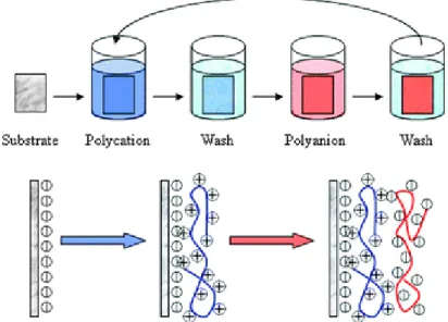
Layer-by-layer (LBL) assembly technology, which can conduct the formation of thin films on distinct substrates by adsorbing oppositely charged polyelectrolytes or other species, has been widely used in the membrane fabrications and surface modifications.
Hammond 2012, reported the review about building biomedical materials layer-by-layer, presenting specific advantages of the use of Layer-by-Layer assembling versus traditional polimeric blends encapsulation. Also presented the impact and potencial of this technique in biomedical areas such as drug delivery or tissue engineering.
This technique is presented as an easy, facile, robust, reproducible, flexible, and efficient way of modifying membrane surfaces and fabricating highly ordered nanostructured thin films and nanocomposites with regular thickness, compositions, structures and properties.
Figure 2.5- Layer-by-Layer film deposition.17
The LBL assembly process is comparatively simpler and environmental friendly as it shown in figure 2.5. The surface (glass, metal…) exhibits negative charge when in solution because surface oxidation and hydrolysis. When immersed in a solution of positive charged polyelectrolyte, electrostatic interaction reactions will occur which
cause the change of liquid surface charge becomes positive (polycation). The next step is to wash the surface for removing the polyelectrolyte that is not adsorbed to ensure the efficiency of the assembly process. Then the substrate is again immersed in a polyelectrolyte solution but now with negative charge and once again, it is washed. Although the LBL assembly process requires many steps, these steps are always repeated and simple, including alternate immersion and washing procedures without chemical reactions. Therefore, this method allows control the number of layers required.
This technique was used in experimental work in two different situation. In first case, the depositions of seven bilayers more one last of chitosan, was realized in a glass support, to then be placed in contact with the enzyme solution in order to verify the mass lost. Weighing of the support was performed in three different times: before deposition, after deposition and finally after the defined degradation time.
The same thing as done but using as support the QCM crystals in order to use the quartz crystal microbalance offline, i. e., using the equipment only for measuring the frequency and dissipation of the crystals in a specific period of time (before deposition, after deposition and after degradation time). This is a innovate form to use QCM technique to study the degradation of very thin films.
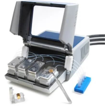
2.5.2 Quartz Cristal Microbalance with dissipation monitoring (QCM-D)
The quartz crystal microbalance with dissipation monitoring (QCM-D) measure mass variations in the nanometric scale, thus allowing study the adsorption variations in layers of ultra-thin films or molecules. It is a very important when it’s necessary to confirm the construction of films. This technique has attracted attention from researchers because it allows in-situ study of obtained results in real time, which is a huge advantage for film construction processes.18
This technique is based on the piezoelectric effect of quartz crystals. This effect is caused by the intrinsic properties of anisotropic and minerals such as quartz, which reacts to
oscillator is pressed on one of its faces, will cause a deformation of the crystal lattice and will cause the rearrangement. This will induce opposite charges on their faces, so generating an electric field within the crystal.19
Figure 2.6 – Quartz Cristal equipment. 20
When the crystal is coupled to the oscillator circuit and is applied an electrical current that has appropriate frequency characteristics and magnitude to the geometry of the crystal, it will oscillate in a manner called shear (wave propagation is perpendicular to the electric field). The range of frequencies that can be used in the crystals can be very high, which ensures high precision in the determination of the variations - Figure 2.6. In 1959, G. Sauerbrey presented a mathematical relationship where a ratio between the mass change rigidly coupled to the crystal and the change in resonance frequency of a quartz crystal when subjected to oscillations by applying alternating current signals is established. According Sauerbrey relation, adsorption of mass on a thin quartz sensor surface, induces a decrease in which for a rigid substance translates into the increase in mass.21
∆𝑚 =−𝐶. ∆𝑓 𝑛
Where, C is the mass sensitivity constant (17.7 ng.cm-2.Hz-1 at 5 MHz) and n is the overtone number. The quartz crystal microbalance with dissipation monitoring allows to simultaneously measuring the changes in the resonant frequency and in the viscoelastic properties (dissipation) when a film is adsorbed at the crystal surface. The measurements can be conducted at the fundamental frequency and at several overtones. When exist any viscoelastic behaviour in adsorbed layer, the Saurbrey model
can’t be applied and the QCM-D response of a viscoelastic film has been modelled using a Voigt based model. In this case the changes in the resonant frequency (Δf) and in the dissipation factor (ΔD) according to Voinova and co-workers22 are:
∆𝑓 ≈ − 1 2𝜋𝜌0ℎ0{ ɲ𝐵 𝛿𝐵+ ℎ𝐿𝜌𝐿𝜔 − 2ℎ𝐿 ( ɲ𝐵 𝛿𝐵) 2 ɲ 𝐿𝜔𝐿 𝜇𝐿2+ 𝜔2𝑛 𝐿 2} ∆𝐷 ≈ 1 𝜋𝑓𝜌0ℎ0{ ɲ𝐵 𝛿𝐵 + 2ℎ𝐿 ( ɲ𝐵 𝛿𝐵) 2 𝜇 𝐿𝜔𝐿 𝜇𝐿2+ 𝜔2𝑛 𝐿 2}
where 𝜔 is the angular frequency of the oscillation and 𝜌0and ℎ0are the density and thickness of the crystal, respectively.
The viscosity of the bulk liquid is ɲ𝐵, 𝛿𝐵 = [(2ɲ𝐵
𝜌𝐵𝜔)
2 ]
1/2
is the viscous penetration depth of the shear wave in the bulk liquid and 𝜌𝐵 is the liquid’s density. The thickness, density, viscosity and elastic shear modulus of the adsorbed layer are represented byℎ𝐿, 𝜌𝐿 , ɲ𝐿 and 𝜇𝐿, respectively.
2.5.3 Scanning Electron Microscope (SEM)
The scanning electron microscope (SEM) is an instrument frequently used in scientific research because it allows producing high-resolution images of a sample surface and when applied in thin films allows to determinate the thickness, identify impurities and defects, or study the adhesion, corrosion and fracture in them.
The images obtained have a very high range of magnification in a nanometric (nm) and micrometer (µm) range.
The equipment consists on an optoelectronic column (electron cannon and reducing electron beam system), a scanning unit, a vacuum chamber (where sample is placed) and a system image viewing as in Figure 2.7.
Figure 2.7 - Scanning electronic microscope equipment.
The samples will be sequentially subjected to an accelerated electron beam, focused by an electromagnetic lens system so that the beam is focused on a small diameter in a small area of the sample. The interaction of the electron beam with the sample, results in the issuance of several types of radiation such as secondary electrons, backscattered electrons, characteristic X-rays, etc. – Figure 2.8. When be captured, these particles give information about the characteristics of the samples, such as composition, crystal structure or the topography of the sample.
Figure 2.8 - Interaction volume in SEM.
For the formation of the final SEM image, some signals, are important such as secondary electrons and backscattered electrons. The first are a result of energy transfer between electrons emitted and the bombarded electrons on the sample surface and allows the detection of the sample surface topography.
Backscattered electrons are originated from elastic interactions, they are a result of the angular deviation of the incident electrons caused by the sample surface. These electrons give information about the sample surface roughness.
To be subject to SEM, samples must meet the following conditions: (1) provide good surface conductivity, if any conductivity is verified shall be applied an ultra-thin coating that can be Au or C; (2) supporting vacuum since this technique utilizes electron beam; (3) Stable physical and chemical interaction with the electromagnetic beam.
2.6 References
1. Croisier, F. & Jérôme, C. Chitosan-based biomaterials for tissue engineering. Eur.
Polym. J. 49, 780–792 (2013).
2. Rinaudo, M. Chitin and chitosan: Properties and applications. Prog. Polym. Sci. 31, 603–632 (2006).
3. Suh, J. K. & Matthew, H. W. Application of chitosan-based polysaccharide biomaterials in cartilage tissue engineering: a review. Biomaterials 21, 2589–2598 (2000).
4. Madihally, S. V. & Matthew, H. W. T. Porous chitosan scaffolds for tissue engineering. Biomaterials 20, 1133–1142 (1999).
5. Ma, J. & Sahai, Y. Chitosan biopolymer for fuel cell applications. Carbohydr. Polym.
92, 955–975 (2013).
6. El-hefian, E. a, Nasef, M. M. & Yahaya, A. H. Chitosan Physical Forms : A Short Review. Aust. J. Basic Appl. Sci. 5, 670–677 (2011).
7. Ren, D., Yi, H., Wang, W. & Ma, X. The enzymatic degradation and swelling properties of chitosan matrices with different degrees of N-acetylation.
Carbohydr. Res. 340, 2403–2410 (2005).
8. Silva, J. M. ; et al. . Tailored Freestanding Multilayered Membranes Based on Chitosan and Alginate. Biomacromolecules Lang. English, Database CAPLUS 15, 3817–3826 (2014).
9. Review, P. C omparison of chitosan , alginate and chitosan / alginate nanoparticles with respect to their size , stability , toxicity and transfection. 4, 372–377 (2014). 10. Gombotz, W. R. & Wee, S. F. Protein release from alginate matrices. Adv. Drug
Deliv. Rev. 64, 194–205 (2012).
11. Lee, Y. K. & Mooney, D. J. Alginate : properties and biomedical applications. Prog
Polym Sci. 37, 106–126 (2013).
12. Tao, X., Chen, H., Sun, X. J., Chen, J. F. & Roa, W. H. Formulation and cytotoxicity of doxorubicin loaded in self-assembled bio-polyelectrolyte microshells. Int. J.
Pharm. 336, 376–381 (2007).
13. Venkatesan, J., Bhatnagar, I., Manivasagan, P., Kang, K.-H. & Kim, S.-K. Alginate composites for bone tissue engineering: A review. Int. J. Biol. Macromol. 72, 269– 281 (2014).
Characterization and Application for Antibody Immobilization. 13046–13052 (2007).
15. Carrillo, W. Lisozima : Actividad antibacteriana y alergenicidad Lysozyme : Antibacterial Activity and Allergenicity. Actual. EN Nutr. 14, 314–326 (2013). 16. Haug A, Larsen B, S. O. Studies on the sequence of uronic acid residues in alginic
acid. 691–704 (1967).
17. Xiang, Y., Lu, S. & Jiang, S. P. Layer-by-layer self-assembly in the development of electrochemical energy conversion and storage devices from fuel cells to supercapacitors. Chem. Soc. Rev. 41, 7291 (2012).
18. Dixon, M. C. Quartz crystal microbalance with dissipation monitoring: enabling real-time characterization of biological materials and their interactions. J. Biomol.
Tech. 19, 151–8 (2008).
19. Fredriksson, C., Kihlman, S., Rodahl, M. & Kasemo, B. The Piezoelectric Quartz Crystal Mass and Dissipation Sensor: A Means of Studying Cell Adhesion.
Langmuir 14, 248–251 (1998).
20. Q-sense. Quartz crystal microbalance with dissipation. Langmuir 27, 2 (2011). 21. Dunér, G., Thormann, E. & Dėdinaitė, A. Quartz Crystal Microbalance with
Dissipation (QCM-D) studies of the viscoelastic response from a continuously growing grafted polyelectrolyte layer. J. Colloid Interface Sci. 408, 229–234 (2013).
22. Voinova, M. V, Rodahl, M., Jonson, M. & Kasemo, B. Viscoelastic acoustic response of layered polymer films at fluid-solid interfaces: continuum mechanics approach. Phys. Scr. 59, 391–396 (1999).
Chapter 3
Study of stability of multilayer films based
Chapter 3 - Study of stability of multilayer films based on natural
polymers
The stability study of films containing biodegradable polymers is essential to optimize their properties envisaging biomedical applications. The present work explores the stability of polysaccharide multilayer films under the action of two different enzymes, lysozyme and alginate lyase. First, multilayer films of chitosan and alginate were prepared using the layer-by-layer technique. In order to investigate the stability of these films to the action of enzymes, in situ tests were made through the microbalance quartz crystal (QCM-D) technique, at distinct pH. Moreover, the degradation for long times (until 30 days) was analyzed for the first time by the QCM-D technique. The morphology of the produced films after degradation was analyzed by SEM. The QCM-D experiments allowed to conclude that the constructed films (CHT/ALG)7+CHT are stable when they were in contact for 3 days in PBS as well as in enzymatic solutions, at either pH 5.5 or 7.4 and at 25ºC. The produced films for LbL, when immersed for 3 days in PBS or lysozyme (0.013 mg.mL-1 and 1mg.mL-1) at either pH 5.5 and 7.4 at 25ºC analyzed by SEM, present more porosity at higher concentration.
Keywords: chitosan, alginate, multilayer films, stability, enzymatic degradation. 3.1 Introduction
The use of natural polymers in substitution of the synthetic ones has been considered very important, since they are based on renewable resources, which are available in large quantities in the environment, and their commercial value is much lower when compared to the synthetic polymers. In particularly, for biomedical applications, natural biodegradable polymers such as gelatin, collagen, chitosan and alginate have been extensively studied for biomedical applications.1
One of the most studied polymers for biomedical purposes is chitosan (CHI), which is a N-deacetylated chitin, poly(D-glucosamine), and presents interesting biological properties such as biocompatibility, biodegradation, antibacterial and wound-healing activity.1In similarity with CHI, biomedical studies on alginate (ALG) have also intensified
due to its non-toxicity, biocompatibility and non-immunogenicity, which are very important for their tissue regeneration applications.2 ALG is comprised of (1→4)-linked β-D-mannuronate (M) and α-L-guluronate (G), and these uronic acids are arranged in block structures, which may be homopolymeric (poly-M block and poly-G block) or heteropolymeric (random sequence with MG random block).3
In the present work films containing CHI and ALG were produced through the layer-by-layer technique (LbL)). Such method has been widely used for the preparation of different multilayer nanostructured systems.4 The basic principle of this technique relies on the fabrication of nanostructured multilayered films by assembling a stratified organization of polyanions, polycations or bipolar amphiphilic on the material surfaces.5 In the LbL assembly, a thin film is grown up from a substrate by its alternating exposure to aqueous solutions containing species with opposite multivalent attractive affinities. Upon each exposure, one component deposits to overcompensate the surface affinity, causing reversal of that affinity and allowing the deposition of a layer of opposite affinity.6 Films created by LbL assembly are inherently two-component composites and it can be designed to provide practically any type of functional performance. 7 Combinations of CHI and ALG have been widely used as drug delivery systems, where ALG beads are coated with CHI.8,9 LbL films containing both polysaccharides could find application in the coating of substrates for different biomedical applications, including in tissue engineering and in the delivery of bioactive agents constructed with each chitosan/alginate multilayer films10 could also offer unique properties for the immobilization of antibodies with retaining its binding activity. 11
The stability study of films containing biodegradable polymers is essential to optimize their properties envisaging biomedical applications. In particular, for chitosan/alginate films, it is important to understand the degradation mechanism of each polymer individually. In case of polysaccharides, the degradation occurs by enzymatic hydrolysis12. The principal agent is lysozyme, that exists in many organisms including in the human body with concentrations from 4 to 13mg.L-1.12–16 This enzyme acts by hydrolysis in β 1-4 glycosidic linkages between N-acetylglucosamine monomers.16,17 In the case of alginate, normally the degradation occurs by action of alginate lyase through β-elimination of glycosidic bonds18. Various alginate lyases have been found and isolated
from algae, marine microorganisms and marine invertebrates.19 However, the enzymatic activity is affected by several factors such as pH, temperature and ionic strength. Attending to these effects, in the present work it was performed a stability study of the multilayer films containing chitosan and alginate, based in enzymatic reactions strictly controlled in terms of pH and temperature. As far as we know, this is the first time that such a study is conducted for CHI/ALG multilayer films. A detailed study of the degradation behavior under distinct conditions was conducted by quartz crystal microbalance with dissipation monitoring (QCM-D). The changes in the film morphology due to distinct degradation conditions were analyzed by scanning electron microscopy (SEM).
3.2 Materials and methods
3.2.1 Materials
Medium molecular weight chitosan (CHT), sodium alginate (ALG), lysozyme from chicken egg white (~100000 units.mg-1), alginate lyase (~10000 units.g-1), sodium chloride, acetic acid and phosphate buffered saline tablets were purchased from Sigma-Aldrich. All reagents were used without further treatment, unless the chitosan which was previously purified by recrystallization following the procedure described in detail by Couto and co-workers20.
3.2.2 Methods
3.2.2.1 Quartz Crystal Microbalance with dissipation monitoring (QCM-D)
The formation of multilayer CHT/ALG was realized in situ by QCM-D (Q-Sense E4 system), for monitoring the alternately adsorption of chitosan and alginate onto the gold-coated crystals.
LbL films were produced using fresh solutions prepared in 0.15M of sodium chloride: 0.5mg.mL-1 CHT solution with 1% (v/v) of acetic acid and 0.5 mg.mL-1 ALG solution. The multilayered systems were assembled at pH of 5.5 adjusted with NaOH and HCl
The construction consisted in sequential adsorptions onto the QCM crystals, which were excited at 15, 25, 35, 45 and 55 MHz. The system started with 0.15M NaCl solution, then, the polyelectrolyte solutions were injected into the cell during 10 min using a peristaltic pump, beginning with CHT. A rinsing step of 5 min with a 0.15 M NaCl solution was included between the adsorptions of each polyelectrolyte, since it is essential for a stable construction of multilayer films. These experiments were performed at room temperature (25ºC) and, for LbL construction, a flow rate of 50 µL.min-1 was used. Then, the injection of enzymatic solutions was performed using a lower flow rate.
After the LbL construction, the enzymatic solutions were injected using a lower flow rate, 6.71 µL.min-1. Lysozyme solutions with two distinct concentrations (1mg.mL-1 and 0.013mg mL-1) were prepared in 0.1M PBS solution for pH values of 5.5 and 7.4. The alginate lyase solution with concentration 0.1 mg.mL-1 was also prepared in 0.1M PBS solution for pH value of 5.5. As a control condition, a 0.1M PBS solution was also injected for pH values of 5.5 and 7.4.
The final graphics obtained were obtained through the Voigt model in the Q-Tools software from Q-Sense.
3.2.2.2 Construction of multilayers films
The construction of [CHT/ALG]7+CHT films onto glass slides was performed using manual dip coating technique. The substrates were dipped alternately in the polyelectrolyte solutions in order to obtain films using exactly the same conditions described for the QCM-D experiments.
Similarly with the QCM-D experiments, after the LbL construction, the multilayer films were immersed in the enzymatic solutions and in the control solution (0.1 M PBS solution), during a certain period of time (up to 30 days). At the end of each period of time, the samples were removed from those solutions and washed with distilled water. Finally, they were dried at room temperature for further characterization.
3.2.2.3 Scanning electronic microscopy
The analysis of the surface morphology of the constructed films on glass substrates, before and after the injection of enzymatic solutions, was performed using scanning electron microscopy, SEM (model JSM-6010LV,JEOL, Japan) with the secondary electron image mode and an accelerating voltage of 10 kV on previously gold coated samples.
3.3 Results and Discussion
3.3.1 QCM-D experiments
The construction of (CHT/ALG)7+CHT films was monitored in situ with QCM-D. In Figure 3.1, it was represented the variation of the normalized frequency Δf5/5 and dissipation, ΔD5, which confirmed a successful build-up of these multilayer films. As expected, the frequency decreases after each injection of polyelectrolyte solution, as a consequence of the increase of the mass deposited onto the surface of the gold-coated quartz crystals. On the other hand, ΔD5 increased due to thenon-rigid adsorbed layer structure of the film that is forming onto the crystal surface. In addition, there is a small change of both Δf5/5 and ΔD5 during the washing step, showing that the adsorbed layers are relatively stable. 0 20 40 60 80 100 120 -750 -650 -550 -450 -350 -250 -150 -50 50 Δ D5 (1E -6) Δ f5 /5 ( Hz ) Time (min) 2 3 1
Figure 3.1 - Normalized frequency (Δf5/5) and dissipation changes (ΔD5). 1) Chitosan injection 2) washing step (NaCl) 3)
Based on these QCM-D results, the cumulative thickness of these films was estimated using a Voigt viscoelastic model instead of the Sauerbrey model, also used in literature to model QDM-D data.
Figure 3.2 shows that the thickness of CHT/ALG multilayers increases as the number of layers increases, presenting a linear growth, as previously reported.10 The linear growth observed indicates that each CHI layer is effectively adsorbing on the top of the previous ALG layer.
Figure 3.2 - Cumulative thickness evolution of polymeric film as a function of the number of deposited layers. The line
represents a linear regression with R2=0.9888.
With the QCM technique, it was also possible to evaluate in situ the degradation of CHI/ALG films by lysozyme. Thus, the enzymatic solution was injected for 72 hours and the degradation of the film was evaluated taking into account the evolution of the normalized frequency and dissipation, and especially, based on the estimated cumulative thickness after the injection of enzymatic solution. All studies were performed at 25 ºC and the effect of pH on enzymatic degradation was also evaluated for two distinct values, 5.5 and 7.4. In order to complement the analysis of the stability of the films, the graphics of thickness for each of the cases are presented. These graphics were obtained through the Voigt model in the Q-Tools software from Q-Sense.22
Initially it was conducted a test with the concentration of 1mg/mL de lysozyme without PBS with pH=5.5 and the result is presented in Figure 3.3. The result didn’t any alteration in films stability to the action of the lysozyme prepared in these conditions.
R² = 0,9888 0 20 40 60 80 100 120 0 5 10 15 20 Th ickn es s (n m ) Layeres
Then and follow the literature, a lysozyme solution with the same concentration was prepared with PBS in order to roughly simulate the hydration conditions in the human body, when the materials are implanted.1,23
Figure 3.4 – 3.5 present the stability of the build-up of (CHT/ALG)7+CHT films to the action of PBS at different pH (5.5 and 7.4) as control. Regarding these results, it can be seen the Δfn/n variation is equal for both conditions. By comparing these results it is possible to say that PBS does not cause any change in the film, i.e., the thickness remains unchanged throughout the injection of PBS.
Figures 3.6 and 3.7 compare the stability of the build-up of (CHT/ALG)7+CHT films to the action of lysozyme with a concentration of 1mg/mL prepared in PBS at a different pH (5.5 and 7.4). The results show similar Δfn/n variation similar in both graphics and the action of this enzyme is not relevant in the thickness of the film.
So, it can be concluded that, for the time period of 72 h, the produced CHI/ALG films are quite stable in the presence of lysozyme. Lin at al. reported that chitosan with a 62% deacetylation degree, when in contact with lysozyme, presented degradation right from the first day and after 28days there was only 75% of the total mass.24 In fact, the combination of ALG with CHI should be responsible for the system stability when compared with chitosan alone.
Figure 3.3 - a) QCM monitoring with normalized frequency (Δf5/5) and dissipation change (ΔD5) during (CHT/ALG)7+CHT build up and degradation
with 1mg/mL of lysozyme solution at pH=5.5, and respective B) cumulative thickness evolution estimated using Voigt viscoelastic model.
b a
00 20 40 60 80 100 120 140 160 0 1000 2000 3000 4000 5000 Th ickn es s (n m ) Time (min) 0 20 40 60 80 100 120 140 0 1000 2000 3000 4000 5000 Th ickn es s (n m ) Time (min)
Figure 3.4 - a) QCM monitoring with normalized frequency (Δf5/5) and dissipation (ΔD5) change during (CHT/ALG)7+CHT build up and degradation of PBS at pH=5.5, and respective b) cumulative thickness evolution estimated using a Voigt viscoelastic model.
Figure 3.5 - a) QCM monitoring with normalized frequency (Δf5/5) and dissipation (ΔD5) change during (CHT/ALG)7+CHT build up and degradation of PBS at pH=7.4, and respective b) cumulative thickness evolution estimated using a Voigt viscoelastic model.
-20 0 20 40 60 80 100 120 -180 -160 -140 -120 -100 -80 -60 -40 -20 0 20 0 1000 2000 3000 4000 5000 Δ D5 (E 1 -6) Δ F5 /5 (H z) Time (min) Frequency Dissipation b a -10 0 10 20 30 40 50 60 70 80 90 -90 -80 -70 -60 -50 -40 -30 -20 -10 0 10 0 1000 2000 3000 4000 5000 Δ D7 (1E -6) Δ f7 /7 (Hz ) Time (min) Frequency Dissipation b a
The study of stability of the build-up (CHT/ALG)7+CHT in situ was also carried out with the alginate lyase enzyme. This was performed under the same condition used with lysozyme. These results are presented in Figure 3.8.
0 20 40 60 80 100 120 140 -120 -100 -80 -60 -40 -20 0 20 0 1000 2000 3000 4000 5000 Δ D7 (1E -6) Δ f7 /7 (Hz ) Time (min) Frequency Dissipation 0 20 40 60 80 100 120 140 160 0 1000 2000 3000 4000 5000 Th ickn es s (n m ) Time (min) -10 0 10 20 30 40 50 60 70 80 90 100 -140 -120 -100 -80 -60 -40 -20 0 20 0 1000 2000 3000 4000 5000 Δ D5 (1E -6) Δ f5 /5 (Hz ) Time (min) Frequency Dissipation 0,00 20,00 40,00 60,00 80,00 100,00 120,00 140,00 0 1000 2000 3000 4000 5000 Th ickn es s (n m ) Time (min)
Figure 3.7 - a) QCM monitoring with normalized frequency (Δf7/7) and dissipation (ΔD7) change during (CHT/ALG)7+CHT build up and
degradation of 1mg/mL lysozyme solution in PBS at pH=5.5, and respective b) cumulative thickness evolution estimated using a Voigt viscoelastic model.
Figure 3.6 - a) QCM monitoring with normalized frequency (Δf5/5) and dissipation (ΔD5) change during (CHT/ALG)7+CHT build up and degradation of 1mg/mL lysozyme solution in PBS at pH=7.4, and respective b) cumulative thickness evolution estimated using a Voigt viscoelastic model.
b b
a a
By looking at Figure 3.8, it can be seen that either Δfn/n or the thickness values didn’t change significantly in the presence of this enzyme. So, CHI/ALG films are also stable in the presence of alginate lyase for the time period of 72 h.
Inoue et al. 2014 reported the effiency of enzymatic degradation of the alginate by action of alginate lyase.25 In case of film (CHT/ALG)
7+CHT it doesn’t happen, being possible to say that the combination of alginate with chitosan improves stability to enzymatic action of this specific enzyme.
Quartz Crystal Microbalance was also used as an innovative technique to measure the frequency and dissipation offline for multilayer films prepared by layer-by-layer on the QCM crystals. The degradation was made with the immersion of these crystals in the enzyme solution during 7, 14 and 30 days and different conditions. The comparison was made in relation to the change in frequency and dissipation of the crystals, before and after the deposition, made by layer-by-layer. The result present refers to 0.013mg.mL-1 lysozyme concentration – Figure 3.9.
-10 0 10 20 30 40 50 60 70 -70 -60 -50 -40 -30 -20 -10 0 10 0 1000 2000 3000 4000 5000 Δ D7 (1E -6) Δ F7 /7 (Hz ) Time (min) Frequency Dissipation 0 10 20 30 40 50 60 70 80 90 100 0 1000 2000 3000 4000 5000 Th ickn es s (n m ) Time (min)
Figure 3.8 - a) QCM monitoring with normalized frequency (Δf7/7) and dissipation (ΔD7) change during (CHT/ALG)7+1 build up and degradation of 0,1mg/mL alginate lyase in PBS, at pH=5.5 b) Thickness change during (CHT/ALG)7+1 build up and degradation of 0,1mg/mL alginate lyase in PBS, at pH=5.5
b a
Figure 3.9 - QCM offline monitoring during (CHT/ALG)7+1 degradation of 0.013mg/mL lysozyme solution in PBS at pH=5.5.
The result present the increase of Δf and decrease of ΔD, showing that the enzymatic degradation of the multilayer film in the presence of lysozyme is only visible after 7 days, growing during all the 30 days wherein the films were in contact with the enzyme solution.
3.3.2 Scanning electronic microscopy
The characterization of the surface of the films built through the LBL technique and degraded under different conditions was analysed by SEM. (CHT/ALG)7+CHT were built using as support glass slides. The obtained films were placed in lysozyme for 3 days with different concentration (0.013 mg.mL-1 and 1 mg.mL-1) and posteriorly, they were washed and dried at room temperature for further SEM analysis. The SEM results obtained for the distinct degradation conditions were illustrated in Figure 3.10
-5000 -4000 -3000 -2000 -1000 0 0 7 14 30 Δ f (Hz ) Days Frequency