__________________________________________________________________________________________ “NOTICE: this is the author’s version of a work that was accepted for publication in BioChip Journal. Changes
resulting from the publishing process, such as peer review, editing, corrections, structural formatting, and other quality control mechanisms may not be reflected in this document. Changes may have been made to this work since it was submitted for publication. A definitive version will be subsequently published in BioChip Journal,
[Vol.8(1), pp.42‐47, (2014)].”
_______________________________________________________________________________________
Extensional flow-based microfluidic device: deformability
assessment of red blood cells in contact with tumor cells
Vera Faustino1, Diana Pinho1, Tomoko Yaginuma1, Ricardo C. Calhelha1, 2, Isabel C.F.R. Ferreira1, 2 &
Rui Lima1, 3
1 Polytechnic Institute of Bragança, IPB, C. Sta. Apolónia, 5301-857 Bragança, Portugal. 2 CIMO, C. Sta. Apolónia, 5301-857 Bragança, Portugal.
3 CEFT, Faculdade de Engenharia da Universidade do Porto (FEUP), R. Dr. Roberto Frias, 4200-465 Porto, Portugal.
* Correspondence and requests for materials should be addressed to R. Lima ([email protected])
Abstract Red blood cell (RBC) deformability has become one of the important factors to assess blood and cardiovascular diseases. The interest on blood studies have promoted a development of various microfluidic devices that treat and analyse blood cells. Recent years, besides the RBC deformability assessment, these devices are often applied to cancer cell detection and isolation from the whole blood. The devices for cancer cell isolation rely mainly on size and deformability of the cells. Howev-er, the examination of deformability of the RBCs mixed with cancer cells is lacking. This study aims at determining the deformation index (DI) of the RBCs in contact with cancer cells using a hyperbolic microchannel which generates a strong extensional flow. The DIs of human healthy RBCs and human RBCs in contact with a tumor cell line (HCT-15, colon carcinoma) were compared by analyzing the flowing RBCs images captured by a high speed camera. The results reveal that the RBCs that were in contact with HCT-15 cells have lower deformability than the normal RBCs.
Keywords: Biomicrofluidics, Microfluidic devices, Blood on chips, Red blood cells, Cell deformability, Deformation index
Introduction
Red blood cells (RBCs) deformability is the ability of RBCs to deform when submitted to certain flow conditions. The major determinants of the deformability include geometry and size of the microvessels, flow rate, mechanical properties of the cell membrane and its cytoskeleton and intracellular viscosity. For example, the RBCs elongate significantly when they pass through microchannels with dimensions smaller than the diameter of the RBCs at rest [1-5]. Decreased RBC deformability is a threat to human health, since it causes a clog in the small capillaries. Therefore, RBC deformability evaluation has become a research interest in a range of research fields.
In biomechanical and microfluidics studies, the clinical significance of RBC deformability has been promoting the development of methods for measuring this phenomenon. Some examples are the RBC filtration [6], laser diffraction ellipsometry [7], rheoscopy [8], micropipette aspiration [9] and cytometry [10]. Recently, by using a soft lithography technique it is possible to fabricate transparent microchannels to investigate the motion and dynamical deformation of cells [3, 11-17].
mechanical properties of healthy and diseased cells, a high efficiency of cell sorting will not be achieved. As mentioned above, there are a number of studies for development of microfluidic devices for cancer cell separation from a mixture of blood cells and cancerous cells but to our knowledge no insightful investigation has been done for characterizing deformability, by using DI, of the RBCs in contact with cancer cells. As RBC deformability is one of the most important properties for designing microfluidic devices for sorting heterogeneous cells, there is a need to examine the deformability of RBCs which were in contact with cancer cells.
In this paper, we examine deformability of human RBCs mixed with HCT-15 (colon carcinoma) using the microchannel having a hyperbolic shape in which the fluid experiences a strong extensional flow with a nearly constant strain rate at the centerline of the microchannel. The deformability is characterized by deformation index (DI) measuring the major and minor axis lengths of the RBCs flowing near the centreline of the microchannel. The influence of the time duration that the RBCs were in contact with the cancer cells is also examined.
Materials and Methods
Working fluids and microchannel geometry
The working fluid used in this study was Hank's Balanced Salt Solution (HBSS) containing 2% Hct of human RBCs and the preparation was done as follows. The blood was collected from a healthy adult volunteer, and ethylenediaminetetraacetic acid (EDTA) was added to prevent coagulation. The RBCs were separated from the plasma and buffy coat by centrifugation (1000 rpm for 10 min). The RBCs were then washed and centrifuged with HBSS twice. Washed RBCs were diluted with HBSS to make several samples with hematocrit levels of ~2% by volume. All blood samples were stored hermetically at 4ºC until the labelling.
A human tumor cell line HCT-15 (colon carcinoma) was used to mix with RBCs. Cells were routinely maintained as adherent cell cultures in culture medium so that RPMI 1640 was supplemented with fetal bovine serum (10% FBS), streptomycin/penicillin antibiotic, 2 mM glutamine, and essential amino acids, at the temperature of 37ºC, in a humidified air incubator containing 5% of CO2. Each cell
Figure 1. Images of cell culturing after 24h of incubation. (a) HCT-15 cell line only, (b) HCT-15 cell line mixed with RBCs. HCT-15 cells are attached on the bottom of the dish so that the suspended RBCs (red color cells) are more visible.
In order to avoid the possibility of the influence other than tumor cells’ contact on RBCs, the samples of RBCs only with HBSS and RBCs only with the medium were also prepared. All the samples prepared are summarized in Table 1.
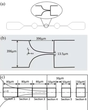
The hyperbolic microchannel geometry and the regions for DI measurement are shown in Fig. 2.
Table 1. Sample preparation
Case Cell sample Incubation time (h) Temperature (ºC) CO2 (%)
1 RBCs with HBSS - Room temperature -
2 RBCs with medium 24h 37ºC 5%
3 RBCs with medium 48h 37ºC 5%
4 RBCs with HCT-15
cell line
24h 37ºC 5%
5 RBCs with HCT-15
cell line
Figure 2. (a) Illustration of the entire microchannel view, (b) schematic diagram and geometries of the microchannel focused on the hyperbolic contraction part, (c) deformation measurement sections with corresponding lengths in microns.
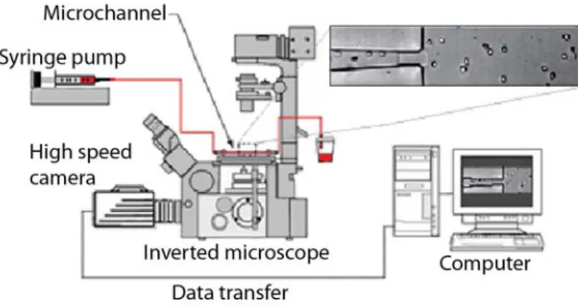
Experimental set-up
Figure 3. Experimental set-up.
Image Analysis
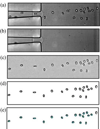
Fig. 4 shows the steps of image processing with the corresponding actual images in each step. Briefly, the original video data was converted to a stack of JPEG images (Fig. 4a). Then by averaging each pixel over the sequence of original images (stack), a background image was created (Fig. 4b). The background image contains the objects that are static in all the images such as microchannel walls, dusts and so on. The next step is to subtract the background image from the stack images. This process eliminates all the static objects from the original images and leaves only RBCs visible (Fig. 4c). The images were then filtered by replacing each pixel with the median of the neighbouring pixel values with a mask size of 3×3 pixels, in order to reduce the artifacts and enhance the image quality. The brightness and contrast of the resulted images were also manually adjusted. Finally, the grey scale images were converted to binary images adjusting the threshold level (Fig. 4d). Well-known Otsu threshold method was used in this case [24]. The resulted images contain RBCs as black edged ellipsoidal objects against a white background. After this segmentation process, the flowing RBCs were measured frame by frame automatically by using Analyze Particles function in ImageJ [25]. This function counts and measures the objects in binary images, by detecting the edge of the objects and outlining them (Fig. 4e). Various parameters for measurement such as area and circularity of the cells were appropriately pre-set so that the values outside the range specified are ignored. For instance, we set the range of area as 17-150 µm2 such that the objects smaller than 17 µm2 and larger than 150 µm2 are eliminated from the results table. Likewise, the circularity range was set to be 0.5-1.0. Here the circularity is defined as 4π × (area) / (perimeter)2. This way, most of the apparent deviant objects (eg. out-of-focus cells, aggregated cells, etc.) were ignored for measurements.
Figure 4. Steps of image analysis. (a) original image, (b) created background image, (c) subtracted image where only RBCs are visible, (d) binarized image by thresholding, (e) RBCs outlined by ImageJ, Analyze Particles function.
Results and Discussion
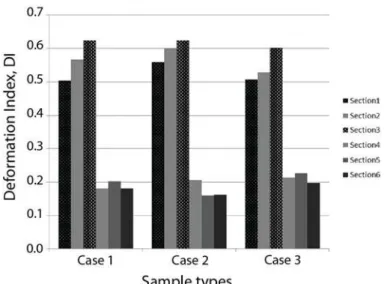
Fig. 5 shows the comparison of DIs among the three control cases (c.f. RBCs with HBSS, RBCs with medium for 24h and RBCs with medium for 48h). For each case, the mean DI values for six pre-defined regions in the microchannel were shown (c.f. Fig.2c).
Figure 5. RBC deformation index for Case 1, 2 and 3. Note that Case 1 is RBCs with HBSS, Case 2 is RBCs with medium for 24h, and Case 3 is RBCs with medium for 48h.
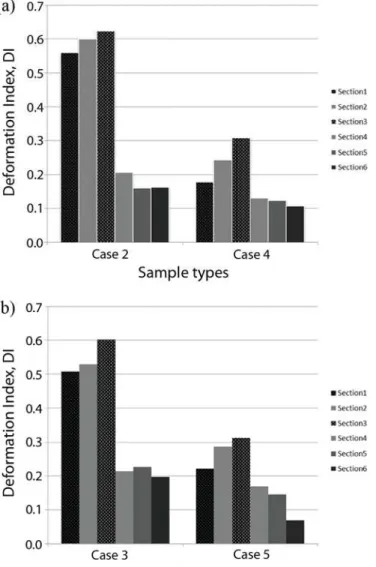
Fig. 6 compares RBC deformation between RBCs with medium and RBCs mixed with HCT-15 as a function of time duration: 24h and 48h. It reveals that the RBCs mixed with HCT-15 show extremely low DI compared with the RBCs with medium in the contraction regions. This implies the RBCs in contact with HCT-15 became more rigid than healthy RBCs. It is also possible to say that an increase of rigidity occurred within 24h after a mixture with HCT-15 and this status did not change with time (see Fig. 6b for 48h case). Although there is a tendency that the DI values increase towards the exit of the contraction part and then drops after the release, the maximum value of RBCs in contact with HCT-15 is almost half of that of the RBCs with medium. The DI values for the Sections 4, 5 and 6 do not show significant differences among the cases shown in Fig. 6.
Figure 6. (a) RBC deformation index for Case 2 and 4, (b) RBC deformation for Case 3 and 5. Note that Case 2 is RBCs with medium for 24h, Case 4 is RBCs with HCT-15 for 24h, Case 3 is RBCs with medium for 48h, and Case 5 is RBCs with HCT-15 for 48h.
Acknowledgment The authors acknowledge the financial support provided by: Student Mobility Placements with the program Lifelong Learning (Erasmus Program), 2007 Global COE Program “Global Nano-Biomedical Engineering Education and Research Network”, Japan. Grant-in-Aid for Science and Technology (PTDC/SAU-BEB/105650/2008, PTDC/EME-MFE/099109/2008 and PTDC/SAU-ENB/116929/2010) from the Science and Technology Foundation (FCT) and COMPETE, Portugal. The authors are also very grateful to Professor Mónica S.N. Oliveira (Strathclyde University), Professor Geyong M. Kim (University of Navarra) and Professor Sergio Arana (University of Navarra) for their discussion and suggestions to this research work.
References
[1] Caro, C., Pedley, T., Schroter, R., & Seed, W. The Mechanics of the Circulation. Oxford Universi-ty Press, (1978).
(1969).
[3] Abkarian, M., et al. Cellular-scale hydrodynamics. Biomed Mater 3, 034011 (2008).
[4] Hardeman, M.R., & Ince, C. Clinical potential of in vitro measured red cell deformability, a myth? Clin Hemorheol Microcirc 21, 277-284 (1999).
[5] Cho, Y.I., Mooney, M.P., & Cho, D.J. Hemorheological disorders in diabetes mellitus. J Diabetes Sci Technol 2, 1130-1138 (2008).
[6] Gueguen, M., et al. Filtration pressure and red blood cell deformability: evaluation of a new device: erythrometre. Biorheology Suppl 1, 261-265 (1984).
[7] Shin, S., Ku, Y., Park, M.S., & Suh, J.S. Measurement of red cell deformability and whole blood viscosity using laser-diffraction slit rheometer. Korea-Australia Rheol J 16, 85-90 (2004).
[8] Dobbe, J.G.G., et al. Analyzing red blood cell-deformability distributions. Blood Cells Mol Dis 28, 373-384 (2002).
[9] Mokken, F.C., Kedaria, M., Henny, C.P., Hardeman, M.R., & Gelb, A.W. The clinical importance of erythtrocyte deformability, a hemorrheological parameter. Ann Hematol 64, 113-122 (1992).
[10] Bow, H., et al. A microfabricated deformability-based flow cytometer with application to malaria. Lab Chip 11, 1065-1073 (2011).
[11] Lima, R., et al. In vitro blood flow in a rectangular PDMS microchannel: experimental observations using a confocal micro-PIV system. Biomed Microdevices 10, 153-167 (2008).
[12] Fujiwara, H., et al. Red blood cell motions in a high hematocrit blood flowing through a stenosed micro-channel. J Biomech 42, 838-843 (2009).
[13] Lima, R., et al. Axisymmetric PDMS microchannels for in vitro haemodynamics studies. Biofabrication 1, 035005 (2009).
[14] Lee, S.S., Yim, Y., Ahn, K.H., & Lee, S.J. Extensional flow-based assessment of red blood cell deformability using hyperbolic converging microchannel. Biomed Microdevices 11, 1021-1027 (2009).
[15] Zhao, R., et al. Microscopic investigation of erythrocyte deformation dynamics. Biorheology, 43, 747-765 (2006).
[16] Yaginuma, T., Oliveira, M.S.N., Lima, R., Ishikawa, T., & Yamaguchi, T. Human red blood cell behavior under homogeneous extensional flow in a hyperbolic-shaped microchannel, Biomicrofluidics 7, 054110 (2013).
[17] Pinho, D., Yaginuma, T., & Lima, R. A microfluidic device for partial cell separation and deformability assessment. BioChip J 7, 367-374 (2013).
[18] Hou, H.W., et al. Microfluidics for applications in cell mechanics and mechanobiology. Cel Mol Bioeng 4, 591–602, (2011).
[19] Tan, S.J., Yobas, L., Lee, G.Y., Ong, C.N., & Lim, C.T. Microdevice for the isolation and enumeration of cancer cells from blood. Biomed Microdevices 11, 883-892 (2009).
[20] Mohamed, H., Murray, M., Turner, J.N., & Caggana, M. Isolation of tumor cells using size and deformation. J Chromatogr A 1216, 8289-8295 (2009).
[27] Sevick EM, & Jain RK. Effect of red blood cell rigidity on tumor blood flow: increase in viscous resistance during hyperglycemia. Cancer Res 51, 2727-30 (1991).
[28] Kuzman, D., et al. Effect of pH on red blood cell deformability. Pflugers Arch 440, R193-194 (2000).
[29] Griffiths, J.R. Are cancer cells acidic? Br J Cancer 64, 425-427 (1991).