Background. The authors conducted an in vivo study to compare a laser fluorescence system with a visual system for occlusal caries diag-nosis in children’s primary and permanent molars.
Methods. The authors selected for evaluation 320 untreated, cavity-free primary and permanent molars in healthy children aged 6 through 14 years. Two of the authors conducted the laser fluorescence evaluation. Another of the authors completed the clinical evaluation. The κ value was 0.68. The authors compared sensitivity, specificity, predictive values, odds ratio and receiver operating characteristic (ROC) curves for the laser fluorescence system.
Results. For the whole sample, the sensitivity and specificity of the laser fluorescence system were 0.79 and 0.87, respectively. The positive and negative odds ratios for the whole sample were 6.33 and 0.23. The positive and negative predictive values for the whole sample were 33.9 percent and 98.1 percent. The value of the area beneath the ROC curve (AUC) was 0.92 for the whole sample.
Conclusions. The laser fluorescence system was more precise than visual evaluation in identifying lesions without cavities and healthy sur-faces in primary and permanent molars.
Clinical Implications. In daily practice, dentists can consider the laser fluorescence system a complementary tool in the visual exploration of occlusal surfaces of primary molars and permanent first molars. Key Words. Caries diagnosis; diagnostic tests; laser fluorescence system; early detection of caries.
JADA 2008;139(5):572-579.
T
ooth decay is a dynamic process resulting from an imbalance between demineralization and remineralization of the dental surface.1It begins whenbac-teria in acidogenic dental plaque— mainly Streptococcus mutans,
Strep-tococcus sobrinus and Lactobacillus acidophilus—ferment
carbohy-drates in the diet,2producing
organic acids such as lactic, formic, pyruvic, butyric, acetic and propi-onic acids. The acids’ hydrogen ions act on hydroxylapatite crystals, freeing the calcium and phosphate mineral content and, thereby, initi-ating the process that forms a cavity.1
Once the diffuse destruction of the hydroxylapatite crystals has begun, the bacteria that invade the lesion in the enamel can reach the deepest layers of the enamel, even in incipient lesions without cavities, all the way to the amelodentinal limit.3This process is generally
slow, and periods of demineraliza-tion alternate with other periods when, if oral conditions change, remineralization predominates.4,5
Preventive programs have decreased the prevalence and inci-dence of dental caries in children and adolescents6-9and have changed
the pattern of caries distribution, with an increase in the proportion of occlusal caries.7-12In addition,
A B S T R A C T
Dr. Barbería is a professor and the director, Dental Care Program for Children, Department of Preven-tion, Pediatric Dentistry and Orthodontics, Faculty of Dentistry, Complutense University of Madrid, Avda. Complutense s/n, 28040 Madrid, Spain, e-mail “[email protected]”. Address reprint requests to Dr. Barbería.
Dr. Maroto is an assistant professor, Department of Prevention, Pediatric Dentistry and Orthodontics, Faculty of Dentistry, Complutense University of Madrid, Spain.
Dr. Arenas is an assistant doctor, Dental Care Program for Children, Department of Prevention, Pedi-atric Dentistry and Orthodontics, Faculty of Dentistry, Complutense University of Madrid, Spain. Dr. Cardoso Silva is an assistant doctor, Dental Care Program for Children, Department of Prevention, Pediatric Dentistry and Orthodontics, Faculty of Dentistry, Complutense University of Madrid, Spain.
A clinical study of caries diagnosis
with a laser fluorescence system
Elena Barbería, MD, DMD, PhD, DDS; Myriam Maroto, DMD, PhD; Marcela Arenas, DMD, PhD; Cristina Cardoso Silva, DMD
on May 8, 2008
jada.ada.org
cavities appear later, and, therefore, occlusal sur-faces that are clinically healthy and apparently intact may hide lesions that penetrate the dentin.
In children, the susceptibility to demineraliza-tion of primary teeth is greater than that of per-manent teeth,13and the fact that the enamel in
primary teeth is thinner means that the progres-sion of decay is faster.14It is difficult to diagnose
the depth of occlusal caries without
cavi-ties,5,6,11,12,15-18and the decision to restore the lesion
or remineralize it varies greatly among dentists. 19-22Thus, early detection and determination of the
carious lesion’s depth are fundamental because they can lead to a shift from surgical intervention to preventive treatment.4,23-28
The use of conventional diagnostic techniques seems satisfactory for the diagnosis of cavities29,30
but inadequate for the diagnosis of lesions without cavities, lesions on the root surface or recurrent caries.31An ideal diagnostic method
should offer a high level of sensitivity—that is, yield a low rate of false-negative findings. It also should be highly specific, yielding a
low rate of false-positive findings. However, these properties are diffi-cult to achieve using the traditional diagnostic methods that are based on visual exploration with a mirror, a probe, a halogen dental lamp and radiographs because they are sub-ject to a broad variety of criteria among those performing the exami-nations.22,32,33
Researchers and manufacturers have been developing instruments that measure the changes in dental tissue resulting from tooth decay by means of detecting the tissue’s
optical properties.34It is possible to contrast
healthy enamel and carious tissue through an evaluation of the fluorescence stimulated by a laser or infrared light.35Red light, like infrared
light, is absorbed less by enamel and is dispersed throughout the enamel to a greater degree than is light with a shorter wavelength. For that reason, infrared light penetrates more deeply, and it is possible to use infrared light to measure the fluo-rescence of dentinal caries located beneath the enamel even when the dental surface is clinically whole.35
Caries diagnosis is based fundamentally on a meticulous examination and the application of the clinical evidence of caries diagnosis.
Never-theless, many studies have demonstrated that the diagnosis of occlusal caries without cavities is dif-ficult, and false-positive and false-negative find-ings occur frequently.31,33,36-43To avoid the
occur-rence of false-positive and false-negative findings, dentists may use complementary tools, such as a laser fluorescence system (for example,
DIAGNOdent, KaVo Dental, Biberach, Germany). Few studies have been performed that compare the results of clinical exploration with those obtained through the use of DIAGNOdent for diagnosis of children’s primary and permanent molars. For this reason, we decided to study the validity of this instrument by measuring the fluo-rescence of occlusal surfaces in a sample of pri-mary molars and to analyze the possible differ-ences in the permanent molars of the same group of children.
METHODS, SUBJECTS AND MATERIALS We performed our study within ongoing investi-gational activities of the Dental Care Program for
Children–Pediatric Clinic of the Complutense University of Madrid (UCM), Spain, and it was approved by the institutional review board of the UCM General Foundation. We obtained consent forms from the children’s parents or legal represen-tatives on which they duly autho-rized the use of collected data for diagnosis.
We designed an in vivo study to use the DIAGNOdent fluorescent laser device (Model D88400) to measure the fluorescence of pri-mary first and second molars and permanent first molars in a conve-nience sample of boys and girls aged 6 through 14 years who sought dental care at the pediatric clinic of UCM from January through October 2004.
The operation of DIAGNOdent is based on the concept of stimulation of fluorescence through the use of laser light. This device produces a red light with a wavelength of 695 nanometers that the user applies to the dental surface.6,32,40,44The light
penetrates the enamel and the dentin, and the fluorescent light’s intensity is measured by a pho-todiode and converted into digits, which appear ABBREVIATION KEY.RSS: Research Support Service. UCM: Complutense University of Madrid.
An ideal diagnostic method should offer
high levels of sensitivity and specificity. However, these properties are difficult to achieve using traditional diagnostic methods.
on May 8, 2008
jada.ada.orgDownloaded from
by Ekstrand and
colleagues39: whole surfaces
that do not require treat-ment, demineralized
occlusal surfaces susceptible to remineralizing treatment or demineralized occlusal surfaces requiring restora-tive treatment.
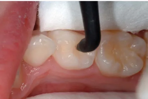
Two examiners (M.M., M.A.) unfamiliar with the data obtained in the clinical exploration examined the same molars by using DIAGNOdent. Following the manufacturer’s instructions, they dried teeth for two sec-onds and took readings in several places on the occlusal surface, noting the maximum value obtained. In accordance with the manufacturer’s criteria, they classified surfaces as follows: values between zero and 4, healthy occlusal surface; values between 5 and 25, lesion limited to the enamel; values of 26 or more, lesion affecting the dentin (Figure 1). They used the same tip shape in all examinations to prevent reading variations (according to the technique suggested by KaVo Dental, written communication, Dec. 10, 2007).
The UCM Research Support Service (RSS) evaluated concordance between examiners through use of the κ statistic. RSS staff members calculated sensitivity, specificity, predictive values and the odds ratio of DIAGNOdent read-ings for the whole sample, as well as separately for primary molars and permanent molars. To determine DIAGNOdent’s diagnostic precision, they traced a curve for the receiver operating characteristic (ROC). They established the level of significance at P ≤ .05 for all cases, and they applied 95 percent confidence intervals (CIs). An analyst from the RSS performed the statistical data analysis by using a statistical software package (SPSS, Version 11.0, SPSS, Chicago). RESULTS
After we applied the inclusion and exclusion cri-teria, the studied sample totaled 320 molars, including primary first (116) and second (127) molars and permanent first molars (77).
Results of clinical exploration. Primary
molars. Of 243 occlusal surfaces, we diagnosed
Figure 1. Use of the fluorescent laser device on the occlusal surface of a primary first molar.
on a screen.
We used a linear scale, as proposed by the manufacturer, whose cutoff points were deter-mined on the basis of previous studies6,32,40,44that
relate histologic variations due to carious lesions with the measurement potential of the filtered light expressed in specific units for DIAGNOdent (KaVo Dental, written communication, Dec. 10, 2007). DIAGNOdent readings between zero and 4 indicate a healthy occlusal surface; readings between 5 and 25 indicate the presence of enamel caries, and readings of 26 or more indicate the presence of dentinal caries (P ≤ .05) (KaVo Dental, written communication, Dec. 10, 2007).
We included in the study all the molars that had erupted completely; did not appear to have cavities, sealants or occlusal restorations; and did not have hypoplastic surfaces, pathological abra-sions or other structural defects. We excluded from the sample molars of children who had sys-temic diseases that could interfere with the diag-nostic process: temporomandibular joint defects that limited the child’s ability to open his or her mouth and syndromes that made it impossible for the child to cooperate.
A single examiner (E.B.) who had broad expe-rience in the clinical diagnosis of caries per-formed the visual exploration. She conducted it with a mirror, a no. 4 probe and a halogen dental lamp; she dried the occlusal surface beforehand for three to five seconds. The examiner classified the fissures according to the criteria established
on May 8, 2008
jada.ada.org
200 (82 percent) as healthy, we classi-fied 24 (10 percent) as susceptible to remineralization treatment and we determined that 19 (8 percent) required restorations.
Permanent molars. Of 77 occlusal
surfaces, we diagnosed 49 (64 percent) as whole or healthy, 23 (30 percent) as needing remineralization treat-ment and 5 (6 percent) as needing restorations.
Of the whole sample of 320 primary and permanent molars, we diagnosed 249 (78 percent) occlusal surfaces as healthy, 47 (15 percent) as needing remineralization treatment and 24
(7 percent) as irreversible lesions requiring resto-rations (Table 1).
Results of evaluation with DIAGNOdent. To measure concordance among the examiners, RSS staff members compared the results
obtained by the two examiners through an evalu-ation of DIAGNOdent’s data regarding molar sur-faces, listed in Table 2 according to the whole sample, and separately for primary molars and permanent molars.
The concordance between examiners obtained through use of the κ statistic was 0.66 for pri-mary molars, 0.71 for permanent molars and 0.68 for the total sample. Given that the concordance between the examiners was strong, we chose to use the data obtained by examiner no. 1 in the rest of the statistical analysis.
Sensitivity and specificity. DIAGNOdent’s sensitivity for the diagnosis of unhealthy surfaces was 0.89 in primary molars and 0.40 in perma-nent first molars (Table 3). In the whole sample, this sensitivity index was 0.79.
The specificity obtained for diagnosing healthy surfaces was 0.89 for primary molars and 0.82 for permanent first molars. In the whole sample, the specificity was 0.87.
Odds ratio. We determined the ratio between the sensitivity and specificity values we obtained by calculating the negative and positive odds ratio. The positive odds ratio obtained was 8.36 in primary molars and 2.20 in permanent molars. The negative odds ratio was 0.11 in primary molars and 0.73 in permanent molars. The posi-tive odds ratio for the whole sample was 6.33 and the negative odds ratio was 0.23.
Predictive values. The positive predictive value for DIAGNOdent’s diagnostic test was 41.5
percent for primary molars and 13.3 percent for permanent first molars. The negative predictive value was 99.0 percent for primary molars and 95.2 percent for permanent molars. In the whole sample, the positive predictive value obtained was 33.9 percent, and the negative predictive value was 98.1 percent.
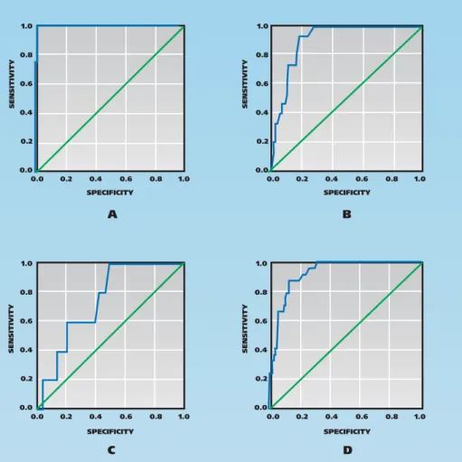
ROC curve. We analyzed the ROC curve for the purpose of relating the readings obtained by DIAGNOdent to the size of the caries, thereby determining the precision of occlusal caries diag-nosis with DIAGNOdent (Figure 2, page 577). The value of the area under the ROC curve (AUC) obtained for the occlusal surfaces of primary first molars was 0.99 (95 percent CI: 0.99-1.00) (Figure 2A) and 0.90 (95 percent CI: 0.85-0.96) for the occlusal surfaces of primary second molars (Figure 2B). In permanent first molars, the value obtained was 0.72 (95 percent CI: 0.58-0.91) (Figure 2C). Finally, the AUC obtained for the whole sample was 0.92 (95 percent CI: 0.88-0.96) (Figure 2D).
DISCUSSION
A great discrepancy can exist among profes-sionals when diagnosing occlusal caries without cavities31,33,36-43; therefore, clinicians’ judgment is
essential. Our study evaluated the clinical applic-ability of the DIAGNOdent laser device as a com-plementary tool in the visual exploration of the occlusal surfaces of primary molars and perma-nent first molars. This device was put on the market in 1998, and various studies, both in
vivo40,42,43,45-47and in vitro,48-51have evaluated its
reproducibility, sensitivity and specificity. The evaluation criteria we used were those defined previously by the manufacturer. In addition, sev-TABLE 1
Results of visual evaluation of primary
and permanent molars, according to
classification.
TYPE OF MOLAR CLASSIFICATION, AS DETERMINED BY VISUAL EVALUATION* (n†[% F‡]) TOTAL (N) Healthy Remineralization RestorationPrimary 200 (82) 24 (10) 19 (8) 243
Permanent 49 (64) 23 (30) 5 (6) 77
TOTAL 249 (78) 47 (15) 24 (7) 320
* The clinical visual evaluation was conducted by a single examiner who classified teeth according to criteria established by Ekstrand and colleagues.39
† N: Number in sample. ‡ F: Frequency.
on May 8, 2008
jada.ada.org
eral authors42,43,52have used the same criteria as
those used in other in vitro studies.41,48,49
The criteria we adopted in this study to deter-mine the validity of carious lesions were those published by Ekstrand and colleagues in 1998,39
which classify the integrity of the occlusal surface according to its histologic validation. We consid-ered clinical exploration essential for confirmation of the laser device’s diagnosis, as did the various authors who also adopted this methodology in their studies.36,40,41,43,53
We found our results for the primary dentition interesting because few studies have been pub-lished that evaluated the use of the DIAGNOdent fluorescent laser device on primary molars. The only study comparable with our research was described by Rocha and colleagues.42The
speci-ficity we obtained (0.89) is lower than theirs (0.95), and the sensitivity we obtained (0.89) is higher than theirs (0.73). This may be because they considered values greater than 21 as
signi-fying dentinal lesions,42
whereas we considered readings of 26 or greater as signifying dentinal lesions.
Comparing the results obtained in this study with those of other authors, we observed that for permanent first molars, the sensitivity level of 0.40 we obtained is higher than the 0.17 obtained by Verdonschot and colleagues4but lower
than the 0.92 obtained by Anttonen and colleagues7
and the 0.81 reported by Angnes and colleagues.45
Our study’s lower sensitivity value for permanent first molars may be because we excluded lesions with cavities, thereby increasing the percentage of healthy teeth (64 percent) and caries limited to enamel (30 percent). Given that this diagnostic method is considered more sensitive for dentinal lesions,54-56it is reasonable that when lesions with
cavities are excluded, a lower value would be obtained.
The difference in the number of false-positive findings we found between permanent and pri-mary molars could be because that dentists find it easier to use visual exploration to diagnose occlusal caries in primary molars, given the char-acteristics of their occlusal morphology and their anterior location in the arch. We found no differ-ences in sensitivity and specificity values between the groups of maxillary and mandibular molars, which is similar to the results obtained by Heinrich-Weltzien and colleagues.40
The positive odds ratio obtained for the whole sample (6.33) indicates a high capacity for dis-crimination in the diagnosis of carious lesions with the laser device. Nevertheless, it was much higher for primary molars (8.36) than for permanent molars (2.20).
The negative odds ratio obtained for the whole sample was 0.23 (0.11 for primary molars and 0.73 for permanent molars). These dissimilar neg-ative odds ratio values point to a higher capacity for discrimination of caries-free surfaces in pri-mary molars than in permanent molars.
The values we obtained for primary molars were similar to those published by Lussi and Francescut,57who used this device in a sample of
95 primary molars, and to the values published TABLE 3
Laser fluorescence system’s
sensitivity and specificity in
diagnosis of healthy surfaces.
TYPE OF TOOTH SENSITIVITY SPECIFICITY Primary Molars 0.89 0.89First 1 0.96
Second 0.87 0.82
Permanent First Molars 0.40 0.82
TOTAL 0.79 0.87
TABLE 2
Results of laser fluorescence evaluation of primary
and permanent molars, according to classification.
TYPE OFMOLAR
EXAMINER CLASSIFICATION, AS DETERMINED BY LASER FLUORESCENCE EVALUATION* (n†[% F‡])
TOTAL (N) Healthy Remineralization Restoration
Primary 1 114 (47) 88 (36) 41 (17) 243 2 118 (49) 88 (36) 37 (15) 243 Permanent 1 18 (23) 44 (57) 15 (20) 77 2 16 (21) 44 (57) 17 (22) 77 TOTAL 1 132 (41) 132 (41) 56 (18) 320 2 134 (42) 132 (41) 54 (17) 320
* The laser fluorescence evaluation was conducted by two examiners who classified teeth according to criteria established by Ekstrand and colleagues.39
† N: Number in sample. ‡ F: Frequency.
on May 8, 2008
jada.ada.org
by López and colleagues55for
permanent first molars. The values we obtained by analyzing the ROC curve in the molar groups we studied indicate that this diagnostic procedure was precise in all of the studied groups. Neverthe-less, the AUC diminished from primary first molars (0.99) to primary second molars (0.90) and permanent molars (0.72). However, when comparing ROC curve values and the posi-tive and negaposi-tive odds ratios obtained for primary and per-manent molars, we found a higher precision for primary molars. This could be due to histologic, morphological occlusal surfaces and eruptive chronology differences between primary and permanent molars.
The results obtained in this study by two different exam-iners for sensitivity and speci-ficity values, odds ratios (posi-tive and nega(posi-tive) and the ROC curve showed that the DIAGNOdent is a valuable complement to clinical exami-nation. However, when we
compared the results we obtained with the statis-tical tests for primary and permanent molars sep-arately, we noted that DIAGNOdent demon-strated a higher precision and higher capacity to discriminate caries in primary molars than in permanent molars.
The reproducibility of our study’s data (κ = 0.68), confirmed by values obtained in previous research,42,56-58indicates that this laser device can
be a tool for the longitudinal evaluation of occlusal carious lesions without cavities in
patients who are at high risk of developing caries. The comparison of at least two consecutive mea-surements can alert the dentist to the existence of a lesion. In an in vitro study of primary molars, researchers demonstrated that the results
obtained with DIAGNOdent by various examiners are more reproducible and homogeneous than those derived from visual exploration.22 The
results of our study affirmed that DIAGNOdent is
accurate in diagnosing lesions that are unde-tectable with a probe. Given our results and the comparative analysis with results published by other researchers, we consider it important to include DIAGNOdent readings among the data necessary for making clinical judgments and ther-apeutic decisions.
CONCLUSIONS
The sensitivity we obtained in the exploration of occlusal surfaces using the DIAGNOdent fluores-cent laser device made it possible to identify 89 percent of the lesions without cavities in the occlusal surfaces of primary molars. This comple-mentary tool also revealed a high level of speci-ficity for the same molars and locations, demon-strating the capacity to identify healthy surfaces in 89 percent of the cases.
The AUC we obtained for primary and perma-nent molars was close to 1.0. This makes it
pos-1.0 0.8 0.6 0.4 0.2 0.0 1.0 0.8 0.6 0.4 0.2 0.0 1.0 0.8 0.6 0.4 0.2 0.0 1.0 0.8 0.6 0.4 0.2 0.0 0.0 0.2 0.4 0.6 0.8 1.0 0.0 0.2 0.4 0.6 0.8 1.0 0.0 0.2 0.4 0.6 0.8 1.0 0.0 0.2 0.4 0.6 0.8 1.0 SPECIFICITY SENSITIVITY SENSITIVITY SENSITIVITY SENSITIVITY SPECIFICITY SPECIFICITY SPECIFICITY A C D B
Figure 2. Receiver operating characteristic curves and areas under the curves. A. Primary first
molars. B. Primary second molars. C. Permanent first molars. D. The whole sample.
on May 8, 2008
jada.ada.org
sible to state that the laser device is a highly pre-cise diagnostic tool for the early diagnosis of occlusal caries in primary and permanent molars, as well as for the verification of the effectiveness of remineralization treatments in incipient lesions. ■
Disclosure. KaVo Dental S.A. (Biberach, Germany) donated the
laser fluorescence system DIAGNOdent.
The authors thank the Research Support Service of the Complutense University of Madrid, Spain, for the assistance given on the statistical analysis.
1. Featherstone JD. The continuum of dental caries: evidence for a dynamic disease process. J Dent Res 2004;83(special no. C):C39-C42.
2. Featherstone JD. Prevention and reversal of dental caries: role of low level fluoride. Community Dent Oral Epidemiol 1999;27(1):31-40. 3. Mejare I, Brannstrom M. Deep bacterial penetration of early prox-imal caries lesions in young human premolars. ASDC J Dent Child 1985;52(2):103-107.
4. Verdonschot EH, Angmar-Mansson B, ten Bosch JJ, et al. Devel-opments in caries diagnosis and their relationship to treatment deci-sions and quality of care. ORCA Saturday Afternoon Symposium 1997. Caries Res 1999;133(1):32-40.
5. Astvaldsdóttir A, Holbrook WP, Tranaeus S. Consistency of DIAGNOdent instrument for clinical assessment of fissure caries. Acta Odontol Scand 2004;62(4):193-198.
6. Alwas-Danowska HM, Plasschaert AJ, Suliborski S, Verdonschot EH. Reliability and validity issues of laser fluorescence measurements in occlusal caries diagnosis. J Dent 2002;30(4):129-134.
7. Anttonen V, Seppa L, Hausen H. Clinical study of the use of the laser fluorescence device DIAGNOdent for detection of occlusal caries in children. Caries Res 2003;37(1):17-23.
8. Biesbrock AR, Chesters RK, Ellwood RP, Smith SR. The chal-lenges of validating diagnostic methods relative to a conventional two-year caries clinical trial. J Dent Res 2004;83(special no. C):C53-C55.
9. Whelton H. Overview of the impact of changing global patterns of dental caries experience on caries clinical trials. J Dent Res 2004;83 (special no. C):C29-C34.
10. Lussi A. Validity of diagnostic and treatment decisions of fissure caries. Caries Res 1991;25(4):296-303.
11. Kidd EA, Joyston-Bechal S, Beighton D. Microbiological valida-tion of assessment of caries activity during cavity preparavalida-tion. Caries Res 1993;27(5):402-408.
12. Pitts NB. Diagnostic tools and measurements: impact on appro-priate care. Community Dent Oral Epidemiol 1997;25(1):24-35.
13. Shellis RP. Relationship between human enamel structure and the formation of caries-like lesions in vitro. Arch Oral Biol 1984; 29(12):975-981.
14. Ashley P. Diagnosis of occlusal caries in primary teeth. Int J Pae-diatr Dent 2000;10(2):166-171.
15. Wenzel A. Bitewing and digital bitewing radiography for detec-tion of caries lesions. J Dent Res 2004;83(special no. C):C72-C75.
16. Sawle RF, Andlaw RJ. Has occlusal caries become more difficult to diagnose? A study comparing clinically undetected lesions in molar teeth of 14-16-year old children in 1974 and 1982 (published correction appears in Br Dent J 1988;164[11]:361). Br Dent J 1988;164(7): 209-211.
17. al-Sehaibany F, White G, Rainey JT. The use of caries detector dye in diagnosis of occlusal carious lesions. J Clin Pediatr Dent 1996; 20(4):293-298.
18. Pastor C, López G, Gómez I, Sánchez R, Llamas R. Valoración de los métodos de exploración de caries oclusales sin cavitación [Evalu-ation of methods for the explor[Evalu-ation of occlusal caries without cavities]. Rev Europea de Odontoestomatol 1998;11(4):214-224.
19. Elderton RJ, Nuttall NM. Variation among dentists in planning treatment. Br Dent J 1983;54(7):201-206.
20. Kay EJ, Knill-Jones R. Variation in restorative treatment deci-sions: application of Receiver Operating Characteristic curve (ROC) analysis. Community Dent Oral Epidemiol 1992;20(3):113-117.
21. Bader JD, Shugars DA. Variation in dentists’ clinical decisions. J Public Health Dent 1995;55(3):181-188.
22. Bengtson AL, Gomes AC, Mendes FM, Cichello LR, Bengtson NG, Pinheiro SL. Influence of examiner’s clinical experience in detecting occlusal caries lesions in primary teeth. Pediatr Dent 2005;27(3):
238-243.
23. Nyvad B. Diagnosis versus detection of caries. Caries Res 2004;38(3):192-198.
24. Pine CM, Ten Bosch JJ. Dynamics of and diagnostic methods for detecting small carious lesions. Caries Res 1996;30(6):381-388.
25. Zandona AF, Zero DT. Diagnosis tools for early caries detection (published correction appears in JADA 2007;138[3]:298). JADA 2006;137(12):1675-1684.
26. Fontana M, Zero DT. Assessing patients’ caries risk. JADA 2006;137(9):1231-1239.
27. Young DA, Featherstone JD. Digital imaging fiber-optic trans-illumination, F-speed radiographic film and depth of approximal lesions. JADA 2005;136(12):1682-1687.
28. Christensen GJ. The advantages of minimally invasive dentistry. JADA 2005;136(11):1563-1565.
29. Hamilton JC. Should a dental explorer be used to probe suspected carious lesions? Yes—an explorer is a time-tested tool for caries detec-tion. JADA 2005;136(11):1526, 1528, 1530 passim.
30. Akarsu S, Köprülü H. In vivo comparison of the efficacy of DIAGNOdent by visual inspection and radiographic diagnostic tech-niques in the diagnosis of occlusal caries. J Clin Dent 2006;17(3):53-58.
31. Horowitz AM. A report on the NIH Consensus Development Con-ference on Diagnosis and Management of Dental Caries Throughout Life. J Dent Res 2004;83(special no. C):C15-C17.
32. Lussi A, Imwinkelried S, Pitts N, Longbottom C, Reich E. Per-formance and reproducibility of a laser fluorescence system for detec-tion of occlusal caries in vitro. Caries Res 1999;33(4):261-266.
33. Bader JD, Shugars DA, Bonito AJ. A systematic review of the per-formance of methods for identifying carious lesions. J Public Health Dent 2002;62(4):201-213.
34. van der Veen MH, ten Bosch JJ. Autofluorescence of bulk sound and in vitro demineralized human root dentin. Eur J Oral Sci 1995; 103(6):375-381.
35. Lussi A, Hibst R, Paulus R. DIAGNOdent: an optical method for caries detection. J Dent Res 2004;83(special no. C):C80-C83.
36. Attrill DC, Ashley PF. Occlusal caries detection in primary teeth: a comparison of DIAGNOdent with conventional methods. Br Dent J 2001;190(8):440-443.
37. Lussi A. Comparison of different methods for the diagnosis of fis-sure caries without cavitation. Caries Res 1993;27(5):409-416.
38. Ekstrand KR, Luzmina I, Bjorndal L, Thylstrup A. Relationship between external and histological features of progressive stages of caries in the occlusal fossa. Caries Res 1995;29(4):243-250.
39. Ekstrand KR, Ricketts DN, Kidd EA, Qvist V, Schou S. Detection, diagnosing, monitoring and logical treatment of occlusal caries in rela-tion to lesion activity and severity: an in vivo examinarela-tion with histo-logical validation. Caries Res 1998;32(4):247-254.
40. Heinrich-Weltzien R, Kuhnisch J, Oehme T, Ziehe A, Stosser L, Garcia-Godoy F. Comparison of different DIAGNOdent cut-off limits for in vivo detection of occlusal caries. Oper Dent 2003;28(6):672-680.
41. Cortes DF, Ellwood RP, Ekstrand KR. An in vitro comparison of a combined FOTI/visual examination of occlusal caries with other caries diagnostic methods and the effect of stain on their diagnostic perform-ance. Caries Res 2003;37(1):8-16.
42. Rocha RO, Ardenghi TM, Oliveira LB, Rodrigues CR, Ciamponi AL. In vivo effectiveness of laser fluorescence compared to visual inspection and radiography for the detection of occlusal caries in pri-mary teeth. Caries Res 2003;37(6):437-441.
43. Sheehy EC, Brailsford SR, Kidd EA, Beighton D, Zoitopoulos L. Comparison between visual examination and a laser fluorescence system for in vivo diagnosis of occlusal caries. Caries Res 2001; 35(6):421-426.
44. Konig K, Hibst R, Meyer H, Flemming G, Schneckenburger H. Laser-induced autofluorescence of carious regions of human teeth and caries-involved bacteria. SPIE 1993;20(8):170-180.
45. Angnes V, Angnes G, Batisttella M, Grande RH, Loguercio AD, Reis A. Clinical effectiveness of laser fluorescence, visual inspection and radiography in the detection of occlusal caries. Caries Res 2005; 39(6):490-495.
46. Costa AM, Bezzerra AC, Fuks AB. Assessment of the accuracy of visual examination, bite-wing radiographs and DIAGNOdent on the diagnosis of occlusal caries. Eur Arch Paediatr Dent 2007;8(2):118-122.
47. Olmez A, Tuna D, Oznurhan F. Clinical evaluation of DIAGNO-dent in detection of occlusal caries in children. J Clin Pediatr Dent 2006;30(4):287-291.
48. Shi XQ, Welander U, Angmar-Mansson B. Occlusal caries detec-tion with KaVo DIAGNOdent and radiography: an in vitro comparison. Caries Res 2000;34(2):151-158.
49. Llamas R, Sánchez Barriga R, Bonilla V, Pastor C, Herrera M.
on May 8, 2008
jada.ada.org
Estudio comparativo in vitro del diagnóstico de las caries de surcos y fisuras de dientes del sector posterior, por examen visual y un sistema de fluorescencia producido por láser [Comparative in vitro study of diagnosis of caries in pits and fissures of teeth in the posterior sector, by visual examination and a laser-produced fluorescent system]. Av Odontoestomatol 2001;17(9):447-464.
50. Mendes FM, Hissadomi M, Imparato JC. Effects of drying time and the presence of plaque on the in vitro performance of laser fluores-cence in occlusal caries of primary teeth. Caries Res 2004;38(2): 104-108.
51. Mendes FM, Nicolau J, Duarte DA. Evaluation of the effective-ness of laser fluorescence in monitoring in vitro remineralization of incipient caries lesions in primary teeth. Caries Res 2003;37(6): 442-444.
52. Stamm JW. The classic caries clinical trial: constraints and opportunities. J Dent Res 2004;83(special no. C):C6-C14.
53. Hamilton JC, Gregory WA, Valentine JB. DIAGNOdent measure-ments and correlation with the depth and volume of minimally
inva-sive cavity preparations. Oper Dent 2006;31(3):291-296.
54. El-Housseiny AA, Jamjoum H. Evaluation of visual, explorer, and a laser device for detection of early occlusal caries. J Clin Pediatr Dent 2001;26(1):41-48.
55. López N, Pérez L, Rocamonde J, Bernal M, Pérez D. Diagnóstico de caries por fluorescencia: alternativa al examen convencional. Odont Pediátrica 2000;8(3):7-14.
56. Lussi A, Megert B, Longbottom C, Reich E, Francescut P. Clinical performance of a laser fluorescence device for detection of occlusal caries lesions. Eur J Oral Sci 2001;109(1):14-19.
57. Lussi A, Francescut P. Performance of conventional and new methods for the detection of occlusal caries in deciduous teeth. Caries Res 2003;37(1):2-7.
58. Pinelli C, Campos Serra M, de Castro Monteiro Loffredo L. Validity and reproducibility of a laser fluorescence system for detecting the activity of white-spot lesions on free smooth surfaces in vivo. Caries Res 2002;36(1):19-24.
on May 8, 2008
jada.ada.org