Braz. Arch. Biol. Technol. v.52 n.5: pp. 1063-1073, Sept/Oct 2009 Vol.52, n. 5: pp.1063-1073 September-October 2009
ISSN 1516-8913 Printed in Brazil BRAZILIAN ARCHIVES OF
BIOLOGY AND TECHNOLOGY
A N I N T E R N A T I O N A L J O U R N A L
High Molecular Diversity of the Fungus
Guignardia
citricarpa
and
Guignardia mangiferae
and New Primers for
the Diagnosis of the Citrus Black Spot
Danyelle Stringari
1, Chirlei Glienke
1*, Daniel de Christo
1, Walter Maccheroni Jr.
2and
João Lucio de Azevedo
21Departamento de Genética; Universidade Federal do Paraná; C. P.: 19071; 81531-990; Curitiba -PR - Brasil.
2
Departamento de Genética; Universidade de São Paulo; C. P.: 9; 13418-900; Piracicaba - SP - Brasil
ABSTRACT
RAPD markers were used to investigate the distribution of genetic variability among a group of Guignardia
citricarpa, G. mangiferae, and Phyllosticta spinarum isolates obtained from several hosts in Brazil, Argentina,
Mexico, Costa Rica, Thailand, Japan, United States and South Africa. Pathogenic isolates G. citricarpa Kiely
(anamorph form P. citricarpa McAlp Van Der Aa) are the etiological agent of the Citrus Black Spot (CBS), a disease
that affects several citric plants and causes substantial injuries to the appearance of their fruits, thus preventing their export. Several previous studies have demonstrated the existence of an endophytic species with high morphological similarity to the causal agent of CBS that could remain latent in the same hosts. Consequently, the identification of the plants and fruits free from the causal agent of the disease is severely hampered. The RAPD
analysis showed a clear discrimination among the pathogenic isolates of G. citricarpa and endophytic isolates (G.
mangiferae and P. spinarum). In addition, a Principal Coordinate Analysis (PCO) based on a matrix of genetic similarity estimated by the RAPD markers showed four clusters, irrespective of their host or geographical origin. An Analysis of Molecular Variance (AMOVA) indicated that 62.8% of the genetic variation was found between the
populations (G. citricarpa, G. mangiferae, P. spinarum and Phyllosticta sp.). Substantial variation was found in the
populations (37.2%). Exclusive RAPD markers of isolates of G. citricarpa were cloned, sequenced and used to
obtain SCARS (Sequence Characterized Amplified Regions), which allowed the development of new specific primers
for the identification of G. citricarpa PCR (Polymerase Chain Reaction) analysis using a pair of primers specific to
pathogenic isolates corroborating the groupings obtained by the RAPD markers, underscoring its efficiency in the identification of the causal agent of CBS.
Key words: Guignardia, SCARS, Citrus Black Spot, Citrus, Diagnosis, RAPD
* Author for correspondence: cglienke@ufpr.br
INTRODUCTION
The orange fruit is currently the most cultivated plant in the world, with an annual production exceeding 60 million tones. Brazil is responsible for nearly a third of the world production of orange, as well as 85% of the control of the
international orange juice market. Therefore, the cultivation of citric fruits is among the most important activities in Brazilian agrobusiness (Agrianual, 2005).
control and the ensuing increases in the costs of
production (Agrianual, 2005). There is
considerable depreciation in the value of fruits
with CBS in the in natura market, with strong
opposition to its import by the European Union, which is currently Brazil’s most important consumer. Therefore, the diagnosis of the fruits destined for export is of fundamental importance.
The CBS is caused by the fungus Guignardia
citricarpa Kiely (anamorphic form is Phyllosticta citricarpa Van Der Aa) and was first described in 1895 in Australia in orange fruits of the ‘Valência’ variety, causing considerable losses. This disease is currently found in several countries, with the maximum impact in South Africa, Japan, Argentina, and particularly in Brazil, where the disease has been reported most frequently after 1995 and is strongly affecting the local citriculture (Feichtenberger, 1996).
Although the identification of the causal agent of CBS can be obtained by the conventional techniques such as culturing and microscopy, these procedures require weeks to be completed. This delay is impractical in the in natura business of the fruit. In addition, there have been reports of the existence of an endophytic species (G. mangiferae) that is very similar morphologically with the causal agent of CBS, inhabiting latently the same hosts (Glienke-Blanco et al., 2002, Baayen et al., 2002). Therefore, the identification of the plants and fruits free from the causal agent of CBS is made even more difficult. Although these two fungus species are morphologically nearly identical, their physiological differences are clear. Baayen et al. (2002) showed that the most
significant morphological difference among G.
citricarpa and G. mangiferae is the thickness of the mucoid conidial sheath, which is smaller than
1.5 µm in the former but ranges between 1.5 and
3.0 µm in the latter. However, the long time
required for the isolation, culturing and pycnidium formation makes this method unsuitable for the quarantine purposes.
Baayen et al. (2002) investigated ITS sequences and showed that the endophytic species isolated in
Citrus spp. was the anamorphic state of P.
capitalensis. Okane et al. (2001) reported the
endophytic occurrence of P. capitalensis
inhabitanting Ericaceae plants, describing the
teleomorphic state as G. endophyllicola. A survey
of the taxonomic literature showed that the oldest name associated with the teleomorphic state of
these isolates probably was G. mangiferae A. J.
Roy, described in 1968. In fact, the endophytic G.
mangiferae is found inhabiting a wide range of
hosts, and probably has been repeatedly described from many different host species, causing extensive several cases of synonymy.
The present work investigated the Brazilian
isolates of Guignardia spp. and Phyllosticta spp.
with the following objectives: 1) to verify the
endophytic occurrence of G. citricarpa and G.
mangiferae in Citrus spp and llex paraguariensis;
2) to assess the genetic variability and population structure of endophytic isolates, as well as those obtained from CBS lesions; 3) to differentiate the
isolates of G. mangiferae and G. citricarpa based
on RAPD markers and to develop SCARS specific to the causal agent of CBS. The development and
validation of an identification method for G.
citricarpa using PCR could expedite the identification process of contaminated fruits and orchards.
MATERIALS AND METHODS
Fungal Material
The isolates investigated in this work were obtained from monosporic cultures. The strains of
G. citricarpa were isolated directly from CBS
injuries. The endophytic strains of G. mangiferae
and Phyllosticta spp. were isolated from healthy
host plants without any apparent lesions (Pimentel et al., 2006). The geographical origin, host plant and nature (endophytic or pathogenic) of all the isolates are listed in Tables 1 and 2.
Three strains of G. citricarpa and three strains of
G. mangiferae that were tested positive and
negative for pathogenicity, respectively
(Baldassari, 2005) were used in the validation of the PCR diagnostic (Table1).
DNA Extraction
Braz. Arch. Biol. Technol. v.52 n.5: pp. 1063-1073, Sept/Oct 2009
water. The DNA solution was treated with RNAse
(50µg/mL) and the concentration of DNA was
measured by agarose gel electrophoresis, followed by staining with ethidium bromide.
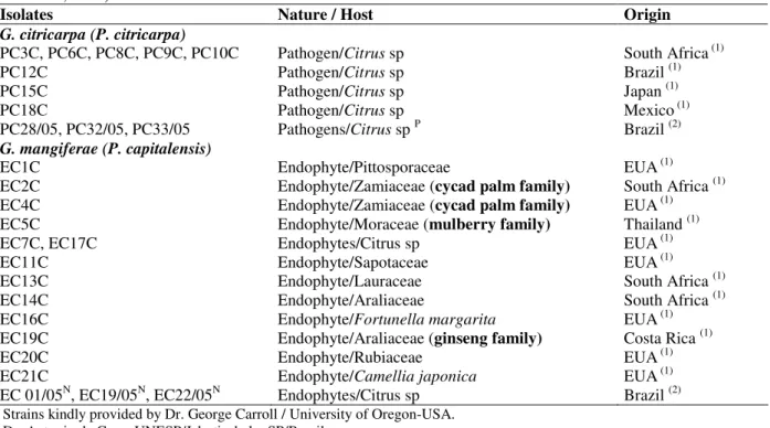
Table 1- Isolates of the citrus pathogen G. citricarpa, the citrus endophyte G. mangiferae, and Phyllosticta species
from other host species used in the RAPD analyses and PCR tests as reference isolates (Baayen et al., 2002; Baldassari, 2005).
Isolates Nature / Host Origin
G. citricarpa (P. citricarpa)
PC3C, PC6C, PC8C, PC9C, PC10C Pathogen/Citrus sp South Africa (1)
PC12C Pathogen/Citrus sp Brazil (1)
PC15C Pathogen/Citrus sp Japan (1)
PC18C Pathogen/Citrus sp Mexico (1)
PC28/05, PC32/05, PC33/05 Pathogens/Citrus sp P Brazil (2)
G. mangiferae (P. capitalensis)
EC1C Endophyte/Pittosporaceae EUA(1)
EC2C Endophyte/Zamiaceae (cycad palm family) South Africa (1)
EC4C Endophyte/Zamiaceae (cycad palm family) EUA(1)
EC5C Endophyte/Moraceae (mulberry family) Thailand (1)
EC7C, EC17C Endophytes/Citrus sp EUA(1)
EC11C Endophyte/Sapotaceae EUA(1)
EC13C Endophyte/Lauraceae South Africa (1)
EC14C Endophyte/Araliaceae South Africa (1)
EC16C Endophyte/Fortunellamargarita EUA(1)
EC19C Endophyte/Araliaceae (ginseng family) Costa Rica (1)
EC20C Endophyte/Rubiaceae EUA(1)
EC21C Endophyte/Camelliajaponica EUA(1)
EC 01/05N, EC19/05N, EC22/05N Endophytes/Citrus sp Brazil (2) 1 Strains kindly provided by Dr. George Carroll / University of Oregon-USA.
2 Dr. Antonio de Goes, UNESP/Jaboticabal – SP/Brazil
P Positive strains in pathogenicity tests for CBS (Baldassari, 2005). N Negative strains in pathogenicity tests for CBS (Baldassari, 2005).
RAPD
Random Amplified Polymorphic DNA (RAPD) analysis was carried out using 50 ng of genomic DNA. The amplification reaction was done in a
final volume of 25 µl containing 1.5 U of Taq
DNA polymerase (Invitrogen, Carlsbad, CA,
USA), 0.4 µM of primer, 0.2 mM of dNTPs
(Pharmacia, Freiburg, Germany), 3.0 mM of
MgCl2 and 1 X PCR Buffer (50 mM of KCl, 200
mM of Tris-HCl, pH 8.4). Negative controls containing all the components, except genomic DNA were included in all the experiments. The primers selected were obtained from Operon Technologies (Alameda, CA, USA): OPX08, OPX12, OPX13, OPX14, OPX17, OPX19. The amplification was performed in a MJ
Research thermocycler (Watertown, MA,
USA) according to the following steps: initial denaturation at 94°C for 2 min, followed by 40 cycles of denaturation for 1 min. at 94°C / annealing for 1.5 min at 37°C / extension for 2
min. at 72°C, and a final extension for 3 min at 72°C.
Data Analysis
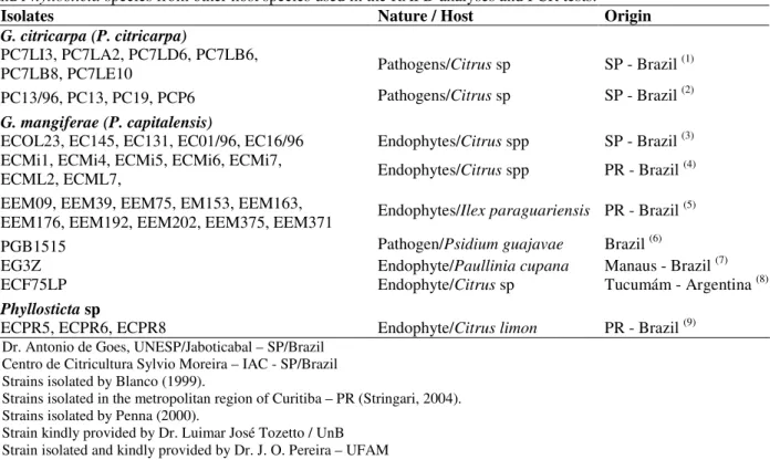
Table 2 - Isolates of the citrus pathogen G. citricarpa, the citrus endophyte G. mangiferae, Phyllosticta spinarum
and Phyllosticta species from other host species used in the RAPD analyses and PCR tests.
Isolates Nature / Host Origin
G. citricarpa (P. citricarpa)
PC7LI3, PC7LA2, PC7LD6, PC7LB6,
PC7LB8, PC7LE10 Pathogens/Citrus sp SP - Brazil (1)
PC13/96, PC13, PC19, PCP6 Pathogens/Citrus sp SP - Brazil (2)
G. mangiferae (P. capitalensis)
ECOL23, EC145, EC131, EC01/96, EC16/96 Endophytes/Citrus spp SP - Brazil (3)
ECMi1, ECMi4, ECMi5, ECMi6, ECMi7,
ECML2, ECML7, Endophytes/Citrus spp PR - Brazil (4)
EEM09, EEM39, EEM75, EM153, EEM163,
EEM176, EEM192, EEM202, EEM375, EEM371 Endophytes/Ilex paraguariensis PR - Brazil (5)
PGB1515 Pathogen/Psidium guajavae Brazil (6)
EG3Z Endophyte/Paullinia cupana Manaus - Brazil (7)
ECF75LP Endophyte/Citrus sp Tucumám - Argentina (8)
Phyllosticta sp
ECPR5, ECPR6, ECPR8 Endophyte/Citrus limon PR - Brazil (9)
1 Dr. Antonio de Goes, UNESP/Jaboticabal – SP/Brazil 2 Centro de Citricultura Sylvio Moreira – IAC - SP/Brazil 3 Strains isolated by Blanco (1999).
4 Strains isolated in the metropolitan region of Curitiba – PR (Stringari, 2004). 5 Strains isolated by Penna (2000).
6 Strain kindly provided by Dr. Luimar José Tozetto / UnB 7 Strain isolated and kindly provided by Dr. J. O. Pereira – UFAM 8 Strain kindly provided by Dr. Estela Duran, Tucumám - Argentina. 9. Strains isolated in the region of Rio Negro - PR, by Blanco (1999).
Development of Specific PCR Primers
Following the detection of RAPD bands that were
specific to strains of G. citricarpa, a new RAPD
reaction was carried out using the DNA from isolate PC13/96 and the OPX14 primer. The DNA from the purified band (p373) was used, together with the OPX14 primer, in a reamplification reaction using the same conditions as before. This
373 bp fragment was purified, cloned in a E. coli
DH5αF using the pGEM-T Easy Vector Systems
kit (Promega®, Madison, WI, USA), and
sequenced using the T7 Sequencing kit (USB,
Cleveland, Ohio, USA) and the sequencing method of double-stranded DNA. The resulting sequence was used to design the following primer
pair for G. citricarpa: GCP1 5’
AAGTGTGAGTGTCGAAGGTGG 3’ and GCP2 5’ GACGACTCGCTTTTCTACGGC 3’, resulting in a 340 bp amplicon (Blanco, 1999). Reaction conditions were: 1 X PCR buffer (50 mM of KCl, 200 mM of Tris-HCl, pH 8.4), 1.5 U of Taq DNA polymerase (Invitrogen, Carlsbad, CA, USA), 0.25
µM of each primer, 0.2 mM of each dNTP
(Pharmacia, Freiburg, Germany), 25 ng of DNA
and 1.5 mM of MgCl2 in a total volume of 25 µl.
Amplification was carried out in a MJ Research ® thermocycler (Watertown, MA, USA) using the following conditions: initial denaturation at 94°C for 2 min; 35 cycles of 1 min at 94°C / 1 min at 69°C / 1 min at 72°C; and a final extension of 3 min at 72°C.
A positive control for the amplification was conducted in a reaction with the primers ITS1/ITS4 and the conditions described in White et al. (1990). Reaction conditions were: 50 ng of DNA, 1 X PCR buffer (50 mM of KCl, 200 mM of
Tris-HCl, pH 8.4), 1.5 U of Taq polymerase
(Invitrogen, Carlsbad, CA, USA), 0.25 µM of each
primer, 0.2 mM of each dNTP (Pharmacia,
Freiburg, Germany), and 1.5 mM of MgCl2 in a
total volume of 25 µl. Amplification was carried
out in a MJ Research® thermocycler (Watertown, MA, USA) using the following conditions: initial denaturation at 94°C for 2 min; 35 cycles of 1 min at 94°C / 1 min at 50°C / 1 min at 72°C; and a final extension of 3 min at 72°C.
Braz. Arch. Biol. Technol. v.52 n.5: pp. 1063-1073, Sept/Oct 2009
tests (Baldassari, 2005). Specificity was also tested against the cultures of the common citrus
endophyte, Colletotrichum gloeosporioides, and
against leaves of orange and lemon trees.
RESULTS AND DISCUSSION
RAPD markers were used in the present study to investigate the distribution of genetic variability among the isolates of G. citricarpa, G. mangiferae and Phyllosticta spp. (Tables 1 and 2), resulting in 141 polymorphic markers. Species-specific bands
were found both for G. citricarpa and G.
mangiferae.
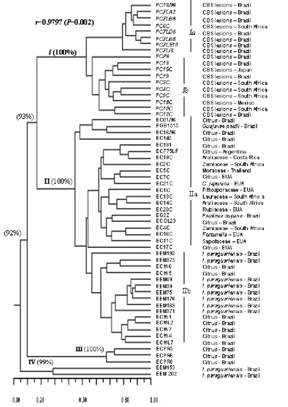
The analysis using RAPD markers allowed for the discrimination of the isolates into four main groups with 5% genetic similarity. The high bootstrap values (P>90%; Fig. 1) confirmed the robustness of the groupings. A Principal Coordinate Analysis
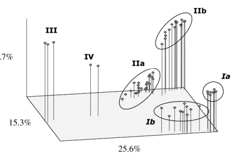
(PCO) based on the genetic similarities estimated with the RAPD markers (Fig. 2) showed high diversity of the isolates of groups I and II, with the clear formation of subgroups that corroborated those observed in Fig. 1.
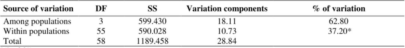
These four groups (I - IV in Figs. 1 and 2) were considered as distinct populations according to the AMOVA, which indicated that 62.8% of the genetic variation was between the populations, although substantial variation was found in populations (37.2% P<0.0001) (Table 3). The high genetic variability observed in and among the groups was surprising, given that all the studied
strains belonged to the genus Guignardia and its
anamorph Phyllosticta. Using the same method, several studies have demonstrated that such low levels of similarity were mainly found among the genera, including the phylogenetically distant ones (Motta et al., 2002; Azevedo et al., 2000; Mehta, 2001).
Table 3 - Analysis of molecular variance (AMOVA).
Source of variation DF SS Variation components % of variation
Among populations 3 599.430 18.11 62.80
Within populations 55 590.028 10.73 37.20*
Total 58 1189.458 28.84
*(P < 0.01)
The low genetic similarity among these groups has also been observed using data from AFLP and ITS sequences. Baayen et al. (2002) suggested the existence of at least three distinct species of Guignardia coexisting in Brazilian citrus plants: G. citricarpa (causing CBS) and two endophytic
species: G. mangiferae (Baayen et al., 2002), and
P. spinarum.
Group I included the G. citricarpa isolated from
the CBS lesions (Fig. 1). The high variability observed in this group did not seem to be associated with the geographical origin of these strains, which were obtained in different continents. Two subgroups could be recognized in this group: Ia and Ib. Subgroup Ia included the
strains isolated in Brazil and one strain isolated in South Africa.
Subgroup Ib was composed of strains obtained in Brazil, Japan, South Africa, and Mexico.
Group II included the isolates of considerable genetic diversity, with broad geographical distribution and considerable host diversity (Table 3). This was shown by the composition of the
subgroup IIa, which included the strains of G.
mangiferae from South Africa, Brazil, Costa Rica, United States, and Thailand obtained from different hosts (Tables 1 and 2). In addition, the
fungus P. capitalensis has already been described
Figure 1 - Dendrogram generated from the genetic similarity matrix obtained using RAPD analysis of the isolates from Tables 1 and 2. Group I: G. citricarpa; Group II: G. mangiferae and
Phyllosticta sp.; Group III: Phyllosticta sp, Group IV: Phyllosticta sp. Bootstrap values
Braz. Arch. Biol. Technol. v.52 n.5: pp. 1063-1073, Sept/Oct 2009
Figure 2 - Principal Coordinate Analysis (PCO) of endophytes based on genetic similarity. Group I: G. citricarpa; Group II: G. mangiferae and Phyllosticta sp.; Group III:
Phyllosticta sp., Group IV: Phyllosticta sp. Ia, Ib, IIa e IIb indicate the subgroups
identified in Figure 1.
Subgroup IIa included all the reference strains of G. mangiferae, six endophytic isolates from
citrus, one endophytic isolate from Paullinia
cupana (Guaraná), and the strain PGB1515 obtained from a lesion in Guajavae psidii (Table
1). The presence of the Guignardia psidii, a
pathogen of the guava tree, in subgroup IIa
indicated that this strain might belong to the G.
mangiferae species and was classified as G.
psidii due to its host plant in another case of synonymy. This reinforced the idea that the same microorganism could behave as an endophyte or as a pathogen depending on the host. This difficulty in establishing the limits between the endophytic and pathogenic species has been underscored by several authors (e.g. Azevedo J.L. et al., 2000; Araújo, et al., 2000). On the other hand, given the high genetic diversity in group II, it was possible that more
than one species of Guignardia similar to G.
mangiferae was endophytic in these plants.
Eight endophytic isolates obtained from Ilex
paraguariensis (erva-mate) in 2000 were
classified in subgroup IIb, as well as seven endophytes isolated from citric plant, all of which in the plants located in the State of Paraná, Brazil. It was noteworthy that although they were obtained from distantly related hosts,
the strains in this subgroup showed genetic similarity higher than those obtained from the same host plants, such as ECMi5 and ECOL23 (Fig. 1). The formation of the subgroup IIb indicated a tendency for grouping strains based on the geographical location of their host plants.
Despite the genetic diversity observed in group II, all the isolates were considered as belonging to the
species G. mangiferae (P. capitalensis). Future
studies including the sequencing of the ITS1-5.8S-ITS2 region of the rDNA might help elucidate this issue.
Group III included three isolates of Phyllosticta sp.
endophytic of C. limon obtained in the municipality of Rio Negro, Paraná, Brazil in 1997. These isolates showed low genetic similarity (<10%) with the remaining isolates. These results not only corroborated the data obtained using other markers by Baayen et al. (2002), but also suggested that these strains could belong to another species of Phyllosticta. Sequencing data (not shown) suggested
that these strains belonged to P. spinarum. The
positioning of these lineages in relation to the remaining groups was clearly shown by the PCO in Fig. 2. Group IV was composed only of two endophytic isolates from Ilex paraguariensis (Figs. 1 and 2), which showed low genetic similarity with the remaining groups (Fig. 1). In addition, they were
25.6%
15.3%
intermediate between group III and the
remaining isolates of G. mangiferae and G.
citricarpa according to the PCO (Fig. 2). Given that it was not possible to identify the species based on the morphological characters alone, the
strain was classified as Phyllosticta sp.
Therefore, there were at least three species that
are morphologically similar to G. citricarpa and
were endophytic in Citrus spp. and Ilex
paraguariensis in Brazil, namely G. mangiferae, P. spinarum and Phyllosticta sp.
It was clear that the isolation from Citrus spp.
that were morphologically similar to G.
citricarpa did not necessarily meant that the host was contaminated with the causal agent of CBS. The identification using the morphological and physiological characters was imprecise and time-consuming, being inadequate for the analysis of fruits to export (Baayen et al., 2002). Thus, it was necessary to develop diagnostic methods that would be rapid and efficient (Bonants et al., 2003).
Results also underscore the efficiency of RAPD markers, not only in the studies of population structure and in the assessment of genetic variability in fungus populations, but also to suggest the presence of distinct species of a given genus in the surveys of the endophytic community of the studied hosts.
Development of Specific PCR Primers
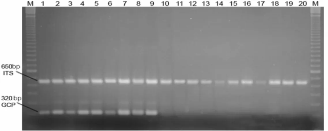
The strains in Tables 1 and 2 were tested using PCRs with the GCP1/GCP2 primer pair. Only strains in Group I (Fig. 1) showed the amplification of a band of approximately 350 bp (Fig. 3).
The primers ITS1 and ITS4 were used as positive controls of the amplification (White et al., 1990). These primers are universal for fungi and allow for the amplification of the ITS1 –5.8S – ITS2 region of the rDNA. All the strains amplified an approximately 650 bp fragment (Fig. 3), which was consistent with the expected fragment sizes ca. 700 bp for ascomycete (White et al., 1990).
Figure 3 - Agarose gel electrophoresis of PCR using the primer pairs GCP1/GCP2 and ITS1/ITS4, and isolates of G. citricarpa and G. mangiferae. Legend: M: 100 bp. DNA Ladder. 1:
Reference strain of G. citricarpa PC28/05 (Table 1); 2 - 9: G. citricarpa (Table 2); 10:
Reference strain of G. mangiferae EC22/05 (Table 1); 11 - 20: G. mangiferae (Table 2).
Therefore, the isolates of the pathogenic fungus G. citricarpa obtained from several different countries could be discriminated from the
endophytic strains of Guignardia spp. isolated
from Citrus spp. using the GCP1 and GCP2
primers. The correct assignment of the strains of
Braz. Arch. Biol. Technol. v.52 n.5: pp. 1063-1073, Sept/Oct 2009
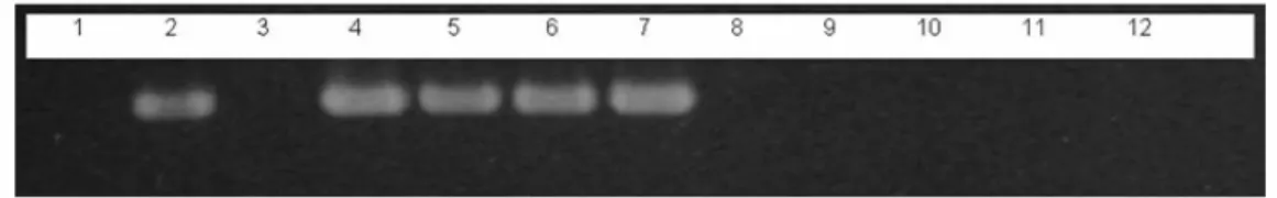
Figure 4 - Agarose gel electrophoresis of PCR using the primer pair GCP1/GCP2 and isolates of
G. citricarpa, G. mangiferae and Phyllosticta sp. 1: Negative control without DNA; 2:
pGEM-T: plasmid containing the 373bp cloned fragment; 3: P. spinarum (ECPR05); 4
- 6: Reference strains of G. citricarpa (PC28/05, PC32/05, PC33/05); 7: Strain
PC13/96 of G. citricarpa; 8 - 10: Reference strains of G. mangiferae (EC 01/05,
EC19/05, EC22/05); 11: Negative control: Colletotrichum gloeosporioides; 12:
Negative control: DNA of citrus leaves.
CONCLUSIONS
The high genetic variability observed among the
groups obtained using RAPD markers
corroborated previous studies (Baayen et al., 2002; Rodrigues et al., 2004) with respect to the existence of at least three distinct species of
Guignardia and its anamorph Phyllosticta that
were very similar morphologically to the pathogen
G. citricarpa, coexisting in Citrus spp. and
hampering the identification of the plants and fruits free from the causal agent of CBS.
The use of RAPD markers allowed for the
classification of isolates into G. citricarpa, G.
mangiferae and Phyllosticta sp.. The fact that most endophytic isolates from different hosts and
regions could be grouped as belonging to G.
mangiferae confirmed that a single fungus species could endophytically colonize several host species. In addition, the high genetic similarity between the
strain of G. psidii, a guava pathogen, with the
strains of G. mangiferae indicated that the same
microorganism might be found acting as an endophyte in some plants and as a pathogen in
others. In other words, G. mangiferae could
colonize certain types of the plants without causing symptoms of CBS (citric plants and other hosts) and became pathogenic in other plant species (guava trees and orchids).
The GPC1/GCP2 was efficient in the identification
of G. citricarpa as the causal agent of CBS
through PCR.
ACKNOWLEDGEMENTS
We thank Dr. Carlos Aguilar-Vildoso (Instituto Agronômico de Campinas/Brazil) and Dr. Antonio
de Goes (UNESP/Jaboticabal, SP - Brazil) for providing some of the fungal strains, and the Brazilian agencies CNPq and FAPESP for financial support.
RESUMO
variação substancial foi encontrada dentro destas populações (37,2%). Bandas de RAPD exclusivas de isolados de G. citricarpa foram clonadas, sequenciadas e utilizadas na obtenção de SCARS (Sequence Characterized Amplified Regions), que permitiram o desenvolvimento de novos primers específicos para a identificação de G. citricarpa. Reações de PCR (Polymerase Chain Reaction) utilizando este par de primers corroboraram os agrupamentos obtidos pelos marcadores de RAPD, revelando sua eficiência na identificação do agente causal da CBS.
REFERENCES
Agrianual 2005: Anuário da Agricultura Brasileira. São Paulo: FNP 2005, p.785.
Araújo, J., Silva, A.C., Azevedo, J.L. (2000), Isolation of endophytic actinomycetes from roots and leaves of maize (Zea mays L.). Braz. Arch. Biol. Technol, 43 (4), 447-451.
Azevedo, J.L. and Costa, S. O. P. (1973), Exercícios
práticos de genética. São Paulo: Nacional / EDUSP.
288p.
Azevedo, J.L., Maccheroni Jr., W., Pereira, J.O., Araújo, W.L. (2000), Endophytic microorganisms: a review on insect control and recent advances on tropical plants. Electr. J. Biotecnol., 3, 40-65.
Azevedo, A.C.S., Furlaneto, M.C., Sosa-Gómez, D.R., Fungaro, M.H.P. (2000), Molecular characterization
of Paecilomyces fumoroseus (Deuteromycotina:
Hyphomycetes) isolates. Scientia Agrícola, 57,
729-732.
Baayen, R.P., Bonants, P.J.M., Verkley, G., Carroll, G.C., van der Aa, H.A., de Weerdt, M., van Brouwershaven, I.R., Schutte, G.C., Maccheroni, W., Glienke-Blanco, C., Azevedo, J.L. (2002), Nonpathogenic isolates of the citrus black spot fungus, Guignardia citricarpa, identified as a
cosmopolitan endophyte of woody plants, Guignardia
mangiferae (Phyllosticta capitalensis). Phytopathol.,
92, 464-477.
Baldassari, R.B. (2005), Patogenicidade, morfologia de colônias e diversidade de isolados de Guignardia
citricarpa e G. mangiferae obtidos de Citrus spp.
PhD Thesis, Faculdade de Ciências Agrárias e Veterinárias, Universidade Estadual Paulista, Jaboticabal, São Paulo, Brasil.
Blanco, C.G. (1999), Guignardia citricarpa Kiely:
Análise genética, cariotípica e interação com o hospedeiro. PhD Thesis, Piracicaba, Escola Superior de Agricultura Luiz de Queiroz, Universidade de São Paulo, São Paulo, Brasil.
Bonants, P.J.M., Carroll, G.C., Weerdt, M., van Brouwershaven, I.R., Baayen, R.P. (2003), Development and validation of a fast PCR-based detection method for pathogenic isolates of the citrus black spot fungus, Guignardia citricarpa. European
J. Plant Pathol., 109, 503-513.
Coelho, A.S.G. (2005), Avaliação de dendrogramas baseados em estimativas de distâncias/similaridades genéticas através do procedimento de bootstrap. Software, V. 3.03. Universidade Federal de Goiás, Goiânia, Brasil.
Excoffier, L., Laval, G., Schneider, S. (2005), Arlequin ver. 3.0: An integrated software package for
population genetics data analysis. Evolutionary
Bioinformatics Online, 1, 47-50.
Feichtenberger, E. (1996), Mancha preta dos citros no Estado de São Paulo. Laranja, 17, 93-108.
Felsenstein, J. (1985). Confidence limits on phylogenies: an approach using the bootstrap.
Evolution, 39, p. 366-369.
Glienke-Blanco, C., Aguilar-Vildoso, C.I., Vieira, M.L.C., Barroso, P.A.V., Azevedo, J.L. (2002), Genetic variability in the endophytic fungus
Guignardia citricarpa isolated from citrus plants.
Genet. Molec. Biology, 25, 251-255.
Mantel N.A. (1967), The detection of disease clustering and a generalized regression approach. Cancer Res.,
27, 209-220.
Mehta, Y.R. (2001), Genetic diversity among isolates of
Stemphylium solani from Cotton. Fitopatol.
Brasileira, 23, 703-709.
Motta, T.R., Moreira-Filho, C.A., Mendes, R.P., Souza., L.R., Sugizak, M.F., Baueb, S., Calich, V.L.G., Vaz, C.A.C. (2002), Evaluation of DNA polymorphisms amplified by arbitrary primers (RAPD) as genetically associated elements to differentiate virulent and non-virulent
Paracoccidioides brasiliensis isolates. FEMS Immun.
Medical Microb., 33, 151-235.
Okane, I., Nakagiri, A., Ito, T. (2001), Identity of
Guignardia sp. inhabiting ericaceous plants. Can. J.
Bot., 79, 101-109.
Penna, E.B., Azevedo, J.L., Glienke, C., Fernandes, J.S.C. (2000), Variabilidade Genética do endofítico
Phyllosticta sp isolado de 5 populações de erva-mate
(Ilex paraguariensis st. Hill).. In: V Encontro
Paranaense de Genética, Maringá, PR, Ed. Universidade Estadual de Maringá, 5, 41.
Pimentel, I.C., Glienke-Blanco, C., Gabardo, J., Stuart, R.M., Azevedo, J.L. (2006), Identification and colonization of endophytic fungi from soybean
(Glycine max (L.) Merril) under different
environmental conditions. Braz. Arch. Biol. Technol.,
Braz. Arch. Biol. Technol. v.52 n.5: pp. 1063-1073, Sept/Oct 2009 Raeder, U. and Broda, P. (1985), Rapid preparation of
DNA from filamentous fungi. Lett. Appl. Microb., 1,
17-20.
Rodrigues, K.F., Sieber, T.N., Grünig, C.R., Holdenrieder, O. (2004), Characterization of
Guignardia mangiferae isolated from tropical plants
based on morphology, ISSR-PCR amplifications and ITS1-5.8S-ITS2 sequences. Mycol. Res., 108, 45-52.
Rohlf, F.J. (2000), NTSYS-PC Numerical taxonomy and
multivariate analysis system. Exeter Software, New
York.
Sneath, P.H. and Sokal, R.R. (1973), Numerical
taxonomy. Freeman, San Francisco, 573p.
Stringari, D. (2004), Estudo da Variabilidade Genética
de Guignardia spp por Meio de Marcadores de
RAPD e Sequências ITS. Ms Sc Thesis, Universidade Federal do Paraná, Curitiba, Paraná, Brasil.
White, T.M., Bruns, T., Lee, S., Taylor, J. (1990), Amplification and direct sequencing of fungal ribosomal RNA for phylogenetics. In: Innis, M.A.,
Gelfand, D.H., Sninsky, J.J., White, T.J. (Eds.). PCR
protocols: a guide to methods and applications, pp.
315-321.