University of Évora
ARCHMAT
(ERASMUS MUNDUS MASTER IN ARCHaeological MATerials Science
)
Mestrado em Arqueologia e Ambiente (Erasmus Mundus – ARCHMAT)
Interdisciplinary study of the dental calculus in skeleton remains from the
cemetery of Santa Maria do Olival (Tomar, Portugal) - 15th to 16th century A.D.
Roshan Paladugu m38416
Anne-France Maurer
(Supervisor - Universidade de Évora) Cristina Maria Barrocas Dias
(Co-supervisor– Universidade de Évora)
Évora, September 2018
University of Évora
ARCHMAT
(ERASMUS MUNDUS MASTER IN ARCHaeological MATerials Science
)
Roshan Paladugu m38416
A R I S T O T L E UNIVERSITY OF THESSALONIKI
Mestrado em Arqueologia e Ambiente (Erasmus Mundus – ARCHMAT)
Anne-France Maurer
(Supervisor - Universidade de Évora) Cristina Maria Barrocas Dias
(Co-supervisor– Universidade de Évora)
O estudo interdisciplinar do cálculo dentário no esqueleto é feito no cemitério de Santa Maria do Olival (Tomar, Portugal) - 15th to 16th século A.D.
Évora, September 2018
Panel of Jury
Dr.Nicola Schiavon (Presidente)
Dr. Ana Cristina Cabaça Manhita (Arguente)
Dr. Anne-France Maurer (Orientadora)
Contents
List of Abbreviations and Terms ... i
List of Figures ... ii
List of Tables ... iii
Abstract ... iv Resumo ... v Acknowledgements ... vi 1. Introduction ... 1 2. Historical Context ... 2 2.1 Tomar as Sellium ... 2 2.2 Visgothic period ... 3 2.3 Paleochristianity ... 3 2.4 Islamic Thomar ... 4
2.5 Reconquista and Order of the Christ ... 4
3. Archaeological Context ... 6
3.1 Necropolis of Santa Maria do Olival ... 6
3.2 Excavation Areas ... 9
4. Diet in Medieval Portugal ... 11
5. Scientific Approach ... 17 5.1 Dental Calculus ... 17 5.1.1 Aetiology ... 17 5.1.2 Archaeological Importance ... 19 5.1.3 Debris ... 20 5.1.4 Incorporation of Debris ... 22 5.2 Microremains ... 23 5.2.1 Phytoliths ... 23 5.2.2 Starch Granules ... 25
5.2.3 Phytoliths and Starches in Dental Calculus ... 26
5.3 Role of Stable Isotopes in Dietary Reconstruction ... 27
5.3.1 Stable Carbon Isotopes ... 28
5.3.2 Stable Nitrogen Isotopes ... 29
5.4 Organic Residues ... 31
5.4.1 Organic Residues in Dental Calculus ... 31
5.5 Trace Elements in Dietary Studies ... 33
5.5.1 Trace Elements in Bones, Teeth and Dental Calculus ... 34
6. Materials and Methods ... 35
6.1 Dental Calculus Sampling ... 35
6.2 Anthropological Information ... 37
6.3 Evaluation and Documentation of Dental Calculus ... 39
6.4 Fourier Transform - Infrared Spectroscopy ... 40
6.5 Demineralisation and Extraction of Microparticles ... 41
6.6 Elemental Analysis – Isotopic Ratio Mass Spectrometry ... 43
6.7 LA – ICP – MS Analysis... 44
6.8 Pyrolysis – Gas Chromatography – Mass Spectrometry ... 46
7. Results and Discussion ... 48
7.1 Structure of the calculus matrix ... 48
7.2 Micro-Debris ... 49
7.3 Carbon and Nitrogen Stable Isotopes in Calculus and Collagen ... 56
7.4 Trace Element Analysis using LA – ICP – MS ... 59
7.5 Biomolecular Analysis using Py – GC – MS... 63
7.6 Implications in Diet ... 73
8. Conclusions ... 74
Bibliography ... 75
Appendix I ……… 88
i
List of Abbreviations and Terms
AD: Anno Domini
AIR: Ambient Inhalable Reservoir
Am/P: Amide I to phosphate ratio (relative collagen content)
ATR-FTIR: Attenuated Total Reflection- Fourier Transmission Infrared Spectroscopy Ba/Ca: Barium to calcium ratio
Ba/Sr: Barium to strontium ratio BCE: Before Common Era C/P: Carbonate to phosphate ratio Ca/P: Calcium to phosphorus index CAM: Crassulacean Acid Metabolism
EA-IRMS: Elemental Analyser- Isotope Ratio Mass Spectrometry IAEA: International Atomic Energy Agency
ICP-MS: Inductively Coupled Plasma- Mass Spectrometry IR-SF: Infrared Splitting Factor
I1: First incisor
LA-ICP-MS: Laser Ablation – Inductively Coupled Plasma- Mass Spectrometry M1: First permanent molar
M2: Second permanent molar mL: Milliliters
NIST: National Institute of Standards and Technology Nm: Nanometer
Ppm: Parts per million REE: Rare earth elements Rpm: Revolutions per minute Sr/Ca: Strontium to calcium ratio SRM: Standard reference materials VPDB: Vienna Pee Dee Belemnite XRD: X-ray Diffraction
ii
List of Figures
Figure 1. Map of the Iberian Peninsula showing the location of Tomar. ... 2
Figure 2. The facade of Santa Maria do Olival, Tomar (DGMN). ... 3
Figure 3. An official document showing the area under the church of Santa Maria do Olival, DGMN Portaria publicada 259. ... 6
Figure 4: Excavation Map showing various areas of the necropolis ... 8
Figure 5. Birds which were cited in medieval Portuguese literature. ... 13
Figure 6. Geo-political situation during the Reconquista period. ... 14
Figure 7. Oral pH and pathology associated with it. ... 18
Figure 8. International Phytolith Nomenclature. ... 24
Figure 9. Starch grain morphology with hilum and maltese cross. ... 25
Figure 10. Carbon and nitrogen isotope fractionation. ... 28
Figure 11. Dental calculus before and after sampling ... 35
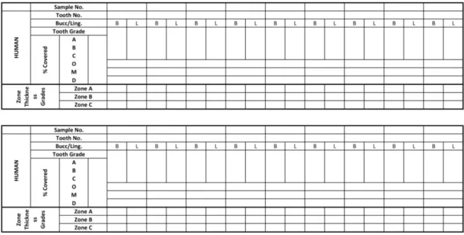
Figure 12. Dobney-Brothwell Human Calculus Evaluation Proforma. ... 39
Figure 13. Infrared Spectra of dental calculus, (Le Geros, 2015). ... 49
Figure 14. Micro-debris from modern sample E2. ... 50
Figure 15. Modern human sample E2 analysed using protocols of Dudgeon (A), Lazzati (B), Tromp (C) and Price (D). ... 51
Figure 16. Aerial particulate material in the laboratory. ... 52
Figure 17. Faunal sample investigated using Hardy et al., 2016 protocol. ... 53
Figure 18. Phytoliths from archaeological sample T2. ... 54
Figure 19. Phytoliths from individual T6 ... 54
Figure 20. A cuneiform bulliform phytolith observed under SEM from individual T2. ... 55
Figure 21. δ13C values of dental calculus and bone collagen comparison. ... 56
Figure 22. δ15N values of dental calculus and bone collagen. ... 57
Figure 23. Ca/P ratios of dental calculus. ... 59
Figure 24. Vanadium and Uranium concentrations in dental calculus samples. ... 60
Figure 25. Sr/Ca and Ba/Ca ratios of dental calculus. ... 61
Figure 26. log (Ba/Sr) ratios of dental calculus. ... 62
Figure 27. Cyclisation reaction of amino acids. ... 65
Figure 28. Side functional group loss of phenylalanine, tyrosine and tryptophan. ... 66
Figure 29. Ring condensation reaction of pyridinic-N. ... 66
Figure 30. NH2*free radical group release and formation of other intermediates ... 66
Figure 31. Amide formation due to fatty acid reaction with amine free radicals or ammonia ... 67
Figure 32. Pyrogram of cleaned sample T9.1 ... 68
Figure 33. Pyrogram of cleaned sample T9.2 ... 69
Figure 34. Pyrogram of uncleaned sample T9.1. ... 70
Figure 35. Pyrogram of uncleaned sample T5.2. ... 71
Figure 36. Pyrogram of sample T5 derivatised using TMAH ... 71
iii
List of Tables
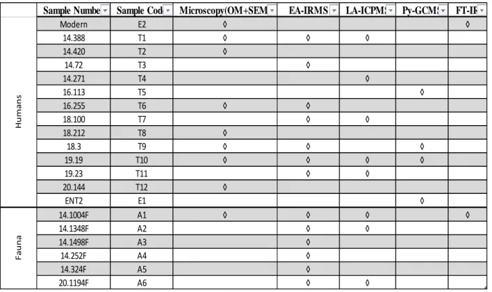
Table 1. List of samples with respective sampled teeth. ... 36
Table 2. List of samples with the techniques applied on them. ... 37
Table 3. List of selected calculus affected individuals ... 38
Table 4. List of fauna chosen for the study ... 38
Table 5. Selected Protocols for Optical Microscopy Investigation. ... 42
Table 6. Standards utilised for measuring isotopic ratios of dental calculus. ... 44
Table 7. Ablation parameters of dental calculus ... 45
Table 8. Parameters of pyrolysis. ... 47
iv
Abstract
14 humans and 6 faunal dental calculus samples from the medieval necropolis of Santa Maria do Olival, Tomar were selected for microdebris, isotopes, trace elements and organic residues analyses. The major aim of the study was to setup methods for analysis of dental calculus to complement information from bones and teeth. The isotopic values from dental calculi of humans and fauna were comparable to those from bone collagen and led to similar conclusions on dietary practices. Trace element values from human and faunal calculi followed similar trends to their respective bones. Microscopy yielded stray phytoliths from the Poaceae family and no silica skeletons or starch grains. Organic residues analysis indicated a diet based on protein and carbohydrates. Evidences of poor air quality due to char substances from incomplete combustion of wood and biomass were found. Since this is a pilot study, sample size is restricted. The direction of future research on dental calculus is to analyse a large quantity of samples to generate standard range of values for isotopes and trace element concentrations.
Keywords: dental calculus, phytoliths, starch grains, microscopy, trace elements, organic
residues, Optical Microscope (OM), Scanning Electron Microscope (SEM), EA-IRMS, LA-ICP-MS, Py-GC-LA-ICP-MS, Tomar.
Resumo
O estudo interdisciplinar do cálculo dentário no esqueleto é feito no
cemitério de Santa Maria do Olival (Tomar, Portugal) - 15th to 16th século
A.D.
Foram selecionadas 14 amostras de humanos e 6 amostras de cálculo dentário da fauna da necrópole medieval de Santa Maria do Olival, Tomar, para análises de microdíbris, isótopos, oligoelementos e resíduos orgânicos. O principal objetivo do estudo foi configurar métodos para análise de cálculo dentário para complementar informações de ossos e dentes. Os valores isotópicos dos cálculos dentários de humanos e fauna foram comparáveis aos do colágeno ósseo e levaram a conclusões semelhantes sobre práticas alimentares. Os valores dos elementos de traço dos cálculos humanos e faunísticos seguiram tendências semelhantes aos seus respectivos ossos. A microscopia produziu fitólitos dispersos da família Poaceae e nenhum esqueleto de sílica ou grãos de amido. A análise de resíduos orgânicos indicou uma dieta baseada em proteínas e carboidratos. Evidências de baixa qualidade do ar devido a substâncias carbonatadas de combustão incompleta de madeira e biomassa foram encontradas. Como este é um estudo piloto, o tamanho da amostra é restrito. A direção de pesquisas futuras sobre cálculo dentário é analisar uma grande quantidade de amostras para gerar uma faixa padrão de valores para isótopos e concentrações de elementos traços.
vi
Acknowledgements
I have enjoyed the challenging and innovative nature of thesis especially during the interpretation of the results. I have had the great fortune of working with many dedicated and experienced people. I would like to express my heartfelt gratitude to my supervisor Anne France-Maurer and co-supervisor Prof. Cristina Barrocas Dias for all the support and guidance they have provided during the period of this study. I would like to thank Ana Margarida Cardoso for her help with FTIR, Pedro Barrulas for all his chemistry pointers and help in trace element analysis, Ana Manhita for helping me run pyrolysis gas chromatography mass spectrometry, Nicasio Tomás Jiménez-Morillo for his help with stable isotope analysis and Tania Rosado for her kind help with microscopy. A special mention to Guillermo Marin Garcia for sparing his time to help me with phytolith identification.
I would like to dedicate a big thank you to Silvia Irene Russo for her constant constructive criticism and company during the entire duration of this thesis. I would also like to thank Sergio Lins, my dear friend for helping me with the Portuguese translation of the abstract. Finally, I would like to thank my parents for providing me with the great opportunity of studying in this masters.
1
1. Introduction
Dental calculus is an emerging source of ancient diet and environment information. The aim of the study was to setup method and to gauge the scope of dental calculus as a complement for teeth and bones in dietary reconstruction. Comparing the isotopic values from dental calculus to the already established values from bones will help in evaluation of the reliability of calculus. The skeletal remains from the necropolis of Santa Maria do Olival gave an excellent opportunity to investigate the potential of calculus. The city of Tomar has constantly evolved through time evolving from one cultural period to another right from Romans, followed by Visigothic and Paleochristian periods and then the brief Islamic period, culminating with the Reconquista establishing Christianity as the major religion. Each cultural period had its own distinctive dietary patterns due to economic and political situations as well as climatic conditions. This provides an excellent testing grounds to see if dental calculus can be relied upon to reconstruct the past diets.
The buried remains from the necropolis showed a relatively large frequency of dental calculi in individuals. Microscopy was carried out to identify biogenic silica such as phytoliths and diatoms as well as starch grains. Phytoliths and starch grains can be utilised to uniquely identify the plant of origin. Trace element studies are also carried out as there are no standard published values for the concentration ranges. Finally, pyrolysis-chromatography mass spectrometry was utilised to understand the organic constituents of the dental calculus.
2
2. Historical Context
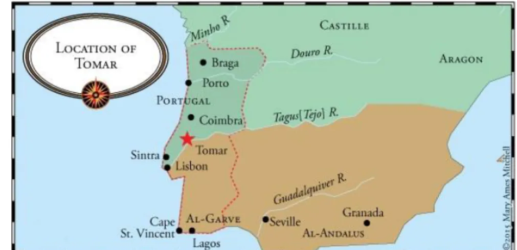
To the north of Lisbon and just below Coimbra, lies Tomar where the church of Santa Maria do Olival is located. The Nabão River, passing in the west, has played the role of a lifeline throughout the human settlement history in the area. There have been neolithic and Iron Age remains discovered in previous excavations (Santos, 2009). The location of the settlement is not based on strategy of military defense or natural resources. Instead, there have been two hypothesis proposed for explaining this fact. The first one is that the Roman city of Sellium was based on the existence of an earlier settlement and the second being that Sellium was built in the beginning of the Empire, as a part of Romanization of Lusitânia where modern day Tomar stands (Figure 1) (Pereira et al., 2009).
2.1 Tomar as Sellium
During the Roman period, Tomar was known as Sellium. It was a part of urbanization reforms carried out by Emperor Augustus leading to establishment and elevation of cities to civitates. Sellium was amongst the ranks of others such as Ammaia, Aritium Vetus, and Eburobritium which were located non-strategically along waterbodies, in this case Nabão River. However what puts Sellium apart from the others is that it served as caput viarium of the route of via Olissipo-Bracara Augusta. The movement of goods was primarily to the north of Sellium on this route. The city further developed due to its centrality with respect to land trade routes and proximity to the Tagus River. In all possible scenarios, the demography was indigenous and homogenous with cultural practices rooted in Lusitânia. In a stark contrast, very little was recorded in historical records about Sellium from the Roman times apart from the existence of a forum amongst many other typical of Roman cities. The tower of Church of Santa Maria do Olival itself, is hypothesized to be built on
3
the foundations of a pre-existing Roman mausoleum after an excavation (Santos, 2009). The construction of the structure was built with limestone slabs bound by mortar, aligned to Roman construction techniques and in sync with structures from Roman town of Sellium.
2.2 Visgothic period
The first migrant populations arrived in the form of Gothic contingents such as Suebi, Alans, Asding Vandala and Siling Vandala etc. to the Iberian Peninsula around 406 A.D. This migration generated instability and metamorphosis in the structure of local cities. This change was mainly invasion and mass destruction due to the disorganized martial populations causing deep damage in the societal structure mainly administrative, social and economic development. The local elite families faced the scenario where their political and socio-economic positions were threatened due to recurrent plundering and conquest of strategic cities. The urban center of Sellium was related to the Germanic tribes since it was located between borders of Suebi and Visigoth territories. The centralization of the Visigothic monarchy in the second half of the 6th century culminated in a civil
war (Batata, 1997). 2.3 Paleochristianity
The Paleochristian mention of Sellium appears in the Parochiale Suevicum in the year 569 A.D. as a church in the diocese of Coimbra and subordinate to the episcopal seat of Braga (Ponte, 1997). In the early 6th century, structures related to paleochristianity have been discovered (Fabião, 2004).
4
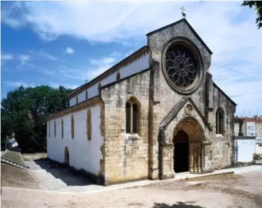
The earliest structures are a set of buildings which served as the parish headquarters of Santa Maria do Selhos and other smaller buildings related to Santa Iria and São Pero de Fins. The location of Santa Maria do Selho is highly suspected to be in the same location of Santa Maria do Olival (Figure 2). The Church of Santa Iria still exists under the same name. The Church of São Pero de Fins seemed to exist between these two buildings, in the current area of Tomar cemetery (Ponte & Miranda, 1991).
2.4 Islamic Tomar
The conflict between the religious authorities and Visigothic monarchy lead to the benefit of the Islamic invader. This is explained by the monumental expansion of the Tariq Ibn Ziyad army in 711. Surprisingly there are no archaeological evidences apart from a single coin in the excavation (Ponte & Miranda, 1991). The Islamic population must have had little artistic and materialistic expressions and probable dispersion in the territory. During this period, the term Sellium was most likely replaced by Thomar, derived from the Islamic name of the Nabão River.
2.5 Reconquista and Order of the Christ
The Reconquista movement provoked by the Christian resistance moved the strategy to fortifications which has never been the specialty of Tomar. The left bank and a part of old Sellium were abandoned and on the opposite bank the Islamic rulers constructed a new fortified stronghold. In the bloody conflict of the Christian North and Muslim South, the borders were constantly redrawn based on the outcomes and flow of military campaigns. The 11th and 12th centuries were
marked by a profound religious intolerance by both Christians and Muslims alike. The initiation of the Portuguese nationality is marked by the religious obligation of the war towards the South against the Muslims. In this scenarios, the religious orders began playing a larger role in military campaigns. The Templar Order of Portugal was given charge of the territories of Zêzere and Tejo. In 1162, the first charter of Tomar was proclaimed by D.Gualdim Pais. On the legal basis of the charter, D.Gualdim Pais chose to undertake the task of building a fortified defensive structure of the Castle of Tomar starting from 1160. Two other structures were also being built along with the defensive constructions which were the Church of Santa Maria do Castelo and Charola. On the other bank of the river, the Church of Santa Maria do Olival was being constructed and developed as the core religious nucleus of religious activity. As the Reconquista progressed and the Islamic territory was pushed even south, urbanization of the settlement started with area around the castle
5
leading the metamorphosis. Commercial activities started gaining pace and attracted more immigrants from different regions to settle around the castle. The location of the church is in the same space as some Paleochristian structures of Sellium. This is supported by the fact that the monastery of Santa Maria do Selho was later designated as Santa Maria do Olival in medieval documentation. The church was most likely reformed under the orders of the Grand Master. From the previous structure only the Gothic façade was preserved with other elements altered to suit the era. The administration of the convent and pantheon passed from the hands of the Templar Order to the order of the Christ in 1307 after the dissolution of the former under the papal bull Pastoralis
praeeminentiae issued by Pope Clement V. The Church of Santa Maria do Olival played an
important role in the field of architecture as it served as a template for other churches built or renovated during the reign of D. Manuel I which concluded in the reign of D.João III (DGEMN, 1942:11).
The Church of Santa Maria do Olival was granted the rank of a Cathedral and Diocese by Pope Calisto III in 1455. In the reign of D. João III, a major reform of the religious order took place where the Order of Christ was subjected to the rigorous rules of cloister and the restrictions of the life in a convent were implemented strictly. The space surrounding the church was partitioned between different properties of the Order of Christ. Many other churches dedicated to St. Michael. St. Peter the Apostle, St. Mary Magdalena and Santo Ildefonso were demolished. An epigraph dating to 1175 reveals that the church was open to worship and burials were also permitted. The Directorate General of National Buildings and Monuments (DGEMN) in 1940s dismantled the ruins which were located around the church.
6
3. Archaeological Context
3.1 Necropolis of Santa Maria do Olival
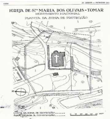
The focus here is the morphing of landscape in the context of the “old cemetery” due to the remodeling works of DGEMN and the construction of access roads to the Municipal Market and Marmelais Road. This funeral area surrounding the Church of Santa Maria do Olival has undergone several changes through the flow of time. The presumption that until the first half of the 19th century burials were made inside the church makes the origin of this funerary space difficult to explain. The burials during the Roman-Visgothic period were located in the immediate space to the structure of the church, more precisely in the churchyards (Santos, 2009). Archaeological interventions in the area began in 1977 after the discovery of tombstones and funerary stelae by Beleza Moreira (Batata, 1997). Major excavations made between 1989 and 1992 by Salete Ponte shed light on the spacio-temporal evolution of the necropolis (Ponte & Miranda, 1991). Ponte also states that the space surrounding the church was converted during the Gothic - Sellio period in to a scared area for praying and looking after the dead which is shown in Figure 3 (Santos, 2009). By the 12th century, the area has become a fixed space for religious activities. Archaeological evidence suggests that the space continues to be used for similar activities until the
Figure 3. An official document showing the area under the church of Santa Maria do Olival, DGMN Portaria publicada 259.
7
end of 16th century. The stratigraphic units documented reveals the specific periods of history when the necropolis was used by the people in the Middle Ages. A part of the exhumed burials were assigned in the chronological period based on the burial type and stratigraphic units. Other major methodology was to identify the typology of burial goods associated with specific exhumations (Ponte, 1997:52). The lowest stratigraphic units which are also chronologically the oldest were dated to 5th-12th centuries which is between the Visgothic and Reconquista. These layers were followed by burials dating to post-Reconquista period. Mass burials were attributed to a period of 300 years where severe famines, bubonic plague and wars were common (Santos, 2009).
To this date the origins of this area are still shrouded in mystery. Under the Romans it was suburban space but later, it evolved to become an urban space of Tomar. The space which has remained constant is the necropolis and its constant role of being a space to honour the dead. Since the early 12th century the space has stood isolated but over the time has started shrinking in size as the space began to be reused. The cemetery of Santa Maria do Olival continues to be sacred sanctum for people’s belief in burying their dead.
8
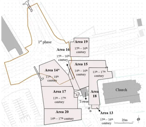
The necropolis of the Church of Santa Maria do Olival was used by the population between the 11th and 18th centuries. The first excavation took place in 2007 and covered a part of the necropolis
which was divided into 11 areas whereas the second phase of the excavations took place in 2008 and covered 8 areas (13-20). Dental calculus is a part of the skeletal artefacts excavated in the Phase-II of archaeological interventions carried out in the area surrounding the church of Santa Maria dos Olivais in Tomar, district of Santarém, under the umbrella of urban requalification and environmental valuation of the Polis program. The interventions were mainly aimed at reducing the negative impact of the urban activities on the necropolis context. The unique characteristic of the site is the rich amount of archaeological remains found in all the numerous previous excavations. All these investigations have contributed to understanding the space of the dead i.e. the necropolis in this area of Tomar. This includes the death rituals starting from the Roman settlement of Sellium to the reconquered city of Tomar until the 18th century. The excavations lasted for 11 months beginning in April 2008 lasting until March 2009. The total excavation area is 4029.672 m2 divided into 8 areas and 6 trenches as shown in Figure 4. The excavation was started in the Area 20 owing to emergency of building a platform access ramp.
9
3.2 Excavation Areas
Area 13 which was also excavated in an emergency is located to the south of the bell tower and Area 18. Sixty stratigraphic units, 15 burials, 6 ossuaries associated with burial and 1 isolated ossage were found. The burials were mostly pits excavated in the geological substrate. Many of the structures have been found violated leading to the belief that there has been reuse of the space for later inhumations. In regard of chronology of the area, it can be postulated that it approaches from late – Roman to upper medieval period. This is supported by the presence of structures oriented east to west, made of limestone and bound by clay which is characteristic of late – Roman period and for the upper medieval period, a single coin from the reign of Afonso V was found in a burial.
Area 14 is located to the north of Area 17 covering an area of 777.82 m2. A sum total of 436 burials and 128 remains were recovered. The burials were found either in soil or in stone graves. Many of the recovered graves were Roman in context, oriented south to northeast (head to feet). Extensive overlapping has made determination of morphology of many pits very difficult. The chronology of the burials ranges from late Roman to Modern age through lower medieval age, which is indicated by burial goods such as coins, copper pins etc. typology.
Area 15 is located north of the Santa Maria do Olivais’s bell tower, between Area 16 and Area 18 covering an area of 641.28 m2. A large collection of remains numbering 235 burials, 145 partial remains associated with burials and 25 isolated bones were recovered. A vast majority of the burials were violated, as only 30 out of 235 skeletons were intact. The chronology of the burials date from the reign of King John I to the reign of Dom.Sebastião. However, the oldest archaeological material dates back to the late Roman period but are not associated with any burial. Area 16 is located between Area 14 and Area 17 to the west and Area 15 to the east covering an area of 121.38 m2. The area revealed an exceptional concentration of burials. A total number of 273 burials, 97 remains associated with burials and 12 isolated burials. The burials were in a position of “dorsal decubitus”, with the head facing west or southwest grave. The chronology of the burials date from the reign of Sancho I to the reign of Dom.António.
Area 17 is located to the north of Area 20, in front of the Santa Maria do Olivais church covering an area of 850,986 m2. A total of 220 burials were recovered from the area with the burials in
10
“dorsal decubitus” position, with the head towards the south. The chronology of burials range between 13th to 17th centuries.
Area 18 is located to the northwest of the Santa Maria do Olivais church covering an area of 570.33 m2. A total of 342 burials, 188 burial related bones and 29 isolated bones were recovered. The burials were in a position of “dorsal decubitus”, with the head towards the southwest or west and facing upwards. In this area there is a predominance of burials in relation to the other areas, with stones, mortars and masonry involved indicating work and monetary investment. This might have to do with the proximity of Santa Maria do Olivais church and the socioeconomic status of the deceased. The chronology of burials ranges from the reign of Dom.Sancho I until the reign of Dom.João IV.
Area 19 is located to the north of the church of Santa Maria dos Olivais and Area 15 with the current cemetery as the northern boundary. A total of 80 burials, 54 osteological remains associated with burials and 10 isolated bones were exhumed from this area. The chronology of the area seems to be between 15th to 16th centuries. However, further studies are required to precisely elucidate the limits of usage of the space.
Area 20 is located in the southern region to the side entrance of the Church of Santa Maria do Olivais with an area of 589.29 m2. The burials are mostly oriented from south to north east direction. The chronology of the area ranges from the 14th to 17th centuries based on the burial goods recovered.
The samples were chosen from all the areas except Area 17 as equally as the sampling strategy allowed to have a representation of all the periods of the necropolis. Area 17 could not be represented as no individual had enough dental calculus to perform multiple analyses.
11
4. Diet in Medieval Portugal
It is essential to understand the variety of foods being consumed as diet is an aspect which abstrusely reflects the economic, political and social order of human society. To understand better the scientific data of the study, it is crucial to collect the contextual specificity and maintain the information about the dietary patterns practiced in Medieval Portugal. The synergy of food with spatiality and time are of the prime focus.
The Roman Empire is the predecessor of the Middle Age European kingdoms and one must trace the evolution of culinary practices from it if one wants to understand the medieval food preferences. The Roman Empire was very uniform with very slight variations in dietary practices. The standardized food customs and practices was based firmly on the classic trinity of wheat, wine and olive oil (Montanari and Bárbaros, 1998; R.Tannahill 1988). The Romans strongly believed that meat was the food of barbarians and practiced culinary customs based on cereals, vegetables, legumes and fruits. The sophisticated culinary techniques developed by the Romans were lost once its influence ended. Seasoning was limited to the essentials. With the flow of time, people developed dietary habits derived from integration of both cultures. The result was that people started consuming agricultural produce in conjugation with incessant intake of meat products such as cattle, game and fish (Montanari and Bárbaros, 1998; Montanari M. R., 1998; Tannahill.R, 1988). Medieval Portugal, like rest of Middle Age Europe made up the lack of quality of food with quantity and was usually a nutrient deficient and monotonous diet. Even though there were no major deficits in the supply of food even to the people in the lowest strata of the society. However, there were differences in the practices of food between social classes (Marques.A, 1987; Ferro.J.P., 1996).
The food in medieval Portugal was based on the new trinity of – cereals, wine and meat. The majority of time production of food was marred by climatic aberrations. This had direct impact on the interaction of people with food with respect to social and economic structures of the era. Peasants usually bore the brunt of the lower food production, as the consequences were increased food prices and lowering of purchase power. This lead to the formation of a restrictive barrier, denying the opportunity to consume a diverse range of food products (Ferro.J., 1996). This conformity lead to deficiencies of vitamin D, vitamin A and vitamin C. The consequence was weak resistance to pathogens leading to frequent epidemics (Marques.A.H.O, 1971). The other main
12
reason was that the cultivated land was not under the ownership of the peasantry but the aristocrats. The peasantry used the cultivated products to pay the rent and only keep a small part for self. The quantities of cereals and wine were just sufficient to fill their stomachs. The consumption of meat such as cattle, game and fish were governed by more sophisticated dynamics of abundance and religion. However, this pattern of absence and presence of foodstuffs along the flow of time shaped the culinary practices of the demography.
In Middle Ages Portugal, cereals occupied the largest share of cultivated area. Amongst the cereals cultivated, different kinds of wheat were the most produced and consumed. All other cereals such as corn, rye, and oats were less preferred (Marques.A.H.O, 1987). Rice was already known and being consumed in the 14th century though it was not widespread ( Marques.A.H.O, 1968, 1987, 1987). Legumes were cultivated on a very small scale and amounted approximately 1% of the total cultivated land (Gonçalves. I, 1984; Marques.A.H.O, 1987). This indicates that meat was the primary source of proteins.
It is crucial however to understand the spatial variation in the cultivation patterns in Portugal. In Ribatejo region where Tomar is located, corn dominated wheat as the largest cultivated crop. However, this was after the discovery of the New World. In Trás-os-Montes region, rye was the most cultivated cereal and other cereals were hardly cultivated. In Beiras and Ribatejo, cereals were very widely cultivated, with an even distribution in the former region whereas in the later wheat dominated maize, barley and rye. In Extremadura and Alentejo, the situation was similar to Beiras as the soils had areas of high agricultural productivity. In a stark contrast, the Algarve region had to deal with inadequate levels of agricultural production (Marques.A.H.O, 1968, 1987).
13
Cereals were mostly consumed in the form of bread (.Marques.A.H.O, 1987; Ferro.J.P., 1996). An individual consumed about 1-2 kilograms of bread a day. The estimate could go up with socio-economic status. The majority of bread was made from wheat as it dominated the cultivation. Bread made from only wheat was white in colour and was consumed by the more well-off with the peasants consuming it on special occasions (Coelho.M.H.C, 1990; Marques.A.H.O, 1968). The lower classes consumed more of dark bread, manufactured from a mixture of flours. With crises in cereal production being frequent followed by soaring prices, people were forced to find replacements (Marques.A.H.O., 1987). In mountainous regions of the north where the climate is not suitable to cultivate cereals, chestnuts, acorn and legumes
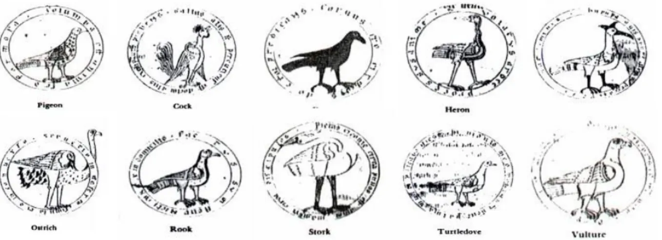
were being consumed. In highly populated regions, legumes such as fava beans, peas, lentils, chickpeas, peas, beans, and tremoço were imported from abroad to substitute cereals (Marques.A.H.O, 1987; Coelho.M.H.C, 1990; Gonçalves. I, 1988; Marques.A.H.O, 1987). The staple diet of the people was predominantly meat and fish (Marques.A.H.O, 1971). Beef, pork, mutton and goat were the commercially available meats. Game and poultry were also consumed frequently. In the middle ages, Portugal was famous for gaming reserves and wilderness. Though hunting was a pastime for the aristocrats, it was an important source of subsistence for the peasantry. Fallow deer, deer, roebuck, hare and even bear were widely sold in the market along with a variety of fowl species such as crane, wild duck, teal, heron, redshank, bald coot, and partridge among many others out of which some are shown in Figure 5. Poultry was almost the same as today including chickens, ducks, geese, pigeons, pheasants and doves. Turkey was included in this list only after the discovery of the America. The peasants utilised the abundant
14
game and poultry accessible to them to cover a good part of the tribute they were obliged to pay such as rent and quitrents to the ruling class. Rabbits however were raised for food rather than being hunted to make various sort of cured meats (Marques.A.H.O, 1971).
Fish however seems to have been less consumed in comparison to the modern times not counting the economically well off class, who considered the partaking of these foods as important. This is shown in the famous cookbook by Domingos Rodrigues where 66% of the dishes were meat based and those based on fish were less than 10% (Rodrigues D., 1987). The content of the book is a reflection of the consumption patterns of the wealthy class. The consumption of the fish by the clergy was based on religion, as 68 days of abstinence from meat was obligatory for Catholics. Church recommended peas, fruit and small fish. In the deprived classes, fish played an important role (Marques.A.H.O, 1971). The peasantry consumed sardines and whiting (peixota). The economically well of preferred other marine and freshwater species such as snapper, tuna, trout, sea bream, shark, sardines, conger, eels, red
snapper, sea bass, shad, surmullet were commonly consumed across all the socioeconomic classes. Crustaceans such as lobster, crab and molluscs such as clams, oysters along with whale and porpoise meat were also eaten (Marques.A.H.O, 1971). Tomar must have been influenced heavily by the presence of the various religious orders present throughout its history.
15
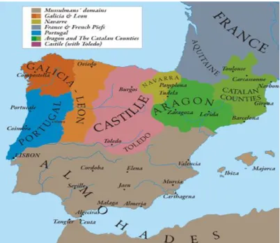
The dietary patterns also varied according to regional variation of religious practices. In the more Christian Coimbra in the twelfth century, pork and mutton were the most expensive whereas in the Muslim prominent Evora, price of beef was twice that of pork and more than twice that of mutton and goat. However, religion was not the only influencing factor at play in determining the price of the meat as pork is considered impure in Islam (Marques.A.H.O, 1987). Figure 6 shows the political situation of medieval Portugal during the Reconquista which influenced the local food prices.
The direct consumption of milk was not very widespread in Medieval Portugal. However, there were varied ranges of dairy products. They were specially used as side dishes or desserts and referred to as “Milk Victuals”. Cheese, cream, butter and various pre-prepared dishes were very commonly consumed (Marques.A.H.O, 1987; Ferro.J.P, 1996). Due to abundance of poultry, eggs were used in large quantities as raw ingredients for more elegant dishes. Desserts were made with milk and sweets were improved upon biscuits and pastries. Honey was used as a sweetener as sugar was exorbitantly priced (Marques.A.H.O, 1987; Ferro.J.P, 1996).
Vegetables were not very preferred by the clergy in medieval Portugal. This is reflected in the cookbook of D. Rodrigues, less than 5% of the recipes are vegetarian (Rodrigues D., 1987). The poor class preferred fresh vegetables. Cabbage, cauliflower, spinach, cucumber, turnip, carrot, onion, broccoli, pumpkin, radish, and mushrooms were commonly consumed (Marques.A.H.O, 1987; Coelho.M.H.C, 1990; Gonçalves. I, 1988; Marques.A.H.O, 1987). Salads, predominantly lettuce based were commonly consumed on the side with meals. Vegetables served to vary the otherwise monotonous diet of the peasants, however they were reliant on the climate and market constraints.
Portugal was always abundant in the production of fruit. Various fruit bearing trees such as figs, pumice, apple, pear, peach, and cherry dotted the Portuguese landscape. Olives and grapes were also abundantly cultivated to produce olive oil and wine (Marques.A.H.O, 1987). Sweet orange became the most common tree after Vasco da Gama brought it back from the East. Fruits were also consumed with wine at night as a refreshment. Seasoning and condiments were really simple in Middle Ages. Usually it consisted of salts and fat - based products to food (Marques.A.H.O, 1987; Ferro.J.P, 1996). Olive oil played a central role as a condiment and base ingredient. Lard was also used as a seasoning by all social classes equally. Various herbs such as coriander, parsley,
16
and mint were used to refine the flavours by the wealthier classes. Spices such as clove, saffron, ginger, and mustard were very expensive and consumed only by the élite.
Medieval Portuguese population suffered periodic shortages of food which was mitigated by imports of non-indigenous agricultural products. The generally consumed food before the discovery of the American continent was the local produce and later started including the vegetables and fruits brought back by the explorers such as potatoes and corn. Investigating the composition of dental calculus may have seems to possess the potential to shed new light on the subsistence patterns of the inhabitants of Tomar.
17
5. Scientific Approach
5.1 Dental Calculus
5.1.1 Aetiology
The multi-dimensional nature of the aetiology of dental calculus was initially thought universally by anthropologists that an alkaline oral environment facilitates the increased precipitation of salts from the oral fluids (Hillson, 1979; Lieverse, 1999). Despite the multitude of studies to elucidate the process of calculus formation, comprehensive mechanism still evades discovery.
The process of formation of calculus begins with the evolvement of pellicle over the enamel surface due to adsorption of highly specific salivary glycoproteins as a preventive measure against the bacterial metabolic acids and the deposition of salivary minerals especially calcium phosphate (Samaranayake, 2006). This is followed by the formation of plaque which is composed by several species of bacteria, including but not limited to streptococci, staphylococci, lactobacilli and corynebacteria (Radini et al., 2017). The first colonisers of the pellicle are the facultative
Streptococcus sanguinis and Streptococcus mutans, which replicate and form micro-colonies in a
short time due to the oxygen rich environment. Streptococcus mutans is especially important as it produces enzymes glucosyltransferase and pyrophosphatase. The enzyme glucosyltransferase converts sucrose into exopolysaccharides (EPS). These EPSs act as adhesive agents for other bacterial species to adhere to the initial colonies, shielding them from acidic environment. The consequence of this phenomenon is that as the thickness of the plaque increases, the availability of oxygen is cut off to the initial colonies (T.M.Roberson, 2006). Starch granules have a higher probability than other substrate as the bacteria in the gingival crevice are principally proteolytic. The metabolism of the bacteria leads to production of compounds such as ammonia increasing the local oral pH (Marcotte & Lavoie, 1998). The process of plaque formation, if not dismissed, causes the surrounding pH to increase even further. This followed by mineralisation of the plaque leads to the formation of dental calculus. In case of the formed plaque not mineralising, which happens when the pH is lowered, caries is formed (Figure 7). The formation of calculus is usually prominent on the lingual surfaces of incisors and canines and the buccal surfaces of the maxillary molars owing to the proximity of the salivary duct openings (Hillson, 1979).
18
Figure 7. Oral pH and pathology associated with it.
A set of four mechanisms were termed to describe the formation of dental calculus out of which two describe the process of mineralization (Mandel, 1990). The first being termed the “Booster Mechanism” that propounds the formation of dental calculus as a crystal growth by spontaneous precipitation, relying on the supersaturation of calcium and phosphate ions in the oral fluids and a number of arbitrating factors such as local pH. This theory of simple precipitation is only feasible when calcium and phosphate ions are in ionised form (Schroeder & Shanley, 1969). The second theory, which is most widely supported, is termed as “Epitactic Concept”. This takes into account that the calcium and phosphate ion concentrations are not high enough for spontaneous precipitation. However, the concentrations are sufficient to reinforce growth of calcium phosphate crystals once a “nucleus” or “seed” has formed. This is very similar to the formation of other ectopic calcifications such as urinary stones, and gall stones (Lieverse, 1999). This nucleation process requires an organic matrix which is available in the form of plaque, a biofilm and an occasional supersaturation of the calcium and phosphate ions to commence and persevere calculus growth (Mandel, 1990). The occurrence of dental calculus at certain sites and not everywhere is explained by the “Inhibition Theory”. It has been established that oral bacteria play a key role in disrupting the inhibiting mechanisms of mineralisation (Scheie, 1989). When the inhibiting mechanism is obstructed, mineralisation occurs (Mandel, 1990). Pyrophosphatase is secreted by
Streptococcus mutans hydrolyses pyrophosphate, a potent mineralisation inhibitor. Many proline
rich proteins also bind with calcium and inhibit mineralisation. Proteolytic enzymes, produced by these bacteria, lyse them into peptides and amino acids (Scheie, 1989). The event of brushite and octocalcium phosphate precipitating first and acting as precursors hydroxyapatite and whitlockite is termed as “Transformation Theory” (Mandel, 1990). The theory is supported by the fact that in
19
immature deposits up to 3 months brushite crystals are the most abundant wherein mature calculus deposits of 6 months and above, hydroxyapatite crystals are the most abundant (Driessens & Verbeeck, 1989; Mandel, 1990).The four theories describe various aspects of the calculus formation process and it is prudent to say that they work in tandem with other factors, which are still unknown.
5.1.2 Archaeological Importance
The use of dental calculus as a proxy of the dietary reconstructions of living organisms from the past is relatively new (Hardy et al., 2015; Hardy, Radini, Buckley, Sarig, et al., 2016; Horrocks, Nieuwoudt, Kinaston, Buckley, & Bedford, 2014; Anita Radini et al., 2017; Tromp & Dudgeon, 2015). Biological matrices such as bones and teeth have been used for a relatively long time for the same purpose (Lee-Thorp et al., 1989; van der Merwe, 1991). This demands that the concernment as well as advantages of dental calculus as a valid proxy for the paleodietary reconstructions must be established.
The first most important facet which is exploited in archaeology is the entrapping of physical and biomolecular debris along with microbial flora in the dental calculus when the organism is alive (Buckley et al., 2014; Hardy et al., 2009; Radini et al., 2016; Wang et al., 2016; Warinner et al., 2014). The second facet is the lack of post-mortem alterations of micro-debris from the environment as the formation process of dental calculus ceases immediately after death (Middleton & Rovner, 1994). However, this does not rule out the alteration of the chemical signal due to diagenesis after death. Starch granule degradation has been reported due to complex diagenetic processes (Barton & Torrence, 2015; Collins & Copeland, 2011). Future crystallography studies can reveal the extent of alteration of the signal due to diagenesis. Finally, the last factor is the variability of the rate of formation of dental calculus from individual to individual which depends on a wide range of parameters, such as genetic factors, phosphate and calcium levels, mineral and silicon levels, salivary flow rate, oral microbiome, local pH, and oral hygiene (Lieverse et al., 2007; Marcotte & Lavoie, 1998). The fourth factor is the span of the lifetime, which the dental calculus represents. Chewing abrasive and fibrous material has the potentiality of mechanically dislodging the calculus deposits from the tooth surface (Gaar et al., 1989). The dental calculus, which is less than 3 months, is an immature deposit whereas a deposit older than
20
6 months is referred to as mature deposit. An immature deposit is not a reliable proxy for dietary reconstruction as it a too short period to represent an individual’s life. One way to date the age of the deposit of calculus is to find the relative concentrations of hydroxyapatite, brushite, whitlockite and octocalcium phosphate as the concentration of brushite is the highest in immature deposits whereas hydroxyapatite is the most abundant in mature deposits. This might be reflected in the difference of carbon isotope ratios obtained from bones and dental calculus as only the former is an average representative of an individual’s life (Salazar-García, Richards, Nehlich, & Henry, 2014). A recent study showed a correlation between the carbon isotope ratios of the inorganic fractions of bone and calculus of an individual (Price et al., 2018).
The advantage of dental calculus over other biological matrices is that it is not a part of the skeletal system. This makes it an ideal material to work with destructive techniques when the skeletal remains are parts of collections, and museum exhibits.
5.1.3 Debris
The first scientific evidences of dietary debris from dental calculus were phytoliths which formed the basis of the conclusions to understand the diet of the herbivore ungulates (Armitage, 1975). Along with phytoliths, other environmental indicative debris were also observed and reported. The first foreign bodies to be categorically identified were silicified oral bacteria (Gonzales & Sognnaes, 1960; Lustmann, Lewin-Epstein, & Shteyer, 1976). The recovery and identification of other debris from the calculus matrix such as charcoal, pollen, and plant fibers from wide periods of historical material by Dobney and Brothwell (1987, 1988) laid the final foundation for the usage of the dental calculus in archaeological dietary studies (Radini et al., 2017).
Any foreign material which enters the buccal cavity is primarily through three sources, with the primary being nutrition intake and the other two being environmental contaminants and debris due to behavioural tendencies. The debris due to nutrition intake usually consists of diatoms from drinking water (Dudgeon & Tromp, 2014), phytoliths from plant based raw material, starch granules from carbohydrate rich food and other trapped physical and biomolecular food components (Henry, Brooks, & Piperno, 2011; Henry & Piperno, 2008; King & Searcy, 2016; Li et al., 2010; Radini, Nikita, & Shillito, 2016; Warinner et al., 2014; Weber & Price, 2016). The environmental contaminants are usually encompassed into the dental calculus matrix by either
21
inhalation (both nasal and oral) or “secondary eating”. Common particulate matter includes, charcoal particles, fibers (Hardy et al., 2015). The oral respiration is carried out while eating, interacting orally, panting and finally when respiratory passage is blocked by nasal mucus. This also includes micro-particles which have lodged or deposited on food or drinks, entering the oral cavity and get embedded in the dental calculus (Hardy et al., 2016; Radini et al., 2017). Sometimes this embedding of soil and grit particles can happen to the food during the initial stages of the raw material synthesis (Tromp & Dudgeon, 2015). A broad range of breathable inorganic and organic particulate material produced in sufficiently high concentrations due to different production activities such as stone tooling, agricultural processing, ceramics, cooking etc. which can cause various respiratory syndromes have been found in dental calculus. These particulate material include soot, micro-charcoal, spores etc. which are highly probable to be embedded into the calculus because of oral respiration (Hardy et al., 2015, 2016; Radini et al., 2017). Finally, the debris capsulated due to behavioural tendencies is usually because of the use of “mouth as third hand” behaviour or practices of introducing non-dietary material into the oral cavity such as the practices of chewing or smoking non-nutritional substances such as tobacco, coca (Yaprak et al., 2017; Klepinger, Kuhn, & Thomas, 1977; Blatt et al., 2011; Charlier et al., 2010; Radini et al., 2016). A more uncommon but interesting behavioural tendency, which has the potential to introduce micro-particles, is gastrophagy. Gastrophagy is the practice of eating the gut contents of hunted animals in some populations as a means of gaining nutrition without having much effort in digestion. The presence of yarrow and chamomile was suggested by Hardy et al., (2012) after finding molecular cursors indicative of them in the dental calculus of El Sidrόn Neanderthals. However, Buck et al. (2015) propounded the possibility of gastrophagy as the mechanism of incorporation of these compounds into the dental calculus and that it should be considered as a potential factor in distorting the signal in dietary studies (Radini et al., 2017). Another widespread cause of non-dietary debris in dental calculus is the extramasticatory use of the teeth. The use of teeth as a force to apply on material which is too small in size to be worked by hands, often results in the material getting embedded in the dental calculus matrix (Blatt et al., 2011). As it has been proved beyond reasonable doubt that the mechanism of debris incorporation in dental calculus is multi-causal, conclusions should be drawn after careful considerations. The possibility of the scientific evidence being embedded by more than one process is highly probable. The attribution
22
of the chemical signal to the source is highly contested as of the current scenario (Radini et al., 2017).
5.1.4 Incorporation of Debris
Incorporation of debris in dental calculus is as multi-causal as it has multiple origins. The dietary debris is incorporated after the mastication process where as the non-dietary debris is incorporated when the mouth acts as a dust trap (Radini et al., 2017). The incisors cut the food into a chewable size and the canines, which tear harder food that cannot be cut by the incisors, carry out a parallel of this function. The food is then mixed with saliva by the tongue and then pushed to the premolars and the molars, which grind it, making it relatively smooth and easy to digest. Because of the positioning of the salivary glands, the formation of the dental calculus is more concentrated on the molars and the incisors (Watson, 2017). The food is then pushed down the throat by the peristaltic motion and it enters the oesophagus, this is called the bolus.
When the pH of the buccal cavity is alkaline and the concentrations of Ca2+ ions and PO4- ions are
high enough for the ectopic formation of the dental calculus nucleus (Mandel, 1990), the food chewed in the mouth gets stuck on the various surfaces of the teeth including the dental calculus surface. The chance of food debris being stuck on the surface of the dental calculus surface is high due to the presence of the EPSs (exopolysaccharides). However, the stickiness of the food also plays a huge role in the amount of debris being incorporated. The subsequent rinsing of the mouth gets rid of the majority of the debris with the exception of very a small amount of debris remaining behind. The biomolecular debris is adsorbed by the calcium phosphate matrix whereas the physical debris is embedded by the next cycle of expansion of the nucleus of the ectopic calculus. This cycle keeps repeating until the dental calculus either dislodges from the surface of the tooth or the calculus formation stops due to the death of the individual. In case of the non-dietary debris, the cycle may or may not begin in the same way as the dietary debris depends on if it is a non-nutritional substance (e.g. tobacco, betel leaves, coca etc.) in which case it undergoes deposition as dietary debris. If it is an inedible substance, it begins with the deposition of the debris on the dental calculus surface. Then the subsequent cycle of the calculus expansion embeds the debris. The amount of debris should be relatively higher at the incisors and the molars than the remaining area of the mouth. The presence of the certain enzymes which hydrolyse inhibiting factors (e.g.
23
Pyrophosphatase) cause calcium phosphate to precipitate more than in other locations (Scheie, 1989).
The incorporation of debris into dental calculus is almost the same in all the scenarios. However due to various enzymes and other factors, the spatial incorporation of the debris is not homogenous. This is caused by the hydrolysis of the debris by suitable enzymes. A good example of this phenomenon is the higher incorporation of starch granules in sub-gingival crevice where the bacteria is proteolytic (Marcotte & Lavoie, 1998). In addition, the debris is relatively shielded by the salivary amylase (α-amylase). The mechanism and the factors controlling the spatial distribution of the debris plays a central role while sampling for scientific analysis.
5.2 Microremains
5.2.1 Phytoliths
The word “phytolith” is taken from two Greek words, “phytos” meaning plant and “lithos” for stone. In the past, this term has been used non-specifically for all forms of siliceous and calcareous mineralized secretions by plants. In current literature, when not mentioned otherwise, the term phytolith refers to siliceous depositions in an intracellular or extracellular locations in a plant. Silica is an omnipresent component of plant and animal microorganisms. Silica is absorbed in to the cellular system in a soluble state from groundwater. Silica enters the plant body in the form of monosilicic acid, Si(OH)4 when the soil pH ranges between 2 and 9 (Barber & Shone, 1966). Silica
is present in underground water in soluble form mainly due to weathering of minerals such as quartz and feldspar (Dunne & Leopold, 1978). The growth of phytoliths in a plant is dependent on several factors such as climate, composition of soil, availability of water, age of plant and taxonomy of the plant. The process of phytolith development begins when the plants take in soluble silica present in groundwater via their roots. This silica is carried up to various organs in the transpiration process by the water conducting tissue, xylem. There are two pathways by which plants absorb water solubilized silica. The two proposed pathways are active transport of monosilicic acid and non-selective passive flow in the transpiration stream. In active transport mechanism, the plant has to expend energy to transport silica whereas it does not in the latter. There is sufficient evidence that passive uptake and active transport of silica occurs in plants based on their taxonomy (Raven, 1983). However, it has also been demonstrated that some species of
24
plants having low concentrations of phytoliths have some mechanism for rejecting silicic acid at the surface of the root.
When the monosilicic acid enters the plant it is polymerized and finally deposited in the form of silicon dioxide (SiO2) in both intracellular and extracellular spaces. Though these deposits are
referred to as opal silica, they behave more like silica gel (Lewin & Reimann, 1969). There are three loci in a plant where phytoliths occur: (1) cell wall deposits (2) infillings of the cell lumen and (3) in the intercellular spaces of the cortex. Patterns of local deposition are repeated in the same species and families of plants, independent of the environment of growth. This fact is exploited to identify families if not species of plants in archaeological context.
The first report on phytoliths in archaeology was by Bryant who observed calcium oxalate crystals in coprolites (Bryant, 1974). Phytoliths being more or less ubiquitously present in archaeological
25
contexts when plant parts are involved. The presence of phytoliths are then subjected to further interpretation based on the surface they are observed. The application of phytoliths on archaeology ranges from economic importance of plant species, agricultural practices, domestication of plant species, dietary practices etc. The advantages of utilizing phytoliths are that they are resilient material, rarely transported by water or wind, extremely easy to evaluate the morphology and cheap analysed. However, phytoliths have their own pitfalls when acting as proxies for reconstruction of the past. Not every family of plant kingdom produce phytoliths. And phytoliths are erodible as well as dissolvable at extreme pH. Phytoliths are also subjected to mobility from surface to surface and also there is a huge dearth in the availability of reference material. Phytoliths are classified as shown in Figure 8 into various categories based on their morphology (Madella et al., 2005). The morphology of the phytoliths is based on the type of cell silicified. The morphometry of the phytolith can be utilized to identify the family of species of plant.
5.2.2 Starch Granules
Starch granules are the end products of plants after the completion of a cycle of photosynthesis. Starch is the stored form of food for plants in seeds, stems and underground organs such as roots, tubers and bulbs. Starch granules are highly utilized in archaeology as they are chemically inert and are morphologically specie specific. They are found preserved on surfaces of ancient artefacts and in paleorecords. Analysis of starch granules is usually used to reconstruct ancient subsistence patterns as well as plants which played a role in staple diet.
Starch granules have a point around which layers of protein are accumulated which is called as a hilum. Tubers and bulbs have an off-center hilum where the growth rings originate out from the hilum. The growth ring pattern is based on the formation geometry. As starch is the product of
Figure 9. Starch grain morphology with hilum and maltese cross. http://archaeobotany.dept.shef.ac.uk/wiki/index.php/Image:PotatoIKI.jpg
26
photosynthesis, its basic unit is glucose. Glucose can be linked into two types of starch chains namely amylose and amylopectin. The layers of amylose and amylopectin called lamellae are tightly packed into each of the rings. The dense packing along with the repeated pattern makes the starch grain partially crystalline. The second most identifying characteristic of a starch grain is its property of birefringence. Birefringence is a property of material where its refractive index is dependent on the direction. A starch grain under plain polarised light exhibits a cross shaped feature ending at its hilum. This is called an extinction cross or a Maltese cross (Figure 9). The third feature of starch granules is the presence of longitudinal fissure. The fissure is a sort of triangular trough originating from the hilum and extending outwards. This is a defining characteristic as some starch granules have it and some do not.
5.2.3 Phytoliths and Starches in Dental Calculus
The most widely used approach to extract the trapped microfossils consists in demineralization of the calcium phosphate matrix using HCl of varying concentrations. These extracted microfossils are then examined for morphology under a light microscope. A large amount of research has been conducted on both human and non – human dental calculus on the identification of phytoliths and starch granules. Usually the non – human samples are herbivores (ungulates) and extinct species such as Gigantopithecus blacki, mastodon etc. (Ciochon, Piperno, & Thompson, 1990; Gobetz & Bozarth, 2001; Middleton & Rovner, 1994). The majority of research has been focused on the extraction and identification of microfossils with specific emphasis on phytoliths and starch granules (Hardy et al., 2012; Henry, Brooks, & Piperno, 2011; Horrocks et al., 2014; Power, Salazar-García et al., 2014; Tromp, 2012; Tromp & Dudgeon, 2015). The other microscopy technique utilised for microfossil identification is the technique of scanning electron microscopy (SEM) coupled with energy dispersive X-Ray spectrometry. The technique has been exploited to study the micro - debris and its association with occupational and dietary habits (Charlier et al., 2010; Power et al., 2014). The technique has also demonstrated potential to identify possible drinking water sources by facilitating easy identification of diatoms (Dudgeon & Tromp, 2014). However, the main issue of the microfossil identification is that it involves the comparison of the extracted entities to be compared with reference collections (Pearsall, 2000). The usual method to explain the taphonomic processes affecting the survival of the microfossils is to compare them with residues extracted from other archaeological substrates (Barton & Torrence, 2015; Li et al., 2010). However, diagenetic alteration of starch granules has been reported and further research is
27
necessary to identify the integrity of dental calculus (Barton & Torrence, 2015; Horrocks et al., 2014).
5.3 Role of Stable Isotopes in Dietary Reconstruction
Isotopes are different atoms of the same element with same atomic number but different mass numbers (Schoeninger & Moore, 1992). In nature, isotopes exist in either radioactive or stable forms with the latter being more abundant. The stable isotopes of the same element possess the same chemical properties but slightly different physical properties. Due to the slight difference in the physical properties, one isotope (usually the lighter one) is favoured over the other isotope in a chemical process. The end product of this reaction is more enriched in the preferred isotope compared to the reactants, a process called isotopic fractionation (Malainey, 2011). The isotopic abundance of an element is measured with the aid of a mass spectrometer by combusting the sample in a controlled environment to liberate the element of interest in the form of a gas. In the case of dental calculus the gases liberated are CO2 and N2 as we are interested in carbon and
nitrogen isotopes. Usually a magnetic sector is used to separate the molecules on the basis of their mass and a detector is used to measure the counts per second. A laboratory standard is also measured along with our samples as the former is calibrated to international standards.
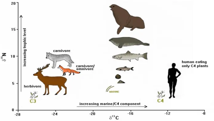
Figure 10 shows the isotope fractionation application of stable isotopes which is quite varied in archaeology such as provenance studies, paleoclimatic reconstructions, diet and mobility of humans and fauna (Craig, Bondioli, Fattore, Higham, & Hedges, 2013; DeNiro & Weiner, 1988; Lee-Thorp et al., 1989; Sealy, Armstrong, & Schrire, 1995; van der Merwe, 1991). In the process of absorption and reuse of elements in the living tissue, the isotopic fractionation is dependent of the biochemical process the element goes through. In the case of dental calculus, the isotopic signal
28
is not subjected to any fractionation from the individual’s metabolic process. Thus, the ratio of carbon and nitrogen isotopes in the dental calculus is the sum of the dietary debris trapped.
5.3.1 Stable Carbon Isotopes
12C and 13C are the stable isotopes of the element carbon where the abundance of the former is a
100 times greater than the latter (Malainey, 2011). In photosynthesis, the rate determining plant enzyme Rubisco (ribulose-1,5-biphosphate carboxylase/oxygenase) favours the lighter 12C in comparison to the heavier 13C (Caemmerer, Ghannoum, Pengelly, & Cousins, 2014; Leary, 2008). There are three different pathways of photosynthesis adapted by plants based on the climatic conditions they grown in. They are the C3, C4 and CAM (Crassulacean Acid Metabolism)
respectively. Due to the difference in pathways, the end products have different 13C/12C ratios (DeNiro & Weiner, 1988; Lee-Thorp et al., 1989; van der Merwe, 1991). The majority of the vegetation of the world’s terrestrial vegetation which includes rice, wheat, legumes and other tree species are C3 in nature. Grasses which are from hot arid regions such as millet, sorghum, maize
etc. are C4 plants (Still et al., 2003). Usually in dietary analysis, osteological remains such as bones
and teeth are measured which reflects the diet of the individual. The intake of C3, C4 and CAM
plants in varying portions results in influencing the 13C/12C ratio reflected (DeNiro & Weiner,
1988; Sealy et al., 1995). The ratios are expressed as δ13C values where the stable carbon isotope