BJRS
RADIATION SCIENCES
07-02A (2019) 01-09ISSN: 2319-0612
Accepted: 2018-11-02
Study of the radiomodifier effect of Pityrocarpa
moniliformis extract
J.V.T., Luna Filho
aR.L.C., Siqueira
a,* W.N., Silva
aH.A.M.F., Pereira
aD.R., Melo
aA.M.M.A.
a Universidade Federal de Pernambuco/Departamento de Biofísica e Radiobiologia, 52171-011, Recife, PE, Brasil
ABSTRACT
Ionizing radiation has been applied in several areas of knowledge, among them the study of the radiomodifier activity of natural substances. These substances can modify the cellular response to the damage induced by the radiation. Therefore, this work aimed to evaluate the radiomodifier action of Pityrocarpa moniliformis extract on Biomphalaria glabrata embryos exposed to 60Co gamma radiation. Initially, toxicity tests were performed on the
extract against the B. glabrata embryos for the choice of concentration that did not cause death and embryonic malformation. Then, the antioxidant activity of the P. moniliformis extract with flavonoids and phenolic compounds was evaluated by means of the ABTS method. To evaluate the radiomodifier activity of the extract, embryos were selected in the blastula stage and irradiated with 7.5 Gy in a 60Co source (gammacell-Co60). Then,
the embryos were exposed for 24 h to the extract of P. moniliformis at a concentration of 250 μg/mL. The results showed that the extract of P. moniliformis presents flavonoids and enzymatic inhibition by ABTS, which demonstrates the presence of antioxidant compounds. However, the tests of the radiomodifier activity did not present radioprotective effect for embryos exposed to ionizing radiation.
Keywords: Biomphalaria glabrata, Pityrocarpa moniliformis, Radiation.
1. INTRODUCTION
Ionizing radiation is the electromagnetic wave or high energy particle that, when interacting with the absorber medium, has the property of transferring, in whole or in part, energy to the atoms and molecules of the medium, resulting in the phenomenon known as ionization [1]. In living organisms, the incidence of ionizing radiation can compromise the functioning of a cell, because, when interacting with the biological system, there is the frequent formation of free radicals that, being unstable and highly reactive, are considered the main responsible for the cellular damage caused by radiation [2].
Since ionizing radiation has the ability to alter the DNA molecule, one of its possible conse-quences would be the induction of mutations in the germ cells that would produce the offspring of the affected individuals. However, there is great difficulty in finding evidence that a given organic change is the exclusive consequence of exposure to ionizing radiation [3]. Another important issue is that the effects of ionizing radiation depend, among other factors, on the total dose and dose rate at which the organism was exposed [4]. In addition, there is a latent period before the detection of any response where it can be prolonged for decades, when exposed to low doses of radiation occurs, or for a very short period of time (minutes or hours), if it occurs in high doses and/or high rates, thus making the radiation effects difficult to observe [5].
Exposure to radiation and its effects have been described as Acute Radiation Syndrome [6]. With the advancement of radiobiology, not only a better understanding of the radio-induced cellular response, but also the development of modern prognostic, diagnostic and therapeutic measures in radioprotection were obtained. Thus, there are many derivatives of plant extracts or genetically modified plant sources which are used for the deleterious effects of ionizing radiation and are ad-ministered prior to irradiation. The interest in new sources of products for treatment after irradiation has been the subject of studies [7]. The radiomodifiers, which are agents that modify biological re-sponses, include natural and chemical compounds [8]. However, the chemical compounds have a high toxicity, which contributed to the realization of research related to new alternatives of products with low toxicity, thus generating the products or compounds isolated from natural sources.
Ionizing radiation has been widely used in different areas of knowledge, medicine (radiodiagno-sis and radiotherapy), agriculture, and industry. With increasing use of radiation, the interest and the need to find substances that can modify the cellular response to the damage induced by radiation. The Biomphalaria glabrata is a mollusk that is considered sensitive to toxic agents present in the environment and has been used as a biological model for the toxicity and embryotoxicity tests of chemical substances [9]. The plant kingdom has contributed significantly to the production of bio-logically active substances and its study has helped discover new compounds useful for various purposes, such as antibiotics or medicines [10].
Therefore, investigating substances of plant origin that may perform functions of pharmacologi-cal interest, such as radiomodifiers, are of great importance because they may present less toxicity and greater efficacy when compared with synthetic substances used for the same purpose [11].
Thus, the objective of this work was to evaluate the radiomodifier action of the aqueous extract of Pityrocarpa moniliformis on embryos of B. glabrata exposed to ionizing radiation.
2. MATERIALS AND METHODS
2.1. Phytochemical analysis
The antioxidant activity of the P. moniliformis extract quantified by ABTS methods, following the methodology of Re at al. [12], the dosage of phenolic compounds by the Folin-Ciocalteau method described by Li et al. [13], and dosage of flavonoids, following the methodology described by Woisky and Salatino [14].
2.2. Bioassays with B. glabrata embryos
2.2.1. Evaluation of the toxicity of the aqueous extract
Embryos of Biomphalaria glabrata in the blastula stage were separated into groups of approximately 100 embryos and exposed for 24 h to the aqueous extract of P. moniliformis (125, 250, 500 e 1000 µg/mL). Two control groups were used: one containing filtered water and the other group with 0.5% DMSO (dimethylsulfoxide) in filtered water. After exposure, the embryos were stored in containers with filtered water until their hatching and were later counted as viable and unviable (dead and malformed). The experiment was performed in triplicate.
2.2.2. Dose determination
The embryos (divided into groups of 100) of B. glabrata were packed in microtubes (Axygen Scientific, Inc., Union City, CA 94587 USA) containing 1 mL of filtered water. Subsequently, the animals were exposed to doses of 5, 7.5, 10, 20, 25, 30 and 35 Gy of gamma radiation of 60Co (model II 200 Excel - MDS
Nordion with dose rate of 3.532 kGy/h) at 25 °C (± 2) for determination of the dose to be used in the radiomodifier test.
2.2.3. Radiomodifier assay
To perform the radiomodifier test, the embryos were exposed to the extract of P. moniliformis at the concentration of 250 μg/mL after being irradiated at the dose of 7.5 Gy to evaluate its radiomodifier activity. The animals were divided into 5 groups: control with filtered and dechlorinated water (C), DMSO, exposed only to extract (E), irradiated (I) and submitted to extract and radiation (E + I).
2.3. Statistical analysis
Statistical analysis was performed using GraphPad Prism 5.0 software. The ANOVA and Student Newman-Keuls tests were used. The data were expressed as a mean ± standard error of the mean, where differences were significant when p < 0.05.
3. RESULTS AND DISCUSSION
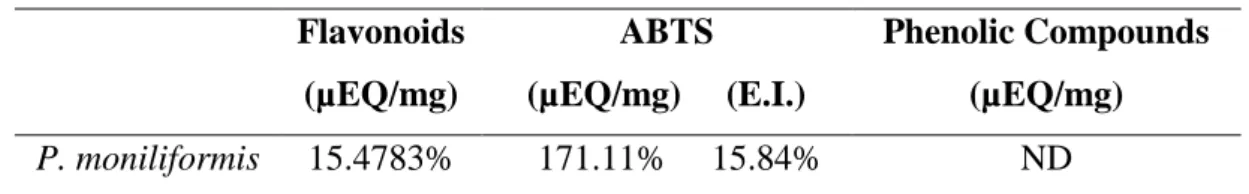
The phytochemical analysis confirmed the presence of antioxidant compounds (Table 1). The quantification of flavonoids was performed, where the presence of the extract of Pityrocarpa
moniliformis (15.4783 µEQ/mg).
Similar results on flavonoid content were observed by Silva [15] in analyses performed with P. moniliformis extract. Through the ABTS method, the aqueous extract of P. moniliformis demonstrated the ability to sequester free radicals. This finding is compatible with those observed by Silva [15], who evaluated the antioxidant activity of P. moniliformis through the DPPH method. However, phenolic compounds were not detected in the extract.
Table 1: Results of the dosage tests of flavonoids, ABTS, and phenolic compounds. Flavonoids (µEQ/mg) ABTS (µEQ/mg) (E.I.) Phenolic Compounds (µEQ/mg) P. moniliformis 15.4783% 171.11% 15.84% ND * Results expressed in μEQ/mg and Enzymatic Inhibition (E.I.). ND = Not detected.
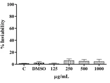
In Figure 1 it is possible to observe the result regarding the exposure of the embryos to the extract of P. moniliformis in different concentrations, where no significant difference was observed in the percentage of inviability between the groups submitted to the extracts when compared to the control groups.
A result similar to that observed by Rocha-Filho et al. [16] after exposing embryos of B. glabrata to the Moringa oleifera extract to evaluate the embryocidal capacity of the substance, however, no
significant differences were observed between the groups of mollusks exposed to the extract when correlated to the animals belonging to the control group.
Figure 1: The graph shows the result of the exposure of the B. glabrata embryos to the aqueous
extract of P. moniliformis in different concentrations.
In Figure 2 it is possible to observe the percentage of embryonic infeasibility of 27, 62, 82.5, 84.5, 92.5, 98.5 and 99% for the exposed embryos at doses of 5, 7.5, 10, 20, 25, 30 and 35 Gy, respectively. The 7.5 Gy dose was selected because it was the closest to LD50 (Lethal Dose to 50%)
for the embryos studied.
Analysis of the results (Figure 2) showed a high percentage of inviability in the groups exposed to ionizing radiation, where even the lowest dose tested (5 Gy) induced a significant percentage (27%) of nonviable embryos.
Figure 2: In the figure, the effect of the 60Co gamma radiation on embryos of B. glabrata can be observed at different doses.
C DMSO 125 250 500 1000 0 20 40 60 80 100 g/mL % I n v ia b il it y C 5 7.5 10 20 25 30 35 0 20 40 60 80 100 * *** *** *** *** *** *** Dose (Gy) % I n v ia b il it y
Okazaki et al. [17], after a study to evaluate the radiosensitivity of B. glabrata embryos, observed that the radiation caused death and development of several malformations, such as head malformations and shell malformations, with non-specific malformations being the most frequent.
Figure 3 shows the results of the radiomodifier test, where a 12% increase in the percentage of nonviable embryos belonging to the I + E group was observed when related to the group exposed to radiation alone, indicating that the P. moniliformis extract damage to embryos of B. glabrata after exposure to ionizing radiation.
Figure 3: The graph shows the result of the radiomodifier test. Where, E = extract of P.
moniliformis (250 μg/mL); I = irradiated (7.5 Gy); I + E = irradiated and later exposed to P. moniliformis extract.
The results found in this work were similar to those demonstrated by Siqueira et al. [18], where it was observed that after exposure to ionizing radiation embryos of B glabrata, in the presence of the aqueous extract of the Anacardium occidentale leaf, an increase in the frequency of unviable
C DMSO E I I + E 0 20 40 60 80 100 ** % I n v ia b il it y
embryos occurred and that this effect possibly was caused by the potentiation of the activity of secondary metabolites of embryotoxic action present in the medium.
4. CONCLUSIONS
The phytochemical analysis showed the presence of flavonoids in the analyzed species, besides showing antioxidant activity through the ABTS test. However, the presence of phenols in their composition was not identified.
The Pityrocarpa moniliformis extract did not show a significant difference in the percentage of inviability for embryos of Biomphalaria glabrata at the concentrations studied making it impossible to determine the LC50 (Lethal Concentration for 50%) for mollusks.
B. glabrata embryos are sensitive to ionizing radiation, presenting a dose-dependent
relationship with their rate of inviability.
The extract of P. moniliformis potentiated the damage caused to B. glabrata embryos after exposure to ionizing radiation.
REFERENCES
[1] KAPLAN, I. Física Nuclear, 1st ed. Rio de Janeiro: Guanabara Dois, 1978.
[2] BITELLI, T. Física e dosimetria das radiações, 2nd ed. São Paulo: Atheneu, 2006.
[3] BOLUS, N. E. Basic review of radiation biology and terminology. JNMT, v. 29, p. 67-73, 2001.
[4] MULLER, H. J. Artificial Transmutation of the Gene. Science, v. 66, p. 84-87, 1927.
[5] HALL, E. J. Radiobiology for the Radiologist, 6th ed. Philadelphia: Lippincott Wilkins & Williams, 2006.
[6] ANNO, G. H., YOUNGBLOOM, R. W. R. M., MERCIER, J. R. Dose response relationships for acute ionizing radiation lethality. Health Phys, v. 84, p. 568-575, 2003.
[7] ARORA, R. Botanicals as potential radioprotective and radiorecovery agents: Current status and emerging directions for clinical applications, In: PROCEEDINGS OF THE WORLD
AYURVEDA CONFERENCE (in press), 2007, Pune, November, 2007, p. 5-12.
[8] JAGETIA, G. C. Radioprotective potential of plants and herbs against the effects of ionizing radiation. J Clin Biochem Nutr, v. 40, p. 74, 2007.
[9] OLIVEIRA-FILHO, E. C. Efeitos de substâncias químicas sobre a reprodução de moluscos de água doce: estudos com caramujos do gênero Biomphalaria. 2003. Tese (Doutorado em Saúde
Pública) - Escola Nacional de Saúde Pública, Fundação Oswaldo Cruz, Rio de Janeiro, p. 138.
[10] PHILLIPSON, J. D., ANDERSON, L. A. Ethnopharmacology and Western Medicine. J
Ethnopharmacol, v. 25, p. 61-72, 1989.
[11] LEE, T. K., O’BRIEN, K. F., WANG, W., JOHNKE, R. M., SHENG, C., BENHABIB, S. M., WANG, T., ALLISON, R. R. Radioprotective effect of American ginseng on human lymphocytes at 90 minutes postirradiation: a study of 40 cases. J Altern Complement Med, v. 16, p. 561-567, 2010.
[12] RE, R., PELEGRINI, N., PROTEGGENTE, A., PANNALA, A., YANG, M., RICE-EVANS, C. Antioxidant activity applying and improved ABTS radical cátion decolorization assay. Free Radic Biol Med, v. 26, p.1231-1237, 1999.
[13] LI, H. B., WONG, C. C., CHENG, K. W., CHEN, F. Antioxidant properties in vitro and total phenolic contents in methanol extracts from medicinal plants. Food Sci Technol, v. 41, p. 385-390, 2008.
[14] WOISKY, R. G., SALATINO, A. Analysis of propolis: some parameters and procedures for chemical quality control. J Apic Res, v. 37, p. 99-105, 1998.
[15] SILVA, M. L. C., COSTA, R. S., SANTANA, A. S., KOBLITZ, M. G. B. Phenolic compounds, carotenoids and antioxidant activity in plant products. Semina: Ciênc Agrár, v. 31, p. 669-682, 2010.
[16] ROCHA-FILHO, C. A., ALBUQUERQUE, L. P., SILVA, L. R., SILVA, P. C., COELHO, L. C., NAVARRO, D. M., ALBUQUERQUE, M. C., MELO, A.M.M.A., NAPOLEÃO, T. H., PONTUAL, E. V., PAIVA, P. M. Assessment of toxicity of Moringa oleifera flower extract to
Biomphalaria glabrata, Schistosoma mansoni and Artemia salina. Chemosphere, v. 132, p.
[17] OKAZAKI, K., ANDRADE JÚNIOR, H. F., KAWANO, T. Effect of 60Co gamma radiation on Biomphalaria glabrata (Mollusca, Gastropoda) embryos: mortality, malformation and hatching. Braz J Med Biol Res, v. 29, p. 1057-1067, 1996.
[18] SIQUEIRA, W. N., SILVA, L. R. S., SILVA, R. C., LACERDA, L. B. N., SILVA, H. A. M. F., SANTOS, M. L. O., SÁ, J. L. F., SILVA, E. B., MELO, A. M. M. A. Radioprotective effect of the extract of Ziziphus joazeiro and Anacardium occidentale on embryos of Biomphalaria
glabrata submitted to ionizing radiation, In: INTERNATIONAL NUCLEAR ATLANTIC