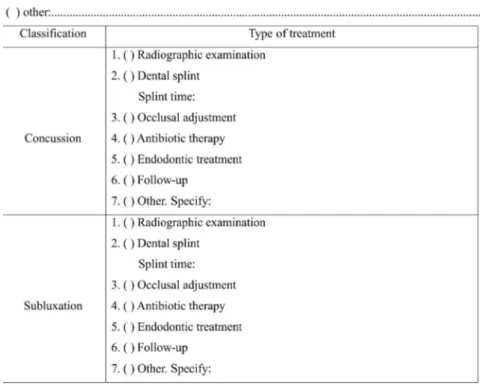
Abstract
Submitted: June 16, 2017
Modification: September 28, 2017 Accepted: October 27, 2017
Evaluation of cases of concussion
and subluxation in the permanent
dentition: a retrospective study
Objectives: This study evaluated the evolution of cases of concussion and subluxation through a retrospective study of 20 years. Material and Methods: Were examined clinical and radiographic records of 1,309 patients who underwent treatment of dentoalveolar trauma in the discipline of Integrated Clinic of the School of Dentistry of Araçatuba, UNESP, of which we selected 137 whose patients had concussion and subluxation injuries, with average age of 23.3 (SD – 10.96). The variables collected were: gender, age, history of previous and actual trauma, treatments performed, the presence of necrotic pulp, and time elapsed until the same trauma. The concussion and subluxation groups were subjected to statistical analyses using the SPSS 16.0 version software (α=0.05), Chi-square, and t-tests. Results: Of the 301 teeth involved, 49 (16.3%) suffered concussion and 252 (83.7%), subluxation, being the upper anterior teeth the most affected (75.1%) for both conditions. Subluxation and concussion traumas were more prevalent in men aged 10 to 20 years, most caused by cycling accidents (36.2%). There was a concomitant presence of crown fracture in 21% of cases of concussion and 34.7% of subluxation. Pulp necrosis was detected in 16.3% (concussion) and 27.1% (subluxation) (p=0.12), and most occurred within 6 months after the trauma (p=0.29). The pulp necrosis shows a positive correlation with motorcycle accidents (p=0.01), direct impact (p≤0.0001), crown fracture with pulp exposure (p≤0.0001), darkening of the crown (p=0.004) and spontaneous pain (p≤0.0001); and negative correlation with indirect impact (p≤0.0001). Conclusions: Although concussion and subluxation traumas are considered of minor degrees, they must be monitored, since the possibility of pulp necrosis exists, and its early treatment favors a good prognosis.
Keywords: Tooth injuries. Periodontal ligament. Wound healing. Denise PEDRINI1
Sônia Regina PANZARINI1 Adelisa Rodolfo Ferreira TIVERON2 Valéria Marisel de ABREU2 Celso Koogi SONODA1 Wilson Roberto POI1 Daniela Atili BRANDINI1
1Univ Estadual Paulista - UNESP, Faculdade de Odontologia de Araçatuba, Departmento de Cirurgia
e Clínica Integrada, Araçatuba, São Paulo, Brasil.
2Univ Estadual Paulista - UNESP, Faculdade de Odontologia de Araçatuba, Programa de
Pós-Graduação em Ciência Odontológica, Araçatuba, São Paulo, Brasil.
Introduction
Of the traumatic dental injuries of the periodontal
tissues, concussion and subluxation present the
less complex clinical resolution, not causing great
emotional impact on patients and their families when
occurring alone. Although the prognosis is favorable
with little pulp and periodontal complications, it must
always be monitored, since the possibility of pulp
necrosis exists9.
Concussion is characterized by an injury of the
tooth support structures without increased tooth
mobility or tooth displacement, but with reaction
to the horizontal or vertical percussion, and may
be associated with crown fracture3,15. The pulp
sensitivity test is usually positive and does not notice
changes radiographically7,9,16. Subluxation already
has increased mobility in the horizontal direction,
and the tooth appears to be sensitive to percussion
and occlusal forces, occurring or not bleeding from
the gingival sulcus. Initially, sensitivity tests may be
negative, but subsequently, they tend to respond
positively and radiographically. Abnormalities are
not found3,7,15,16, although a slight thickening of the
periodontal ligament may be detected in cases of sharp
mobility9. General characteristics of concussion and subluxation include edema and bleeding and breaking of some fibers of the periodontal ligament; moreover, the neurovascular pulp supply may be affected leading
to necrotic pulp3. These injuries can go unnoticed by parents, and seeking care can occur only after the sequel have been installed, mainly when associated
to severe traumas.
The literature has shown that dental trauma has
increased and become a public health problem1. Retrospective studies can contribute to the knowledge of its repair process and possible complications, in
addition to improve the prognosis. Thus, this study
aimed to evaluate the evolution of cases of concussion
and subluxation through a retrospective study of 20
years.
Material and methods
The research protocol for this study was reviewed
and approved by the local Research Ethics Committee
(CAAE – 45716615.6.0000.5420). All participants
received information about the objectives of the study
and provided written informed consent regarding
their participation. Were examined clinical and
radiographic records of 1,309 patients who underwent
dentoalveolar trauma treated by the group of the
discipline of Integrated Clinic, School of Dentistry of
Araçatuba, UNESP - Univ Estadual Paulista, over the
period of 1992−2011. The study was conducted with
a sample of 137 records from patients diagnosed with
concussion injury and/or subluxation, average age of
23.3 (SD – 10.96).
All data were assessed by one trained dentist. The file assessment consisted of the following components: a) patient identification (gender and age); b) history of previous trauma; c) history of the actual trauma (etiology, type of trauma, clinical signs, and symptoms associated with dental trauma); d) treatments performed and; e) the presence of necrotic pulp and time elapsed until the same trauma (Figure 1).
The diagnosis of pulp necrosis was obtained
through clinical and radiographic analysis. Clinical
analysis was performed in all cases of both traumas,
in addition to the monitoring of all patients using
percussion testing and pulp sensitivity for a period of at
least 24 months. Radiographic examination evaluated the increase of thickness of the periodontal ligament, radiolucent area compatible with periapical lesion, and
presence of external root resorption.
Only permanent teeth were included in this sample.
Were excluded the teeth that presented other causes
than trauma, as caries and deep restoration, that
could interfere in pulp status, pre-existing periodontal
disease, and cases that had missing data.
After collection of data, the teeth were divided
into concussion and subluxation groups and subjected
to statistical analyses using the SPSS 16.0 version software (SPSS, Chicago, IL, USA) (α=0.05). Group differences were analyzed using the Chi-square
test for quantitative variables, Fisher’s exact test
for categorical variables, and independent t-tests
for continuous variables. Nonparametric correlation (Spearman’s rank correlation coefficient) was used to refer to a linear relation between two variables.
Results
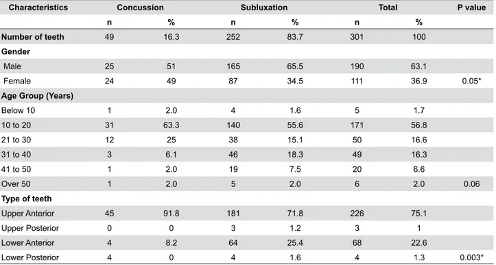
subluxation (65.5%) (p=0.05) was the most prevalent
among men aged 10 to 20 years (Table 1), most
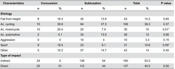
caused by cycling accidents (36.2%) (Table 3).
The occurrence of concussion and subluxation
affected the anterior teeth more, but among the upper
anterior teeth, concussion (91.8%) was the most
prevalent type of injury, while subluxation (25.4%)
(p=0.003) was the most prevalent among the lower
teeth (Table 1). Presence of previous dental trauma
occurred only in cases of subluxation (p=0.04) (Table
2).
Motorcycle accidents represented the etiology of
20.4% of cases of concussions, and 7.9% (p=0.01)
of cases of subluxation (Table 3), showing a positive association with concussion (correlation coefficient rank=0.128, p=0.027) and negative association with
subluxation (correlation coefficient rank=-0.0124, p=0.032).
Among the cases studied, 18.4% of concussion and
9.1% of subluxation cases happened during sports
activities (p=0.05) (Table 3). Sporting accidents were also directly and significantly related to concussion (correlation coefficient rank=0.115 and p=0.047), but not with subluxation.
Thermal sensitivity (41.9%) and occlusion (30.6%)
are the most common symptoms of concussion and
subluxation. Sensitivity to occlusion (p<0.0001) and
mobility (p=0.03) were more related to the cases of
subluxation than concussion (Table 4).
In 8 (16.3%) cases of concussion and 68 (27.1%)
of subluxation, pulp necrosis was diagnosed, and
68.4% occurred within 3 months (Table 5). However,
the Spearman correlation test shows that the more
time passes, the greater the occurrence of pulp necrosis (p≤0.0001). The upper teeth are those at greatest risk of necrosis (correlation coefficient rank -0.0190, p=0.001).
The presence of pulp necrosis was positively
correlated with motorcycle accidents (p=0.014), the occurrence of direct impact (p≤0.0001), crown fracture with pulp exposure (p≤0.0001), darkening of the crown (p=0.004), and spontaneous pain (p≤0.0001); and negatively correlated with the occurrence of indirect impact (p≤0.0001), completion of antibiotic therapy, dental splint, and occlusal adjustment.
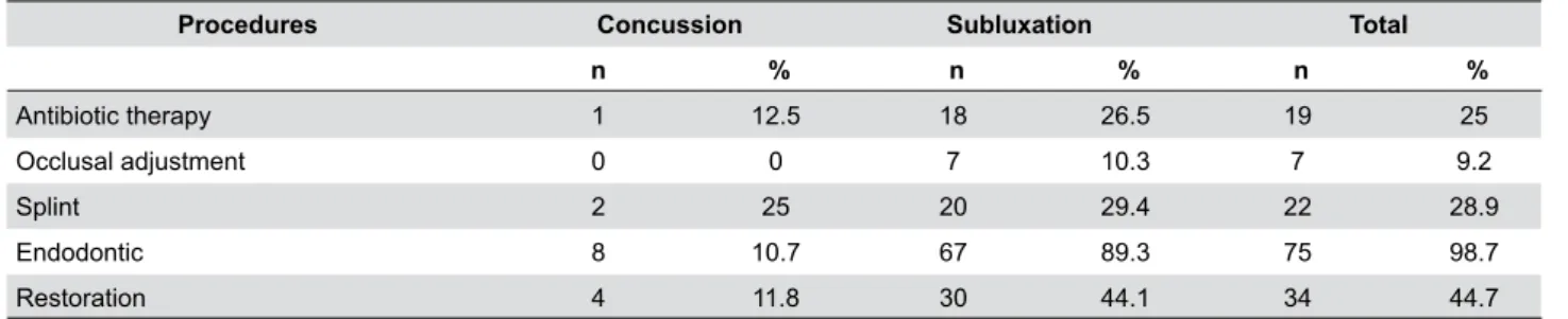
Table 6 shows the main treatments applied in
this sample. Endodontic treatment was necessary
for 16.3% and 26.6% of cases of concussion and
subluxation, respectively.
Discussion
This retrospective research evaluated the evolution of concussion and subluxation cases; pulp necrosis appears more often from 0 to 6 months after the trauma. Direct impact, darkening of the crown, presence of spontaneous pain, and complicated crown
Characteristics Concussion Subluxation Total P value
n % n % n %
Number of teeth 49 16.3 252 83.7 301 100
Gender
Male 25 51 165 65.5 190 63.1
Female 24 49 87 34.5 111 36.9 0.05*
Age Group (Years)
Below 10 1 2.0 4 1.6 5 1.7
10 to 20 31 63.3 140 55.6 171 56.8
21 to 30 12 25 38 15.1 50 16.6
31 to 40 3 6.1 46 18.3 49 16.3
41 to 50 1 2.0 19 7.5 20 6.6
Over 50 1 2.0 5 2.0 6 2.0 0.06
Type of teeth
Upper Anterior 45 91.8 181 71.8 226 75.1
Upper Posterior 0 0 3 1.2 3 1
Lower Anterior 4 8.2 64 25.4 68 22.6
Lower Posterior 4 0 4 1.6 4 1.3 0.003*
* denotes statistically significant result
P values are for comparisons between the two groups, and χ2 or Fisher’s exact test for appropriate categorical variables
Table 1- Demographic characteristics and type of teeth in the cases of concussion and subluxation
History Concussion Subluxation Total P value
n % n % n %
Presence of previous dental trauma
No 48 100 229 92.3 277 93.6
Yes 0 0 19 7.7 19 6.4 0.04*
Associated dental trauma
No 32 65.3 198 78.6 230 76.4
Crown fracture without pulp exposure 10 20.4 40 15.9 50 16.6
Crown fracture with pulp exposure 7 14.3 13 5.2 20 6.6
Root fracture 0 0 1 0.4 1 0.3 0.06
* denotes statistically significant result
P values are for comparisons between the two groups, and χ2 or Fisher’s exact test for appropriate categorical variables
fracture show a greater association with pulp necrosis
in these cases.
Traumatic dental injuries, in some findings, are more common in men aged between 11 and 20
years4,10,11,16,18-20, whose etiology factor include the
practice of more aggressive sporting activities because
of their more violent behavior17,21. This study showed
that among the cases of concussion and subluxation,
the most affected age group was 10-20 years for both
genders, the most predominant etiologies among men
were bicycle accidents (41.6%) and sporting events (13.5%); while for women were bicycle accidents (41.4%) and fall from height (13.8%).
Among all participants, the cycling accident was
the most common etiology followed by fall from
height, car accident, sports and motorcycle accidents,
Characteristics Concussion Subluxation Total P value
n % n % n %
Etiology
Fall from height 8 16.3 35 13.9 43 14.3 0.65
Ac. cycling 15 30.6 94 37.3 109 36.2 0.37
Ac. motorcycle 10 20.4 20 7.9 30 10 0.01*
Ac. automotive 2 4.1 34 13.5 36 12 0.06
Aggression 0 0 10 4 10 3.3 0.15
Sport 9 18.4 23 9.1 31 10.6 0.05*
Other 5 10.2 37 14.7 42 14 0.40
Type of impact
Indirect 24 0 136 54 160 53.2
Direct 25 51 112 44 137 45.5 0.50
* denotes statistically significant result
P values are for comparisons between the two groups, and χ2 or Fisher’s exact test for appropriate categorical variables
Table 3- Etiology and type of impact occurred regarding concussions and subluxations
Clinical signs Concussion Subluxation Total P value
n % n % n %
Spontaneous pain 4 8.2 33 13.1 37 12.3 0.33
Mobility 5 10.2 77 30.6 82 27.2 0.003*
Sensitivity to occlusion 4 8.2 88 34.9 92 30.6 0.0001*
Thermal sensitivity 24 49 102 40.5 126 41.9 0.27
Sensitivity to percussion 15 30.6 72 28.6 87 28.9 0.77
Dental darkening 1 2.0 4 1.6 5 1.7 0.59
* denotes statistically significant result
P values are for comparisons between the two groups, and χ2 or Fisher’s exact test for appropriate categorical variables
Table 4- Frequency of clinical signs in cases of concussion and subluxation
Time (months) Concussion Subluxation Total P value
n % n % n %
Less than 1 4 50 15 22.1 19 25
1 to 2 1 12.5 32 47.1 33 43.4
3 to 6 2 25 11 16.2 13 17.1
7 to 12 0 0 5 7.4 5 6.6
Over 12 1 12.5 5 7.4 6 7.9
Total 8 100 68 100 76 100 0.67
Table 5- Time between the injury and the diagnosis of pulp necrosis
* denotes statistically significant result
probably because of the great number of bicycles in
the region18 and participants’ age. For other studies,
the most prevalent etiologies were domestic accidents, sports and fights, falls, and automobile accidents10,11,16 probably because of the different characteristics of
the population.
E a r l i e r r e p o r t s d e s c r i b e d t h a t m a x i l l a r y
incisors10-12,16,21 are the most traumatized teeth
because of their prominence, which sometimes present
themselves in a protrusive position11, confirming the
data found.
Little attention is sometimes given to the kind of force that caused the trauma. Direct impacts cause a significant number of crown fractures as well as the roots associated with concussion and subluxation.
This result shows a greater association of dental
fractures with concussion than subluxation, even
though there is a similar prevalence of direct impacts
in both groups. These fractures absorb most of the
forces that affect the tooth, reducing the damage
to the periodontium insertion, somehow preventing
tooth avulsion. However, young patients are more
susceptible to traumas of dislocation than fractures
due to the greater resilience of bone tissue, being
generally the most affected population3,15.
Motorcycle accidents showed a direct correlation
with concussion and not with subluxation, what can
be explained, in most cases, by the indirect impacts
resulting from the use of safety equipment, such as helmets, known to produce high impact trauma. This shows the importance of educational campaigns for greater traffic safety.
The clinical signs most observed in both injuries
were sensitivity to thermal stimuli, occlusion and
percussion, as well as mobility and spontaneous pain.
According to Andreasen and Pedersen2 (1985), pain
is usually reported during occlusion and mastication.
Injury to periodontium and pulp by concussion and
subluxation is usually small, transient, and without
serious consequences2,5,6,8,9,16. However, when there
is concomitant crown fracture, mainly in teeth with
complete root formation, the possibility of necrotic pulp
increases, considering that, in general,the patients
are young people with large dentinal tubules, which,
when exposed, may lead to contamination of the pulp,
especially when pulp exposure occurs3,14.
Regarding the diagnosis of pulp necrosis, there are
different chronological standards for various types of
dislocation. In cases of concussion and subluxation, pulp necrosis can be diagnosed in the first six months after the trauma3,15. In this study, we interpreted that the occurrence of indirect impact decreases the risk of pulp necrosis, as well as antibiotic therapy and
splint and occlusal adjustment, although there was no statistically significant correlation.
Dental darkening has as etiologic factor the obliteration of the root canal and pulp necrosis6,13,16. In this study, the number of teeth with dental darkness was small (6.6%) when compared to the number of
endodontically treated teeth (98.7%), emphasizing
that this clinical sign must not be used as a unique
parameter for the indication of endodontic treatment.
More effective tests, such as for sensitivity and/or
cavity, assist in the early diagnosis of pulp pathologies.
Radiographic examination does not replace the tests
abovementioned for the diagnosis and monitoring of
trauma, being very suitable for the diagnosis of pulp
necrosis and external root resorption, which is rare in
this type of injury9.
We observed that dental splint and occlusal
adjustment decrease the chance of experiencing tooth
necrosis. Dental splint is dispensable for the cases of
concussion and may be employed in subluxation for a short period of time (2 weeks); it is better to maintain the repositioned tooth in correct position, provide
patient comfort and improved function7. In this study,
we found that it was used for the concussion and
subluxation due to more severe injuries occurring in
the neighboring teeth.12 Thus, antibiotic therapy was
also used because of other traumas in the neighboring
Procedures Concussion Subluxation Total
n % n % n %
Antibiotic therapy 1 12.5 18 26.5 19 25
Occlusal adjustment 0 0 7 10.3 7 9.2
Splint 2 25 20 29.4 22 28.9
Endodontic 8 10.7 67 89.3 75 98.7
Restoration 4 11.8 30 44.1 34 44.7
teeth.
Occlusal adjustment is an important step, because
premature contacts may occur even in small shifts,
causing unwanted additional trauma3,18.
The association of subluxation and the presence
of previous dental trauma may be explained by individual behavior and habits (i.e. kind of transport, sport practice), not by a deficiency in the repair of a previous injury.
This retrospective study highlighted the importance of knowledge of the dental trauma repair process in the diagnosis, treatment, monitoring, and prognosis
of cases.
Possible limitations of this study include the reliance
of the method on medical records, completed by
different dental students and dental surgeons along
these years, although the instructions were the same.
Conclusion
Although concussion and subluxation traumas are
considered of minor degrees, they must be followed to
decrease the possibility of pulp necrosis and its early
diagnosis and treatment in a timely manner favors a
good prognosis.
References
1- Andersson L. Epidemiology of traumatic dental injuries. Pediatr
Dent. 2013;35(2):102-5.
2- Andreasen FM, Pedersen BV. Prognosis of luxated permanent teeth
- development of pulp necrosis. Endod Dent Traumatol.
1985;1(6):207-20.
3- Andreasen JO, Andreasen FM, Andersson L. Textbook and color atlas of traumatic injuries to the teeth. Oxford: Blackwell; 2007.
4- Andreasen JO, Andreasen FM, Mejàre I, Cvek M. Healing of 400
intra-alveolar root fractures. 1. Effect of pre-injury and injury factors such as
sex, age, stage of root development, fracture type, location of fracture
and severity of dislocation. Dent Traumatol. 2004;20(4):192-202.
5- Bruno KF, Alencar AH, Estrela C, Batista AC, Pimenta FC.
Microbiological and microscopic analysis of the pulp of non-vital
traumatized teeth with intact crowns. J Appl Oral Sci. 2009;17(5):508
-14.
6- De Cleen M. Obliteration of pulp canal space after concussion
and subluxation: endodontic considerations. Quintessence Int.
2002;33(9):661-9.
7- DiAngelis AJ, Andreasen JO, Ebeleseder KA, Kenny DJ, Trope M,
Sigurdsson A, et al. International Association of Dental Traumatology
guidelines for the management of traumatic dental injuries: 1. Fractures
and luxations of permanent teeth. Dent Traumatol. 2012;28(1):2-12. 8- Emshoff R, Gerhard S, Ennemoser T, Hächel O, Scherl M, Strobl H.
The use of likelihood ratio methodology to find predictors of treatment
outcome in patients with dental injury diagnoses. J Oral Rehabil.
2010;37(2):107-15.
9- Epstein JB, Klasser GD, Kolbinson DA, Metha AS, Johnson BR.
Orofacial injuries due to trauma following motor vehicle collisions:
part 1. Traumatic dental injuries. J Can Dent Assoc. 2010;76:a171. 10- Fariniuk LF, Sousa MH, Westphalen VP, Carneiro E, Silva Neto UX, Roskamp L, et al. Evaluation of care of dentoalveolar trauma. J Appl Oral Sci. 2010;18(4):343-5.
11- Ivancic Jokic NI, Bakarcic D, Fugosic V, Majstorovic M, Skrinjaric I.
Dental trauma in children and young adults visiting a University Dental
Clinic. Dent Traumatol. 2009;25(1):84-7.
12- Koyuturk AE, Kusgoz A. Multiple dentoalveolar traumatic injury: a case report (3 years follow up). Dent Traumatol. 2008;24(4):e16-9.
13- Malhotra N, Mala K. Calcific metamorphosis. Literature review and clinical strategies. Dent Update. 2013;40(1):48-50.
14- Moule AJ, Moule CA. The endodontic management of traumatized
permanent anterior teeth: a review. Aust Dent J. 2007;52(1
Suppl):S122-37.
15- Moule AJ, Moule CA. Minor traumatic injuries to the permanent
dentition. Dent Clin North Am. 2009;53(4):639-59.
16- Oginni AO, Adekoya-Sofowora CA. Pulpal sequelae after trauma
to anterior teeth among adult Nigerian dental patients. BMC Oral
Health. 2007;7:11.
17- Onetto JE, Flores MT, Garbarino ML. Dental trauma in children
and adolescent in Valparaiso, Chile. Endod Dent Traumatol.
1994;10(5):223-7.
18- Panzarini SR, Pedrini D, Poi WR, Sonoda CK, Brandini DA, Monteiro
de Castro JC. Dental trauma involving root fracture and periodontal
ligament injury: a 10-year retrospective study. Braz Oral Res.
2008;22(3):229-34.
19- Toprak ME, Tuna EB, Seymen F, Gençay K. Traumatic dental injuries in Turkish children, Istanbul. Dent Traumatol. 2014;30(4):280-4.
20- Zaleckiene V, Peciuliene V, Brukiene V, Drukteinis S. Traumatic
dental injuries: etiology, prevalence and possible outcomes.
Stomatologija. 2014;6(1):7-14.
21- Zerman N, Cavalleri G. Traumatic injuries to permanent incisors.