Occurrence and structure of extrafloral nectaries
in
Pterodon pubescens
Benth. and
Pterodon
polygalaeflorus
Benth.
(1)Élder Antônio Sousa e Paiva(2), Helena Castanheira de Morais(3),Rosy Mary dos Santos Isaias(2),
Dulce Maria Sucena da Rocha(3) and Paulo Eugênio Oliveira(4)
Abstract – Extrafloral nectaries (EFNs) are structurally variable and widely spread among the an-giosperms. The occurrence of EFNs in leaves of Pterodon polygalaeflorus Benth. and Pterodon pubescens Benth. (Fabaceae: Papilionoideae) were detected in adult specimens, at the time of produc-tion of new buds and flowers. The goals of the present study are to register the occurrence of the EFNs in P. pubescens and P. polygalaeflorus, and provide comparative data on the anatomical structures. The EFNs occur in the rachis and are located under the insertion of each petiolule. Each nectary consists of a small elevation whose apical portion is deeply invaginated, resulting in a depression (secretory pole), a common characteristic of both species. Unicellular, non-glandular trichomes occur along the rachis, being less numerous in P. polygalaeflorus while in P. pubescens they cover the EFNs. The secretory tissue consists of parenchyma cells with dense cytoplasm compactly arranged. The nec-tar reaches the surface of the EFNs by rupturing the thin cuticle which covers the secretory pole, since both species lack stomata or any other interruption at the epidermis. The basic difference between the two species, in relation to the EFNs, is the density of the pubescence, which is always greater in P. pubescens. Structural and dimensional modifications may be observed, even between basal and apical nectaries in the same rachis, so it does not constitute a taxonomical tool.
Index terms: nectaries, plant anatomy, plant secretions, nectar, plant protection, herbivores, plant ani-mal relations.
Ocorrência e estrutura de nectários extraflorais
em Pterodon pubescens Benth.e em Pterodon polygalaeflorus Benth.
Resumo – Nectários extraflorais (NEFs) são estruturalmente variáveis e de ampla ocorrência entre as angiospermas. A ocorrência dos NEFs nas folhas de Pterodon polygalaeflorus Benth. e Pterodon pubescens Benth. (Fabaceae: Papilionoideae) foi detectada em espécimes adultas, durante a produção de novas gemas e flores. Os objetivos deste estudo foram registrar a ocorrência de NEFs em P. pubescens e P. polygalaeflorus, e fornecer dados comparativos sobre a estrutura anatômica destas estruturas. Os NEFs ocorrem na raque e estão localizados sob a inserção de cada peciólulo. Cada nectário consiste de uma pequena elevação cuja porção apical é fortemente invaginada, resultando em uma depressão (o pólo secretor), característica comum a ambas as espécies. Tricomas tectores unicelulares ocorrem ao longo da raque, sendo menos numerosos em P. polygalaeflorus, enquanto em P. pubescens eles co-brem todo o NEF. O tecido secretor consiste de células parenquimáticas com citoplasma denso. O néctar alcança a superfície dos NEFs pela ruptura da fina cutícula que cobre o pólo secretor, uma vez que ambas as espécies não apresentam estômatos ou qualquer outra interrupção da epiderme neste local. A diferença básica entre as duas espécies, em relação aos NEFs, é a densidade da pubescência, que é sempre maior em P. pubescens. Modificações estruturais e de dimensões podem ser observadas até mesmo entre os nectários basais e apicais de uma mesma raque, e portanto tais modificações não apresentam valor taxonômico.
Termos para indexação: nectários, anatomia vegetal, secreção vegetal, néctar, proteção das plantas, herbívoros, relação planta-animal.
(1)Accepted for publication on January 28, 2000.
(2)Universidade Federal de Minas Gerais (UFMG), Dep.
de Botânica, Caixa Postal 486, CEP 31270-901 Belo Horizonte, MG. E-mail: [email protected], [email protected]
(3)Universidade de Brasília (UnB), Dep. de Ecologia,
CEP 70910-900, Brasília, DF. E-mail: [email protected], [email protected]
(4)Universidade Federal de Uberlândia, Dep. de Biociências,
Introduction
Pterodon pubescens
Benth. and
Pterodon
polygalaeflorus
Benth. are common species from
Central Brazil vegetation. Their distribution is
dis-junctive, and mixed populations were not observed,
which may indicate some soil characteristic as a
lim-iting factor.
Recently, some researches have tried to establish
distinctive characteristics between the two species,
which were taxonomically situated as synonyms for
P. emarginatus
(Lewis, 1987).
Extrafloral nectaries are extremely variable in
structure and may occur all over the shoots (Elias,
1983; Oliveira & Leitão Filho, 1987). The EFNs
dif-fer from hydatoids, resin glands and other secretory
structures by their sugar watery secretion, where
amino acids are common (Bentley, 1977). The great
structural variability and wide occurrence of the EFNs
are evidenced by several authors (Keeler, 1980;
Oliveira & Leitão Filho, 1987; Morellato & Oliveira,
1991; Oliveira & Oliveira-Filho, 1991; Schupp &
Feener, 1991; Fiala & Linsenmair, 1995).
The EFNs function as defensive traits against
her-bivores and their efficiency vary spatially and
tem-porarily (Pickett & Clark, 1979; Keeler, 1980; Heads
& Lawton, 1985; Barton, 1986).
Mutualistic relationship involving EFNs and
in-sects have been studied. Ants feed on the nectar, which
is rich in carbohydrates and amino acids, and protect
the plants from other insects (Pickett & Clark, 1979).
The flora of the “Cerrados” is rich in plant species
with EFNs and many ant species have been found
associated to the EFNs (Oliveira & Brandão, 1991;
Oliveira et al., 1995).
Qualea grandiflora
and
Q. multiflora
(Vochysiaceae) are typical “Cerrado”
species and the ants that visit their EFNs actually act
as antiherbivores agents (Costa et al., 1992; Del-Claro
et al., 1996).
Beyond the antiherbivore action, some researches
have demonstrated that the insects which visit the
EFNs are responsible for an increase in seed
produc-tion by significantly reducing the predaproduc-tion of the
reproductive structures (Inouye & Taylor, 1979;
Keeler, 1981).
The occurrence, morphology, density and
dispo-sition of the EFNs in plants may have taxonomical
value (Metcalfe & Chalk, 1979). Among the Fabaceae
from "Cerrado", the EFNs are more frequent in the
Mimosoideae (Oliveira & Leitão Filho, 1987).
Ac-cording to Elias (1983), the EFNs are also common
in the Caesalpinioideae, but not frequent in the
Papilionoideae. Oliveira & Oliveira-Filho (1991)
studied the distribution of the EFNs in woody plants
of the “Cerrado”, including areas of occurrence
of
P. pubescens
and
P. polygalaeflorus
, but these
spe-cies were not included in the lists of spespe-cies
with EFNs.
The goals of the present study were to register
the occurrence of the EFNs in
P. pubescens
and
P. polygalaeflorus
, and provide comparative data on
the anatomical structures.
Material and Methods
Leaves of P. pubescens and P. polygalaeflorus were collected during the end of the dry season, in five speci-mens of each species which were producing new flowers and buds, in Brasília, DF, Brazil. Nectar production was observed, allowing the detection and localization of the nectaries. The sugar watery secretion of the EFNs was tested with Fehling reagent (Kraus & Arduin, 1997).
The leaves collected were completely expanded and free from injuries, and were fixed in FAA50 Johansen
(1940). In the laboratory, they were submitted to the usual processes for studies of the anatomical organization of extrafloral nectaries.
Some of the leaves were dissected in the stereomicro-scope. Rachis portions, including the EFNs, were dehy-drated in butanolic series (Strasburger, 1924; Johansen, 1940; Sass, 1951) and embedded in Paraplast. Transverse and longitudinal sections, 12 µm thick, were done in a rotative microtome – Reichert Jung. Staining procedures were done in safranin and astrablue (Johansen, 1940) and/or astrablue and basic fuchsin (Kraus & Arduin, 1997). Slides were mounted in Entellan.
Results and Discussion
Extrafloral nectaries occur in the rachis, under the
insertion of each petiolule (Figures 1 and 2). They
consist of a small elevation which apical portion is
deeply invaginated, forming a depression, where the
secretory pole is located (Figures 3 and 4). These
characteristics are common to
P. pubescens
and
P. polygalaeflorus
.
Figure 1. Longitudinal section of the rachis of P. polygalaeflorus through the EFN (bar = 100 µm; bp: base of petiolule; spr: secretory parenchyma; vb: vascular bundle).
P. pubescens
and scarcely distributed in
P. polygalaeflorus
. Because of these characteristics
the EFNs are difficult to visualize in
P. pubescens
,
where they are covered under a dense layer of
tri-chomes.
P. polygalaeflorus
exhibit trichomes on the
margins of the EFNs, which protect the aperture.
Nev-ertheless, the number of these structures is variable
and they can be lacking, especially, in the superior
portion of the rachis.
In the basal portion of the leaf, the EFNs are
slightly larger than those on the apical portion, in
both species. This difference is also reflected in
nec-tar production, which can be registered through field
observations.
The EFNs in both species present compactly
ar-ranged parenchymatous cells with dense cytoplasm
and large nucleus, which constitute the secretory
tis-Figure 2.Longitudinal section of the rachis of P. pubescens through the EFN (bar = 100 µm; bp: base of petiolule; spr: secretory parenchyma; vb: vascular bundle).
sue (Figures 5 and 6). Sclereids are observed in the
margins of the depression, around the secretory pole.
These cells confer mechanical protection to the
parenchyma, therefore, they are not always present,
especially in
P. pubescens
. According to Elias (1983),
these sclereids are related to protection against
suck-ing insects. In the EFNs studied, this mechanical
bar-rier may not act as a protection against insects, once
it does not involve the secretory tissue completely,
and is concentrated mainly in the margin of the
secretory pole.
Crystals of calcium oxalate may be present in the
vicinity of the parenchyma, associated to the phloem.
Even though these crystals are more abundant in
P. polygalaeflorus
specimens, this difference may be
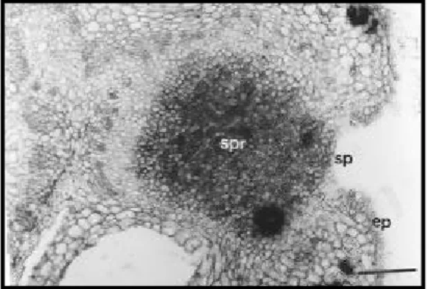
Figure 3. Transverse section of the rachis of P. polygalaeflorus through the EFN (bar = 100 µm; ep: epidermis; sp: secretory pole; vb: vascular bundle).
Figure 4. Transverse section of the rachis of P. pubescens through the EFN (bar = 100 µm; ep: epidermis; sp: secretory pole; vb: vascular bundle).
calcium oxalate in plant tissues
(Zindler-Frank, 1995).
The secretory pole, in both species, is covered by
simple epidermis, lacking trichomes and stomata
(Figures 5 and 6). The cuticle is thin, when compared
to other parts of the leaf. In some transverse sections,
cuticular fragments are observed detaching from the
epidermis, which suggests that the secretion is
accu-mulated between the cuticle and the epidermis.
There-fore, to reach the surface of the EFN, it causes the
rupture of the cuticle.
The nourishment of the nectaries is provided by
the main bundles disposed in a circle, at the center of
the rachis, and by two other vascular bundles,
lo-Figure 6. Transverse section of the rachis of P. pubescens through the EFN (bar = 100 µm; ep: epidermis; sp: secretory pole; spr: secretory parenchyma).
Figure 5. Transverse section of the rachis of P. polygalaeflorus through the EFN (bar = 100 µm; ep: epidermis; sp: secretory pole; spr: secretory paren-chyma).
cated laterally to the rachis in
P. pubescens
as well
as in
P. polygalaeflorus
. The vascular system of the
rachis is similar in both species; the lateral bundles
are present all along the rachis, being involved in the
vascularization of the leaflets.
The vascular pattern and the anatomical structure
of the EFNs in
P. pubescens
and
P. polygalaeflorus
is similar. The differences encountered are related to
the dimensions and density of trichomes near the
secretory pole and may be observed within each
spe-cies and even within the nectaries in the same leaf.
Therefore, in
P. pubescens
, the density of the
tri-chomes in the rachis is considerably higher than that
in
P. polygalaeflorus
, covering completely the EFN.
This characteristic constitutes a relevant
taxonomi-cal tool in the distinction of these two species.
Oliveira & Leitão-Filho (1987) include
P. pubescens
among the species with no EFNs, which
lead to the conclusion that the activity of the EFNs
in this species, as well as in
P. polygalaeflorus
may
be concentrated in a short period of time, during the
production and expansion of new leaves. This fact
should limit the distinction of the EFNs in field
ob-servations. The register of EFNs in
P. pubescens
and
P. polygalaeflorus
should represent the first step to
include them as potential species for ecological and
interactional studies in the “Cerrados”.
Conclusions
1. This is the first register of the occurrence of
the EFNs in
P. pubescens
and
P. polygalaeflorus
.
2. Difficulties in the identification of the EFNs,
in the field, may be due to the fact that the
produc-tion of nectar is practically restricted to the period of
leaf expansion and differentiation.
3. The restricted period of the production of
nec-tar represents an efficient protection against
herbi-vores during the critical period of susceptibility of
the leaves.
4.
P. pubescens
has a higher number of trichomes,
not only over the EFNs, but all over the primary plant
body.
5. The anatomical characteristics of the EFNs are
not confident for the taxonomical identification of
the two studied species.
Acknowledgements
To Dr. Evelyn S.A.M. Frazier for the English
re-view and valuable criticisms on the manuscripts.
References
BARTON, A. M. Spatial variation in the effect of ants on an extrafloral nectary plant. Ecology, Washington, v. 67, n. 2, p. 495-504, 1986.
BENTLEY, B. L. Extrafloral nectaries and protection by pugnacious bodyguards. Annual Review of Ecology, Palo Alto, v. 8, p. 407-428, 1977.
COSTA, F. M. C. B.; OLIVEIRA-FILHO, A. T.; OLIVEIRA, P. S. The role of extrafloral nectaries in Qualea grandiflora (Vochysiaceae) in limiting herbivore: an experiment of ant protection in cerrado vegetation. Ecological Entomology,Oxford, v. 17, p. 363-365, 1992.
DEL-CLARO, K.; BERTO, V.; REU, W. Effect of herbi-vore deterrence by ants on the fruit set of an extrafloral nectary plant, Qualea multiflora (Vochysiaceae). Journal of Tropical Ecology, Cambridge, Great Britain, v. 12, n. 6, p. 887-892, 1996.
ELIAS, T. S. Extrafloral nectaries: their structure and dis-tribution. In: BENTLEY, B.; ELIAS, T. S. (Ed.). The bi-ology of nectaries. New York : Columbia University Press, 1983. p. 174-203.
FAHN, A. Secretory tissues in plants. New York : Aca-demic, 1979. 302 p.
FIALA, B.; LINSENMAIR, K. E. Distribution and abun-dance of plants with extrafloral nectaries in the woody flora of a lowland primary forest in Malaysia. Biodiversity and Conservation, London,v. 4, p. 165-182, 1995.
HEADS, P. A.; LAWTON, J. H. Bracken, ants and extrafloral nectaries. III. How insect herbivores avoid ant predation. Ecological Entomology, Oxford, v. 10, p. 29-42, 1985.
INOUYE, D. W.; TAYLOR, O. R. A temperate region plant-ant-seed predator system: consequences of extrafloral nectar secretion by Helianthella quinquenervis. Ecology, Washington,v. 60, n. 1, p. 1-7, 1979.
JOHANSEN, D. A. Plant microtechnique. New York : McGraw Book, 1940.523 p.
KEELER, K. H. Distribution of plants with extrafloral nec-taries in temperate communities. American Midland Naturalist, Notre Dame, v. 194, p. 274-280, 1980.
KRAUS, J. E.; ARDUIN, M. Manual básico de métodos em morfologia vegetal. Seropédica : Editora da UFRRJ, 1997. 198 p.
LEWIS, G. P. Legumes of Bahia. Kew : Royal Botanic Gardens of Kew, 1987. 369 p.
METCALFE, C. R.; CHALK, L. Anatomy of the dicoty-ledons. 2. ed. Oxford : Clarendon, 1979. v. 1.
MORELLATO, L. P. C.; OLIVEIRA, P. S. Distribution of extrafloral nectaries in different vegetation types of Ama-zonian Brazil. Flora, Salisbury, v. 185, p. 33-38, 1991.
OLIVEIRA, P. S.; BRANDÃO, C. R. F. The ant commu-nity associated with extrafloral nectaries in the Brazilian cerrados. In: HUXLEY, C. R.; CUTLER, D. F. (Ed.). Ant-plant interactions. Oxford : Oxford Science, 1991. p. 198-212.
OLIVEIRA, P. S.; KLITZKE, C.; VIEIRA, E. The ant fauna associated with the extrafloral nectaries of Oureatea hexasperma (Ochnaceae) in an area of cerrado vegetation in central Brazil. Entomologist’s Monthly Magazine, Wallingford, v. 131, p. 77-82, 1995.
OLIVEIRA, P. S.; LEITÃO FILHO, H. F. Extrafloral nec-taries: their taxonomic distribution and abundance in the woody flora of cerrado vegetation in Southeast Brazil. Biotropica, Washington, v. 19, n. 2, p. 140-148, 1987.
OLIVEIRA, P. S.; OLIVEIRA-FILHO, A. T. Distribution of extrafloral nectaries in the woody flora of communities in Western Brazil. In:PRICE, P. W.; LEWINSOHN, T. M.; FERNANDES, G. W.; BENSON, W. W. (Ed.). Plant-ani-mal Interactions: evolutionary ecology in tropical and temperate regions. New York : J. Wiley, 1991. p. 163-175.
PICKETT, C. H.; CLARK, W. D. The function of extrafloral nectaries in Opuntia acanthocarpa (Cactaceae). American Journal of Botany, Columbus,v. 66, n. 6, p. 618-625, 1979.
SASS, J. E. Botanical microtechnique. Ames : Iowa State College Press, 1951. 228 p.
SCHUPP, E. W.; FEENER, D. H. Phylogeny, life form, and habitat dependence of ant-defended plants in a Pana-manian forest. In: HUXLEY, C. R.; CUTLER, D. F. (Ed.). Ant-plant interactions. Oxford : Oxford Science, 1991. p. 175-197.
STRASBURGER, E. Handbook of practical botany. London : G. Allen&Nonwin, 1924.533 p.