UNIVERSIDADE DE LISBOA
FACULDADE DE CIÊNCIAS
DEPARTAMENTO DE BIOLOGIA ANIMAL
Contribution to the functional analysis of genes involved in
cyclic GMP signaling in bacteria of the Burkholderia cepacia
complex
Sofia Patrão Neves Alves
Mestrado em Biologia Humana e Ambiente
Dissertação
UNIVERSIDADE DE LISBOA
FACULDADE DE CIÊNCIAS
DEPARTAMENTO DE BIOLOGIA ANIMAL
Contribution to the functional analysis of genes involved in
cyclic GMP signaling in bacteria of the Burkholderia cepacia
complex
Sofia Patrão Neves Alves
Orientação:
- Professor Doutor Jorge Humberto Gomes Leitão (IST) - Professora Doutora Deodália Maria Antunes Dias (FCUL)
Mestrado em Biologia Humana e Ambiente
Acknowledgments
A Master's thesis is a long journey with many obstacles along the way. This work would not have been possible without the help of those who never stopped supporting and encouraging me, who never let me put down my arms and give up.
First I would like to express my gratitude to my supervisor, Professor Doctor Jorge Leitão for accepting me into his group, for the availability and concern with the achievement of my work and whose expertise, understand, and patience made this work possible.
To Professor Doctor Deodália Dias, Professor and coordinator of the Master in Human Biology and Environment, for agreeing to be co-advisor of this thesis, the availability and sympathy showed whenever I needed help and advice.
A very special thanks to Dr. Sílvia Sousa who helped me whenever I needed; teaching, suggesting and clarifying all the doubts that emerged throughout this work, showing great availability and creating a good working environment.
To Joana Feliciano which proved to be very helpful and always available to answer any questions.
To Miguel Rosa, Inês Nunes, Sara Gomes and friends for all the support and motivation during this process, being always present in good and bad times.
Finally, I would also like to tanks my family, mother, father and brother for the unconditional support gives during this last year.
ii
Sumário
O complexo Burkholderia cepacia (Bcc) é constituído actualmente por 18 espécies de bactérias patogénicas oportunistas, capazes de causar infecções respiratórias graves e por vezes letais doentes com fibrose quística (FQ). A FQ é uma doença genética autossómica recessiva mais comum em caucasianos e é causada por mutações no gene CFTR (Cystic Fibrosis Transmembrane Conductance Regulator), que está localizado no cromossoma 7 e codifica uma proteína de 1480 aminoácidos que funciona como canal transportador de iões cloreto (Cl-) nas membranas epiteliais.
Vários estudos têm sido realizados com o objectivo de entender quais os mecanismos moleculares associados aos processos de colonização e persistência destas bactérias nos doentes com FQ. Normalmente, os organismos pertencentes ao Bcc começam por colonizar o tracto respiratório dos doentes com FQ, podendo em alguns casos invadir o epitélio e sobreviver dentro de células epiteliais e de macrófagos.
As infecções causadas pelo Bcc em doentes com FQ têm um desenvolvimento clinico imprevisível, variando desde assintomático até ao “síndroma cepacia”, uma pneumonia necrotizante rápida e fatal, que pode levar à morte do paciente num período de tempo de cerca de uma semana. Estas bactérias possuem ainda uma elevada resistência a múltiplos antibióticos, o que dificulta a sua erradicação.
Vários factores de virulência têm sido descritos para as bactérias do complexo
Burkholderia cepacia, tais como sistemas de “quorum-sensing” (QS), produção de
biofilmes, produção de exopolissacáridos (EPS), ilhas genómicas e a multirresistência a antibióticos, entre outros
Sabe-se actualmente que o mensageiro secundário di-guanilato cíclico (c-di-GMP) desempenha um papel importante na biologia das bactérias. Devido à sua interacção com várias moléculas efetoras, o c-di-GMP tem uma função regulatória a nível dos processos de transcrição e tradução e na actividade e estabilidade de proteínas. Este permite controlar uma grande variedade de funções relacionadas com o comportamento da comunidade, a secreção, adesão e mobilidade, contribuindo para a virulência de organismos patogénicos. No entanto, ainda pouco se sabe sobre os mecanismos reguladores envolvidos.
A caracterização dos mecanismos associados à regulação pelo c-di-GMP representa um dos principais desafios na compreensão das suas funções. No género
de biofilmes e na invasão e citotoxicidade para as células humanas. Os níveis intracelulares de c-di-GMP são controlados pelas enzimas diguanilato ciclase (DGC) e fosfodiesterase (PDE). As DGC canalizam a sintetize c-di-GMP a partir da condensação de duas moléculas de GTP, enquanto que a actividade de PDE cataliza a hidrolise do c-di-GMP em pGpG. As proteínas com actividades DGC e PDE apresentam, respectivamente, os motivos GG(D/E)EF e EAL. Vários estudos com o objectivo de encontrar genes que codifiquem para proteínas DGC com domínio PAS revelaram a presença de 3 genes no genoma de B. cenocepacia J2315. Estes genes são conservados nos genomas de outras estirpes do Bcc
Diversos estudos têm vindo a demonstrar que concentrações elevadas de c-di-GMP podem suprimir a mobilidade e estimular a formação de biofilmes e a produção de componentes superficiais, tais como a celulose. Estas consequências no comportamento bacteriano devido a elevados níveis de concentração de c-di-GMP podem levar a um bloqueamento das células num estado séssil. Em contraste, baixos níveis de
concentração de c-di-GMP vão ter consequências opostas, inibindo portanto a formação de biofilmes e a produção de componentes superficiais, estimulando a mobilidade. Assim, é possível estabelecer a ligação entre os níveis de c-di-GMP e o comportamento e adaptação bacteriana a ambientes específicos.
A concentração intracelular de c-di-GMP pode ser rapidamente modulada através do aumento da expressão de proteínas com domínios GGDEF e EAL, alcançando assim respectivamente a saturação ou o esgotamento, do conteúdo intracelular de c-di-GMP na célula.
O trabalho desenvolvido teve como principal objectivo o estudo do papel da sinalização pelo mensageiro secundário c-di-GMP na adaptação de bactérias do Bcc a condições ambientais típicas dos pulmões de doentes com Fibrose Quística. Com este objectivo, foram seleccionados 2 genes (BCAM0580 E BCAM2836) de B. cenocepacia J2315 que codificam para DGCs.
Com este trabalho foi possível estabelecer uma associação entre a presença de níveis elevados de proteínas com os domínios conservados EAL e GGDEF e a produção de EPS e virulência. No entanto, não foram detectadas diferenças significativas com a superexpressção dos genes BCAM0580 e BCAM2836, indicando que é necessário mais trabalho para adquirir mais conhecimentos sobre os papéis desempenhados pelo mensageiro secundário c-di-GMP na virulência do Bcc.
iv Este trabalho contribuiu para esclarecer o papel do mensageiro secundário c-di-GMP como um factor de virulência, o que permitiu um conhecimento mais profundo da biologia destes patogénicos. Este estudo contribuiu para a identificação de novos alvos para a identificação de novos agentes antimicrobianos ou para a implementação de novas e melhores estratégias terapias para a irradicação destes patogénicos.
Abstract
The Burkholderia cepacia complex (BCC) currently comprises 18 species of opportunistic pathogenic bacteria capable of causing and often lethal respiratory infections in patients with cystic fibrosis (CF). Several studies have been performed envisaging the understanding of the molecular mechanisms underlying the processes associated with the colonization and persistence of these bacteria in the respiratory tract of patients with CF.
It is now well known that the action of the second messenger cyclic di-guanylate (c-di-GMP) is involved in the adaptive process to new environmental conditions. Due to their interaction with various effector molecules, c-di-GMP has a regulatory function at the levels of transcription and translation processes, and on the activity and stability of proteins. The intracellular levels of c-di-GMP are controlled by the enzyme activities diguanylate cyclase (DGC) and phosphodiesterase (PDE). The DGC enzyme activity synthesizes c-di-GMP by condensing two GTP molecules, while the PDE enzyme activity catalises the hydrolysis of c-di-GMP into pGpG.
Proteins with DGC and PDE activities have, respectively, the GG(D/E) EF and EAL domains. A bioinformatic study was initiated to find genes which encode for DGC proteins. This analysis revealed the presence of three genes enconding putative DGC proteins in the genome of B. cenocepacia J2315
The studied of the role of the second messenger signaling c-di-GMP in the biology of Bcc bacteria involved the functional characterization of two B.cenocepacia J2315 genes encoding DGCs domain.
In this work we were able to establish an association between the presence of increased levels of proteins with the conserved domains EAL and GGDEF and the production of EPS and virulence. However, no significant differences were detected when overexpressing the BCAM0580 or BCAM2836, indicating thet further work is necessary to gain knew knowledge on the roles played by the secong messenger c-di-GMP on Bcc virulence.
vi
Palavras chave
Complexo Burkholderia cepacia (Bcc),
Mensageiro secundário di-guanilato cíclico (c-di-GMP), Diguanilato ciclase (DGC),
Fosfodiesterase (PDE), Burkholderia cenocepacia
Keywords
Burkholderia cepacia complex
Second messenger cyclic di-guanylate (c-di-GMP) Diguanylate cyclase (DGC)
Phosphodiesterase (PDE)
List of abbreviations
(v/v) – Volume per Volume A – AdenosineApr – Resistance to ampicillin
Bcc – Burkholderia cepacia complex
BLAST – Basic Local Alignment Search Tool bp – base pairs
BSA – Bovine Serum Albumin C – Cytosine
CF – Cystic Fibrosis
CFTR – Cystic Fibrosis Transmembrane conductance Regulator CGD – Chronic Granulomatous Disease
DGC - diguanylate cyclase DNA – DeoxyriboNucleic Acid
dNTP’s – 2’-DeoxyNucleoside 5’-Triphosphate EPS – Exopolysaccharide IPTG – Isopropyl-β-D-thiogalactopyranoside kb – 103 base pairs kDa - 103Dalton LB – Luria Broth LPS – Lipopolysaccharide nm - nanometer ºC – degrees celcius OD – Optical density
ORF – Open Reading Frame
PAGE – PolyAcrylamide Gel Electrophoresis PCR – Polymerase chain reaction
PDE - phosphodiesterase
PIA – Pseudomonas isolation agar Ppi – Pyrophosphate
QS – Quorum Sensing Rpm – rotations per minute TAE – Tris Acetate EDTA buffer
TEMED – N, N, N’, N’ – tetramethylethylenediamine Tpr – Resistance to trimethoprim
V – volt W – Watt
INDEX
1. Introduction ... 1
1.1. Burkholderia cepacia complex ... 1
1.2. Bcc pathogenesis ... 2
1.3. Burkholderia cenocepacia: Taxonomy and epidemiology ... 5
1.4. Virulence in Bcc ... 8
1.4.1. Caenorhabditis elegans infection model ... 9
1.5. Cyclic diguanylate ... 12
1.5.1. C-di-GMP metabolism: the GGDEF and EAL domains ... 12
2. Aims of the thesis ... 16
3. Materials and Methods ... 17
3.1. Bioinformatics analysis ... 17
3.2. Strains and plasmids used ... 18
3.3. DNA amplification and Polymerase Chain Reaction (PCR) conditions ... 20
3.4. Insertion of foreign DNA in bacterial cells. ... 22
3.4.1. Preparation of bacterial cells for elecroporation ... 22
3.4.2. Electroporation ... 22
3.5. Protein overexpression and purification ... 23
3.5.1. Production and purification of His-tagged BCAM0580 ... 23
3.5.1.1. Superexpression conditions ... 23
3.5.1.2. Purification conditions used for BCAM0580 ... 24
3.5.2. Production and purification of His-tagged BCAM2836 ... 24
3.5.2.1. Superexpression conditions ... 24
3.5.2.2. Purification conditions used to BCAM2836 ... 25
3.6. Denaturing polyacrylamide gel electrophoresis (SDS-PAGE)... 26
3.7. In vitro phosphodiesterase (PDE) activity ... 27
3.8. Determination of C-di-GMP intracellular levels ... 27
3.9. Expolysaccharide production ... 29
3.10. Virulence assays in the nematode Caenorhabditis elegans ... 29
4. Results and discussion ... 31
4.1. Bioinformatics analysis ... 31
4.2. Protein Overexpression and purification ... 33
4.3. In vitro phosphodiesterase (PDE) activity ... 34
4.5. Expolysaccharide quantification ... 38
4.6. Bacterial virulence assays ... 40
5. Final discussion ... 41
6. Conclusion and future perspectives ... 43
1. Introduction
1.1.
Burkholderia cepacia complex
Burkholderia cepacia bacteria were first described in 1950 by William Burkholder
as Pseudomonas cepacia, the phytopathogen responsible for the sour skin disease in onions[1]. Recently some properties of impact in agriculture and industry were described for this species, including the ability to degrade aromatic compounds pollutants, and to promote plant growth [2, 3]. Burkholderia cepacia was initially described as a single species, but is now divided in at least species, forming the
Burkholderia cepacia complex (Bcc) [4].
Burkholdecia cepacia complex (Bcc) is a group currently composed of at least 18
closely-related species, however, it is possible that the number of Bcc species will increase due to ongoing taxonomic studies [5-7]. Members of the Bcc are gram-negative bacteria of the β-proteobacteria subdivision and include plant, animal and human pathogens, with a widespread distribution in natural and man-made inhabitats [8].
The increasing number of studies on Bcc bacteria in the last few years is due to their evolvement as important and problematic opportunistic Human pathogens, infecting mainly immunocompromised individuals, patients suffering for the chronic granulomatous disease (CGD), and more importantly, cystic fibrosis (CF) patients [5, 9].
Although isolates belonging to all the Bcc species have been obtained from CF patients, it has been shown that B. cenocapacia and B. multivorans are the prevalent species among CF patients. These two species have been associated with higher mortality rates among CF patients [5]. For that reason, these two species have been the main focus of research groups studying the molecular biology, pathogenesis, and epidemiology of Bcc bacteria [4, 10].
2
1.2.
Bcc pathogenesis
Although Bcc bacteria can cause a wide range of infections in healthy and debilitated individuals, CF patients are the group of humans most affected by respiratory infections caused by Bcc [11]
Cystic Fibrosis is the most common autosomal recessive disorder among Caucasians, which affects about 1 in 2500 live newborns [12]. Despite the impressive advances in understanding the molecular basis and pathophysiology of this genetic disorder, CF remains the most common life-threatening single gene disorder in the Caucasian population [13].
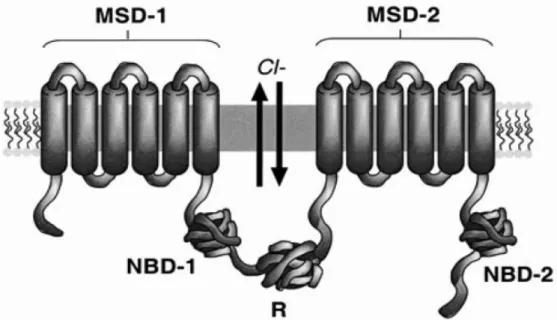
CF is a disease caused by mutations in a 250 kb gene, located on the long arm of chromosome 7, encoding a 1480 amino acid residue polypeptide, known as the Cystic Fibrosis Transmembrane Conductance Regulator (CFTR) (Figure 1) [14, 15]. The CFTR gene encodes a transmembrane glycoprotein, which acts as an electrolyte (chloride-ion) transporter at the apical membrane of epithelial cells and is a member of the ABC transporter family that forms Cl- channel [16].
Figure 1 - Schematic diagram of the CFTR structure. CFTR is a member of the ABC family and consists
of a tandem repeat of the ABC motif. CFTR is composed of five domains: two membrane-spanning domain (MSD-1 and MSD-2), two nucleotide-binding domain (NBD-1 and MBD-2) and a regulatory domain (R). Each MSD motif is composed of six transmembrane stretches of amino acids, followed by an NBD. In CFTR, the two occurrences of this motif are separated by the R domain. Each NBD is able to bind and hydrolyze ATP to operate the chloride (Cl-) channel: hydrolysis of ATP by NBD-1 opens the chloride channel, while ATP hydrolysis by NBD-2 closes the chloride channel. Channel function is further regulated by phosphorylation of serine residues in the R domain. Adapted from [16]
The membrane spanning domains (MSDs) appear to contribute to the formation of a chloride channel pore, since the mutation of specific residues inside the first MSD alters the anion selectivity of the channel [17]. The NBD is responsible for the binding and hydrolysis of ATP and provide the energy necessary for channel activity. The regulatory domain modulates the channel activity of CFTR and can have both inhibitory and stimulatory effects [17].
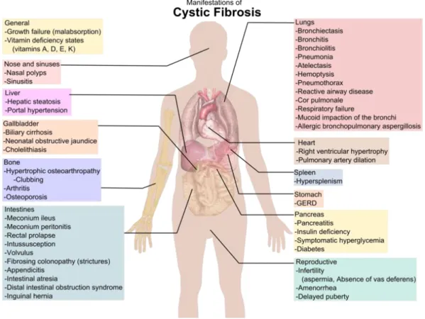
Mutations on CFTR affects the epithelial cells of several organs, including the respiratory tract, exocrine pancreas, intestine, vas deferens, hepatobilirary system and the exocrine sweat gland. This results in multi-organ disease, characterized by the buildup of sticky mucus in the lungs (which predisposes to infection and compromises the respiratory function), pancreatic insufficiency, male infertility and high sweat electrolyte loss [16, 18]. Chronic microbial colonization of the major airways lead to debilitating exacerbations of the pulmonary function and are the major cause of morbidity and mortality among CF patients [19]. The major phenotypic characteristics of CF patients are presented in Figure 1.
Figure 2 - Phenotypic features consistent with a diagnosis of CF. Adapted from:
http://upload.wikimedia.org/wikipedia/commons/thumb/e/e1/Cystic_fibrosis_manifestations.png/79 4px-Cystic_fibrosis_manifestations.png
4 Exocrine pancreatic insufficiency is present in 85% of patients with CF and is the only clinical feature well correlated with genotype [20]. Clinical manifestations such as steatorrhoea and poor nutrition normally are not deadly if overcome with diet and pancreatic enzyme replacement. The Pancreatic sufficient patients is present in 15% of the cases of CF and normally is present at an older age, with these patients usually having a milder lung disease and normal or borderline sweat electrolyte values [21].
Despite all the symptoms, the pulmonary consequences of CF present the most serious danger to CF patients. Chronic microbial colonization of the major airways leading to debilitating exacerbations of pulmonary function are the major cause of mortality and CF patients [19, 21]. On the other hand, in a healthy individual, inhaled bacteria are directed onto the mucus blanket lining the major airways, moved upward by mucociliary clearance and eliminated to the digestive tract. The pathogens that are able to overcome this barrier are faced with the immunological system [22]. In CF patients, those barriers are partially or totally impaired, enabling opportunistic pathogens to establish. Therefore usually the organisms belonging to the Bcc begin to colonize the respiratory tract of CF patients, and in some cases are able to escape the innate immune system, due to the capacity of overproducing extracellular enzymes and consequently to invade the epithelium and survive within macrophages and epithelial cells, making difficult it’s eradication [23, 24].
The Bcc infections are particularly important because they are associated with the lack of a single standard of the clinical evolution, ranging from asymptomatic carriage to chronic infection or the “cepacia syndrome”. The “cepacia syndrome” is a rapid and fatal necrotizing pneumonia, often associated with a rapid decline of lung function, and accompanied by marker pyrexia, multi-organ failure and positive blood cultures, which can lead to death of the patient in less than one week [5, 25]. Briefly, CF patients that are infected with Bcc strains have a higher probability of nosocomial transmission and death, and require longer hospitalizations.
In the last few years, it has been a prevalence of transmissions and infection replacement associated with epidemic strains of the B. cenocepacia species, with an increased virulence and a greater difficulty to eradicate by antibiotic therapy [5, 18]. B.
cenocepacia pathogenesis is thought to be related with the ability of the bacteria to
adapt and survive under changing conditions, which is likely necessary for establishing chronic infections [26].
Since Bcc bacteria are resistant to many clinically useful antibiotics, the study of virulence determinants is important to indentify bacterial processes that could be targeted by novel antibiotics or alternative anti-infective therapies [27].
1.3.
Burkholderia cenocepacia: Taxonomy and epidemiology
Nowadays, the taxonomic characterization of Bcc is based on a polyphasic approach which includes whole-cell protein profiling, whole-cell fatty acid analysis, and conventional biochemical tests, DNA-DNA hybridization, 16S rRNA and recA genes sequencing [28]. More recently, a multilocus sequence typing (MLST) scheme and a public data-base of MLST sequences from Bcc (http://pubmlst.org/bcc/) has been successfully implemented. The MLST scheme for Bcc bacteria is based on the nucleotide polymorphism of seven genes the atpD, gltB, gyrB, recA, lepA, phaC, and
trpB encoding, respectively, the housekeeping enzymes ATP synthase β-chain, the
glutamate synthase large subnit, the DNA gyrase B, the recombinase A, the GTP-binding protein, the acetoacetyl-CoA reductase and the tryptophan synthase [1]
The use of all of these techniques have demonstrated that Bcc bacteria are phenotypically similar, but with significant differences at the genomic level, leading to their classification in several species [28].
The Bcc bacteria have unusual genomes with large dimensions (about 8 Mb), organized in 3 chromosomes and often several plasmids (Table 1). Burkholderia species genomes have a high G+C content (68%), characterized by multireplicon structure and the occurrence of numerous gene duplications, insertion sequences, and mobile elements. All these elements contribute to the plasticity of Burkholderia genomes and their ability to acquire a wide range of metabolic pathways. The
Burkholderia genomes can also mutate rapidly when the organisms are subject to in vitro stress conditions or during infections [26, 29].
6 Table 1-Genomefeatures of Bcc strains with their genome projects completed and in public access. Chr: Chromossome, p: plasmid, nt: nucleotides, NA: information not available. Adapted from: [1]
Species/strain Genome size (nt) Replicons Protein coding genes G + C (%)
B. ambifaria MC40-6 7642536 3 Chr+1 p 6878 66.4 B. cenocepacia J2315 8055782 3 Chr+1 p 7229 66.9 B. dolosa AUO158 6247594 NA 5441 66.8 B. lata 383 8676277 3 Chr+1 p 7725 66.8 B. multivorans ATCC 17616 7008622 3 Chr+1 p 6290 66.7 B. ubonensis Bu 6932532 NA 7192 67.3 B. vietnamiensis G4 8391070 3 Chr+1 p 7775 65.7
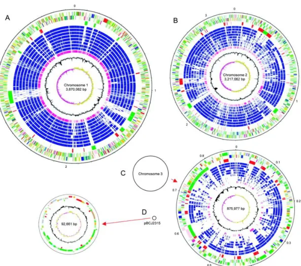
The genome of B. cenocepacia J2315 (also known as the Edinburgh-Toronto strain, responsible for the death of several patients with CF) is already sequenced, and has a total of 8.056 Mb making it one of the largest observed in Gram-negative bacteria [30]. It comprises three circular chromosomes of 3.870, 3.217 and 0.867 Mb and a plasmid of 92.7 kb, with a G+C content of approximately 66.9% (Figure 3) [31] . This genome encodes a broad array of functions typical of this metabolically versatile genus, as well as numerous virulence and drug resistance functions. Although B.
cenocepacia strains can be isolated from soil and can be pathogenic to both plants and
animals, B. cenocepacia J2315 is representative of a particularly epidemic lineage of B.
cenocepacia rarely isolated from the environment and which spreads between CF
patients. The B. cenocepacia J2315 genome contains evidence that its unique and highly adapted genetic content has played a significant role on its success as an epidemic CF pathogen [26, 30]. The interest in this strain is due to these characteristics, its high prevalence worldwide and the high number of genomic islands, the high genomic plasticity and it’s metabolic versatility [30].
Figure 3- Schematic circular diagrams of the B. cenocepacia J2315 genome. The circular diagrams for
chromosomes 1 (A) and 2 (B) are drawn to scale, whereas those for chromosome 3 (C) and plasmid pBCJ2315 (D) are not drawn to scale. Black circles representing these replicons are drawn to scale. The key for the three chromosomal circular diagrams (A, B, and C; outside to inside), with scale in Mb, is as follows. Annotated CDSs are colored according to the predicted function represented on a pair of concentric circles, representing both coding strands. CDSs in genomic island regions are indicated in green, and other RODs defined by pairwise genome comparisons with other BCC are indicated in red. CDSs with matches identified by reciprocal FASTA with other Burkholderia species—B. cenocepacia HI2424, B. cenocepacia AU1054, B. contaminans 383, B. ambifaria AMMD, B. vietnamiensis G4, B. xenovorans LB400, B. pseudomallei K96243, and B. thailandensis E264—are indicated in dark blue. Orthologues shared with Ralstonia solanacearum GMI1000 are indicated in purple. For the G+C content plot, the GC bias (G−C/G+C) is indicated as >1% in khaki and <1% in purple. (D) The key for the circular diagram for plasmid pBCJ2315 is as described for the chromosomes but lacks the orthologue matches. Color coding for CDS functions: dark blue for pathogenicity/adaptation, black for energy metabolism, red for information transfer, dark green for surface associated, cyan for degradation of large molecules, magenta for degradation of small molecules, yellow for central/intermediary metabolism, pale green for unknown, pale blue for regulators, orange for conserved hypothetical, brown for pseudogenes, pink for phage and IS elements, and gray for miscellaneous. Adapted from [30].
8
1.4.
Virulence in Bcc
Several virulence factors have been described for Bcc bacteria, such as lipopolysaccharides (LPS), “quorum-sensing systems, biofilm production, exopolysaccharides (EPS) production, genomic islands, and the multidrug resistance to multiple antibiotics, among others [1, 26, 32].
The LPS of Bcc induces a strong immune response that can contribute to host cell damage [1]. It can increase the inflammatory nature of Bcc infections in CF patients and contributes to the resistance of B. cenocepacia to the antimicrobial peptide polymixin B and human neutrophil peptide 1 (HNP-1) in vitro[33, 34]. The structure of Bcc LPS differs from the LPS from other gram-negative bacteria, in the sense that contains less phosphate, the unusual sugar Dglycero-α-D-talo-oct-2 ulopyranosylonic acid (KO) is present in the inner core oligosaccharide, and 4-amino-4- deoxyarabinose residues are bound to phosphates of the lipid A. These modifications lower the anionic charge of the Bcc cell surface, inhibiting the binding of polymyxin and of cationic peptides antibiotics [1, 5]. Bcc strains also present several distinct O-antigen structures.
So far at least five quorum-sensing (QS) systems have been described in Bcc bacteria: CepIR , CciIR, CepR2, HHQ and BDSF [1, 35].
The first QS system for Bcc bacteria is denominated CepIR. The B. cenocepacia CepIR system has been show to be critical for full virulence in rodent and worm models of infection, being related to virulence phenotypes such as toxin, protease, lipase, siderophores and phenazine production, swarming motility and biofilm formation [5, 36, 37]. The CepIR system allows the regulation of gene expression based on the bacterial population density, due to the accumulation of extracellular compounds secreted by bacteria (in this case N-acyl-homoserine lactones or AHLs), which when reached a particular concentration is altered resulting in gene expression of the whole population in a coordinated manner [38, 39]
Another important feature of Bcc is their ability to form biofilms. Biofilms are complex, multicellular bacterial communities that can protect bacteria from antibiotics and the host immune system [40]. The formation of biofilms seems to be associated with the ability of the bacteria to produce and secrete acyl-homoserine lactones (AHLs), which are signaling molecules involved in quorum sensing system response. Bcc bacteria growing in biofilms show decreased susceptibility to ceftazidime and
ciprofloxacin, contrasting to planktonic cells, which are more susceptible to these antimicrobials [41].
Biofilm formation is also affected by exopolysaccharide synthesis, mobility and iron availability [26]. These characteristics indicate that biofilm formation is a complex process, involving numerous B. cenocepacia virulence determinants, making it a target for the development of new antibiotics [26]
Genomic islands (GI) are thought to contribute to the genome plasticity and metabolic diversity of the Bcc bacteria, acquired by horizontal gene transfer. Several studies suggest that these represent more than 10% of the genome in some species of Bcc. The acquisition of these GI appears to play an important role in the evolution of epidemic strains, by introducing new functions that promote the survival and pathogenesis of these bacteria in the CF lung [8].
Although EPS production by Bcc was initially considered a rare phenomenon, the majority of Bcc clinical isolates from CF patients were found to produces variable amounts of EPS [8, 26, 42]. The chemical structure of EPS is composed of glucose, manose, rhamnose, galactose and glucuronic acid. EPS production by B. cenocepacia clinical isolates is thought to contribute to the overall virulence of the organism through inhibiting both neutrophil chemotaxis and neutriphil generation of H202 and O2- [8, 26]. Production of EPS results in slower clearance of bacteria in lungs.
EPS production is highly stain-dependent and even virulent strains such as B.
cenocepacia J2315 produce few or no EPS.
1.4.1. Caenorhabditis elegans infection model
An important factor for the difficulty in fighting infections by Bcc is its intrinsic multidrug resistance to antibiotics. This has been attributed to the action of the various efflux pumps that efficiently way remove antibiotics from the cell, the formation of biofilms which reduce the contact between the cell surface and the antibiotics, and changes in the cell envelope which decrease the permeability of the cell wall to antibiotics [43].
Several models of infection by Bcc bacteria have been developed to gain a deeper knowledge on the virulence and pathogenesis of these bacteria. This include including mammals, fishes, nematodes, insects, protozoa and plants. With the exception of mammals, none of the others has adaptive immunity [5]. This is the case of the
10 nematode C. elegans, which has a innate immunity to fight infections, like many other infection models [44]. Caenorhabditis elegans is a small, free-living soil nematode that lives in many parts of the world. The nematode survives by feeding on microbes, especially bacteria. The use of this nematode as a model of infection is very advantageous due to its characteristics like its short life cycle, compact genome, stereotypical development, ease of propagation and small size (the adult bodyplan is composed with about 1000 somatic cells) [45]. Another useful feature of C. elegans as model of infection is that it is relatively straightforward to test pathogenesis. As the worm feeds on pathogenic bacteria, the worm will suffer an infection and then the survival rate and other pathogenic traits of the infection can be easily monitored [46].
Despite its simple anatomy, this nematode displays a large repertoire of behavior including locomotion, foraging, feeding, defecation, egg laying, dauer larvae formation, sensory responses to touch, smell, taste and temperature as well as some complex behaviors like male mating, social behavior, learning and memory [47, 48].
The C. elegans life cycle is about 3 days long under optimal conditions and the worms can be maintained in the laboratory in agar plates and with E. coli as food source [45]. The nematode can also endure harsh environmental conditions by switching to a facultative diapause stage called dauer larva that can survive 4 to 8 times longer than the normal 3-week life span [49].
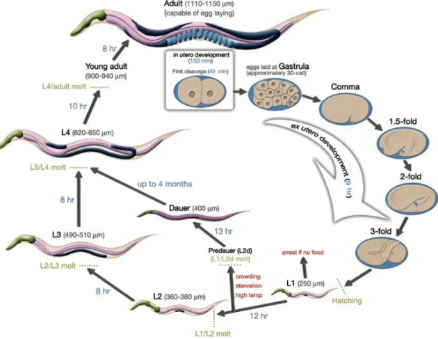
Similar to other nematodes, the life cycle of C. elegans is comprised of the embryonic stage, four larval stages (L1-L4) and adulthood (Figure 3). The end of each larval stage is marked by a molt where a new stage-specific cuticle is synthesized and the old one is shed. Molting is accomplished in three steps: first is the separation of the old cuticle from the hypodermis (apolysis), then the formation of new cuticle arising from the hypodermis, and at last the shedding of the old cuticle (ecdysis) [45, 50].
Figure 4- Life cycle of C. elegans at 22ºC. 0 min indicates the fertilization event. Numbers in blue along the arrows indicate the length of time the animal spends at a certain stage. First cleavage occurs at about 40 min. postfertilization. Eggs are laid outside at about 150 min. postfertilization and during the gastrula stage. The length of the animal at each stage is marked next to the stage name in micrometers. Adapter from [45].
One of the main problems of using models of infection is that some virulence factors are not universal, being specific to the host used as a model of infection as observed when a pathogenicity test using multiple hosts was performed [1, 26, 44].
12
1.5.
Cyclic diguanylate
Cyclic diguanylate or c-di-GMP is a small molecule that was first described in 1987 as an allosteric activator of a bacterial cellulose synthase [51].
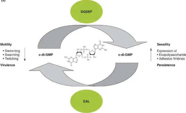
C-di-GMP is now recognized as a bacterial second messenger, since it has shown to have a more global role as a signaling molecule in bacteria. C-di-GMP has been shown to bind to a variety of receptors (proteins and riboswitches) with the consequence in the control of a variety of functions related with secretion, adhesion and mobility, contributing to the virulence of pathogens[52]. C-di-GMP signaling contributes to the regulation of a wide spectrum of phenotypes. The most investigated phenotypes are motility, sessility and virulence. These bacterial phenotypes are often interconnected as they can promote or inhibit each other. However, little is known about the regulatory mechanisms involved [53].
The characterization of the mechanisms associated with the regulation by c-di-GMP is a major challenge in understanding their functions [53-55]. It has been proven that in Burkholderia species c-di-GMP is involved in the synthesis of flagella, mobility, biofilm formation and invasion and cytotoxity to human cells [55, 56]. It was also recently demonstrated in B. cenocepacia the interaction between the “quorum sensing” BDSF system and the c-di-GMP signaling system [57].
The intracellular concentration of the second messenger c-di-GMP is the result of the action of two enzyme activities, diguanylate cyclase (DGC) and phosphodiesterase (PDE), which are responsible for the biosynthesis and hydrolysis of c-di-GMP, respectively. C-di-GMP has been shown to bind to a variety of receptors (proteins and riboswitches) with the consequence of physiological changes previously enumerated [53].
1.5.1. C-di-GMP metabolism: the GGDEF and EAL domains
C-di-GMP is synthesized from two molecules of GTP, via a two-step reaction with 5’-pppGpG as a reaction intermediate, with the concurrent release of pyrophosphate (PPi) [58]. The two GTP molecules are substrates of DGC enzyme activity and these proteins contain a domain GGDEF. Genetic evidence suggested that the GGDEF domain may be sufficient for the DGC enzyme activity[52].
On the other hand, the c-di-GMP is hydrolyzed by enzymes with PDEs activity. The PDE enzymes are characterized by having the EAL or the HD-GYP domains, which hydrolyze molecules of c-di-GMP into pGpG or in two pGs respectively (Figure 4) [52, 59, 60].
Figure 5- Basic biochemistry of c-di-GMP synthesis, degradation, and interactions within c-di-GMP
receptors. The diagrams show the protein domains involved in c-di-GMP metabolism and signaling. Enzymatically active GGDEF, EAL, and HD-GYP domains are shown on a white background.
Enzymatically inactive domains involved in substrate binding are shown in light gray, and domains that are no longer associated with c-di-GMP are shown in dark gray. Adapted from [52]
14 GGDEF and EAL domains proteins are widespread in bacterial genomes and have been found in proteins of all known bacteria, being often linked to regulatory domains, such as phosphorylation receptors or oxygen detection domains [61].
Genetic and biochemical analysis of proteins with a GGDEF domain revealed that this domain is responsible for the diguanylate cyclase activity [58]. In general, GGDEF domains are approximately 170 amino acids long. Stand-alone GGDEF domains are usually not enzymatically active, and require activation through an N-terminal domain signaling domain for activation [62].
The EAL domains are approximately 250 amino acids long. The EAL domains require Mg2+ or Mn2+ for activity, but it is strongly inhibited by Ca2+ or Zn2+. Usually, EAL domains show significant enzymatic activity without N-terminal allosteric activation [62].
Very often one bacterial genome contains more than one GGDEF and EAL domain protein, raising the question of the specificity of the c-diGMP signaling pathway(s). When a protein contains both GGDEF and EAL domains one of the three situations can occur: both domains can be enzymatically active [63], only one domain possesses enzymatic activity, while the other domain remains enzymatically inactive and can serve a regulatory function [64, 65], or none of the domains has enzymatic activity or proteins with only one of the domains don’t work neither as cyclases or phosphodiesterases [66] .
In bacteria, the intracellular concentration of GTP is approximately 100 times higher the concentration of c-di-GMP. Therefore, when c-di-GMP is produced in large quantities, the GTP metabolism is not altered [53]. However, the presence of proteins within EAL motifs and PDE activity in bacteria can lead to high intracellular concentrations of c-di-GMP that suppress motility and stimulate biofilm formation and the production of surface components, such as cellulose [67]. These effects in the bacterial physiology can be due to high c-di-GMP concentration levels and can promote sessility [53, 68].
In contrast, proteins with GGDEF motifs and high DGC enzyme activity can lead to low intracellular concentration levels of c-di-GMP and will have the opposite effects in the bacteria. That is, inhibition of biofilm formation and the production of surface components and stimulating mobility [53, 68].
Thus, it is possible to establish a link between the levels of c-di-GMP and the behavior and bacterial adaptation of bacteria to specific environments (Figure 6) [53].
Figure 6- Regulatory concept of GMP metabolism and signaling on the population level. High
c-di-GMP levels promote sessility aided by the production of adhesive extracellular matrix components such as polysaccharides. Environmental and host persistence is also predicted to be promoted by high c-di-GMP levels. Conversely, low c-di-c-di-GMP levels promote motility behavior (swimming, swarming and twitching motility and virulence). Imagne adapted from [53]
The need to adapt to an environmental that is constantly changing led to the development of sophisticated systems by the bacterial cell with the purpose of detecting these environmental changes. To adapt to new conditions, the cells have multiple sensory that systems activate a chain of intracellular signaling, allowing cells to adjust to the changing environment. The major class of protein domains is represented by the sensing Per-Arnt-Sim, known as PAS domain [69, 70].
PAS domains are important sensory modules that monitor changes in light intansity, redox potential, oxygen availability, small ligands, and the overall energy level of a cell (ratio ATP/ADP) [54, 69, 70]. PAS domains can also sense environmental factors that cross the cell membrane and/or affect cell metabolism. Unlike most of the other sensor modules, the PAS domains are located in the cytosol [71].
16
2. Aims of the thesis
The aim of this work is to the contribute to understanding the role of signaling by the second messenger c-di-GMP in the adaptation of bacteria of the Burkholderia
cepacia complex to environmental conditions typical of the lungs of patients with cystic
fibrosis. For this purpose, two different genes of B. cenocepacia J2315 were selected: one is encoding a putative DGC and another is encoding a putative PDE. Both genes presented an encoded the PAS sensor domain. The bacterial strain used was selected because is a particularly virulent opportunistic human pathogen, having been isolated from CF patients.
This work also intended to contribute to the knowledge of the genes, it’s mechanism of action as well as the effects at the cellular and molecular level, and their involvement in the virulence of this particularly virulent strain belonging to the
Burkholderia cepacia complex (Bcc)
For this purpose was made a detailed bioinformatic analysis for the selection of two genes encoding DGCs activities and their cloning and functional characterization. It was realized the overexpression and purification of the proteins modified with histidine tail to facilitate purification, as well as their biochemical characterization of phosphodiesterase and cyclase activity
The genes were also characterized in their ability to produce exopolysaccharide and survival rate using the nematode Caenorhabditis elegans a model of infection.
3. Materials and Methods
3.1.
Bioinformatics analysis
In this experimental work, we analyzed the DNA sequence and the proteins using bio-computational tools with the purpose of comparing nucleotide and amino acid sequences stored in databases to indentify genes and their functions and to determinate de structure of the proteins in study.
The deduced amino acid sequences of BCAM0580 and BCAM2836 were submitted to the Basic Local Alignment Search Tool (BLAST) from the NCBI (http://blast.ncbi.nlm.nih.gov/Blast.cgi?PAGE=Proteins) for the detection of homologies and identification of the probable protein function.
Sequence motifs and signatures of protein families were analyzed by the MOTIF tool.
The amino acid sequences of the BCAM0580 and BCAM2836 genes were aligned with sequences of homologous proteins retived from the IMG (Integrated Microbial Genomes) databases using the program ClustalX (http://www.clustal.org/)
BCAM0580 and BCAM2836 nucleotide sequences were used to construct specific primers for the amplification of the BCAM0580 and BCAM2836 coding sequences. Both primers were designed with the program OLIGO Primer Analysis Software (http://www.oligo.net/). The NEBcutter software was used (http://tools.neb.com/NEBcutter2/) to obtain the restriction map of BCAM0580 and BCAM2836. The restriction map was used to clone the genes.
The prediction of various physical and chemical parameters for the two different proteins, such as the molecular weight, theoretical pI, amino acid composition, atomic composition, instability index and grand average of hydropathicity were performed using the ProtParam tool able in the Expasy website (http://web.expasy.org/protparam/)
For the automatic prediction of secondary structure based on the amino acid sequence was used de Psipred program.
18
3.2.
Strains and plasmids used
The highly virulent strain Burkholderia cenocepacia J2315 and three different
Escherichia coli strains were the bacterial strains used as host to plasmids or for
overexpress recombinant proteins (Table 2).
Burkholderia cenocepacia J2315 and Burkholderia cepacia IST408 were grown in
Pseudomonas Isolation Agar (PIA) medium at 30ºC for 72 hours and maintained at 4ºC. When appropriate, the PIA plates were supplemented with 300 µg/ml of Trimethoprim. The E. coli strains were maintained in LB solid medium supplemented with antibiotics at the following concentrarions: 100µg/ml of Trimethoprim or 150µg/ml of Ampicillin. When not in use, bacterial strains were kept at -70º in 40% (v/v) glycerol.
Liquid cultures of the Burkholderia and E. coli strains was carried out in Lennox broth (LB) medium, at 37ºC for 24 hours within agitation incubator (250rpm).
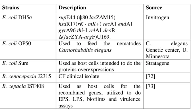
Table 2- Bacterial strains used.
Strains Description Source
E. coli DH5α supE44 (ф80 lacZΔM15)
hsdR17(rK - mK+) recA1 endA1 gyrA96 thi-1 relA1 deoR
Δ(lacZYA-argF)U169.
Invitrogen
E. coli OP50 Used to feed the nematodes
Carnorhabditis elegans
C. elegans
Genetic center, U. Minnesota
E. coli Sure Used as host cells intended to do the proteins overexpressions
Stratagene
B. cenocepacia J2315 CF clinical isolate [72]
B. cepacia IST408 Used as host cells for the recombined genes, utilized to do EPS, LPS, biofilms and virulence assays
[73]
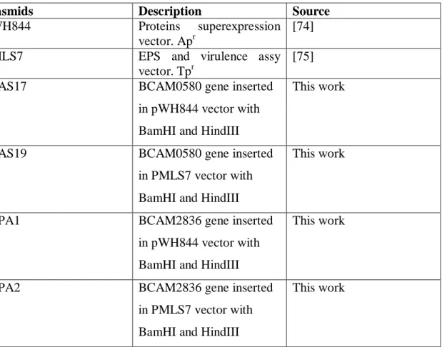
For the studies and manipulation of the BCAM0580 and BCAM2836 genes were used a total of 5 plasmids. Plasmids used in this work are described in Table 3.
Table 3- Plasmids used in this work.
Plasmids Description Source
pWH844 Proteins superexpression
vector. Apr
[74]
pMLS7 EPS and virulence assy
vector. Tpr
[75]
pSAS17 BCAM0580 gene inserted
in pWH844 vector with BamHI and HindIII
This work
pSAS19 BCAM0580 gene inserted
in PMLS7 vector with BamHI and HindIII
This work
pSPA1 BCAM2836 gene inserted
in pWH844 vector with BamHI and HindIII
This work
pSPA2 BCAM2836 gene inserted
in PMLS7 vector with BamHI and HindIII
This work
To construct plasmids for the overproduction of His6-tagged proteins pSAS17 and pSPA1 was constructed by lighting the BCAM0580 and BCAM2836 respectively to the vector pWH844 digested with BamHI and HindIII.
To construct plasmids for the virulence assays, EPS production and for the quantification of intracellular c-di-GMP levels Psas19 and pSPA2 was constructed by lighting BCAM0580 and BCAM2836 respectively to the vector PMLS7 digested with BamHI and HindIII
After digestion, the DNA fragments were precipitated with ethanol and the restricted fragments was the ligated directionally to the expression vector. The ligation mixture contained 250ng of the amplification restricted product, 50ng of the restricted expression vector, 1µl of T4 DNA Ligase and 1X T4 DNA Ligase Reaction Buffer. The reaction was incubated for 30 minutes at 22ºC and the enzyme was inactivated incubating the reaction for 5 minutes at 70ºC. The ligation mixtures were introduced into E. coli DH5 by electrotransformation and the transformants were selected by growing on LB solid medium supplemented with adequate antibiotics and incubated overnight at 37ºC overnight.
20 Plasmid DNA was extracted from overnight cultures of the E. coli host strains grown in LB medium supplemented with appropriate antibiotics using a ZR plasmid MiniprepTM – classic kit, which is designed for efficient isolation of plasmid DNA from
E. Coli.
The recombinant plasmids constructs were confirmed by enzyme restriction analysis.
3.3.
DNA amplification and Polymerase Chain Reaction (PCR) conditions
BCAM0580 and BCAM2836 genes were amplified by Polymerase chain reaction (PCR) using the B. cenocepacia J2315 genomic DNA as template and the specific primers listed in Table 4. The PCR amplification was performed in a GeneAmp® PCR System 2700. The conditions used for the amplification of the two genes were: 100ng template DNA, 0.02U/µl Phusion® Hight-Fidelity DNA polymerase enzyme, 1x of Phusion GC buffer, 200µM of each Deoxynucleotide Triphosphates (dNTPs), 0.5 µM of each primer. 20% glycerol and 6% DMSO were added to the reaction mixtures to amplify BCAM0580 and BCAM2836 respectively.The samples were subjected to an initial denaturation at 98ºC for 3 minutes, followed by 30 cycles of: denaturation, annealing and elongation. At the end it will be submitted to 7 minutes at 72ºC for elongation and samples were stored at 4ºC until used.
The PCR amplifications conditions are presented in table 4.
Table 4- PCR conditions for the amplification of BCAM0580 and BCAM2836 using as template DNA from B. cenocepacia J2315.
Primers Denaturation (Time in seconds) Annealing temperature Elongation Product size BCAM058 0 580-UP 580-LW 98º → 15s 55º → 30s 72º → 65s 2020pb BCAM283 6 2836-UP 2836-LW 98º → 10s 62º → 30s 72º → 30s 963pb
The oligonucleotide primers (Table 5) were design based on the genome sequence of B. Cenocepacia J2315 (http://www.sanger.ac.uk/Projects/B_cenocepacia) and were synthesized as a paid service by MWG Biotech AG (Germany).
Table 5 – Oligonucleotide primers used for PRC amplification. Abbreviations used: A, Adenine; C, Cytosine; G, Guanine; T, Timine
Primer DNA sequence
UP-0580 5’-TTGGATCCATGGATGACGAA-3’, BamHI LW-0580 5’-TTAAGCTTTCAGGCGATCAG-3’, HindIII UP-2836 5’-TTGGATCCCGCGCTATGACG-3’, BamHI LW-2836 5’-TAAGCTTCCGTCACGCGTAC-3’, HindIII
The PCR products were separated by agarose gel electrophoresis (0.8% (wt/t) in TAE 1x buffer). The gel was stained with 1µg/ml GelRedTM in TAE 1X buffer and the DNA was visualized under short wave UV light in a transilluminator (Bio-Rad)
After confirmation of the PCR is specific, the DNA was extracted from the agarose gel using NZYGelpure commercial kit (NZYTech).
22
3.4.
Insertion of foreign DNA in bacterial cells.
Plasmids of interest were inserted into B. cepacia IST408 and E. coli by Electroporation using the procedure described by [76]
3.4.1. Preparation of bacterial cells for elecroporation
The appropriate bacterial host strain was inoculated in 30 ml of LB liquid medium and incubated overnight with orbital agitation (250 rev./min.) at 37 °C for E.
coli or 30 ºC for B. capacia IST408. Aliquots from overnight cultures were used to
inoculate 100 ml of LB liquid medium at a final OD640nm of 0.1 and incubated at 37 ºC or 30 ºC, with orbital agitation (250 rev./min.), until the bacterial culture reached the mid-log phase (OD640nm of 0.8). The cells were harvested by centrifugation at 8000 rev./min. (rotor J2-21, Beckman) for 15 minutes at 4 ºC, in sterile centrifuge bottles. To eliminate all present salts, the cell pellets were washed three times with sterile ice-cold distilled water and centrifuged as described above. The volumes used in the sequential washes were 100 ml, 60ml and 20 ml of ice-cold distilled water. The pellets were then resuspended in 4 ml of 10% (wt/v) glycerol and centrifuged at 5500×g for 15 minutes at 4 ºC. The resulting cell pellets were resuspended in 2 ml 10% (wt/v) glycerol and 110 µl aliquots were frozen at -70 °C, until used
3.4.2. Electroporation
Aliquots of 110µl E. coli sure and B. cepacia IST408 electrocompetent cells prepared as described above were slowly defrozen and 10µl plasmid DNA (previously extracted with the ZR plasmid MiniprepTM) were added to the cells. This mix was added in electro-transformations cuvettes and an short electric pulse was applied. The electotransformation conditions used were: capacitance of 25µF, voltage 2.5 kV and resistant 200 Ohms for the Bcc strains or 400 Ohms for E. coli. After the electric pulse, the cells were incubated with 1ml of LB liquid at 37ºC in the orbital shaking incubator (250rpm) for 1hour or overnight (depending of the strain) for E. coli or Bcc cells recovery, respectively. After incubation, the cells were plated in appropriate selective solid media.
3.5.
Protein overexpression and purification
Recombinant proteins BCAM0580 and BCAM2836 were overproduced in E. coli SURE after transformation with Pwh844 plasmid (pSAS17 and pSPA1 respectively). The proteins were overproduced after induction of plasmids with isopropyl-β-D-thiogalactopyranoside (IPTG)
For the purification it was used a HisTrapTM FF columms (GE Healthcare). These columns retain histidine-tagged fusion proteins when charged with Ni2+ ions. These proteins can be desorbed from the column with buffers containing imidazole, which preserve the antigenicity and functionality of the purified proteins.
The protein concentration of the samples was estimated by with BSA as standard.
3.5.1. Production and purification of His-tagged BCAM0580 3.5.1.1. Superexpression conditions
E.coli sure cells with the plasmid Pwh844 were pre-inoculated in LB liquid medium
containing ampicillin (150 µl/ml) and incubated overnight at 37ºC with orbital agitation (250 rpm). On the next day, 100ml of ampicillin supplemented LB liquid medium were inoculated with cell suspensions equivalent to an initial OD640 nm of 0.1 an incubated at 37ºC with orbital agitation (T0). When the OD640 nm reached 0.6-0.8, the T5 promoter of plasmid pWH844 was induced by the addition of 0.4mM of IPTG. The cells were incubated for an additional period of 16 hours at 16ºC with orbital agitation 160rpm (Tf).
Samples from T0 and Tf were prepared by centrifuging an appropriate volume of culture (volume is equal to 0.6/ OD640nm) and the resulting pellet was ressuspended in 40µl of gel loading buffer h solution [100mM Tris.base (pH 6.8), 4% (wt/v) SDS, 20% (v/v) glycerol, 0.2% (wt/v) bromophenol blue, 200 mM DTT]
After incubation period, inoculums were centrifuged for 5 minutes at 7000rpm at 4ºC. The supernatant was discarded and the retrieved pellet was ressuspendend in 8ml of sonication buffer (Phosphate buffer 1X with pH 7.4 and 10 mM Imidazole) and stored at -80ºC until its use.
24
3.5.1.2. Purification conditions used for BCAM0580
The cells were disrupted by ultrasonic vibration on ice with a Branson 250 sonifier, applying 6 cycles of 25 pulses each at 50% duty cycle and output control 6. Before the last two sonications 2% Tripton and 0.5mM of phenylmethylsulfonyl fluoride(PMSF) were added. The sonicated cells were centrifuged at 12000 rmp for 1 hour at 4ºC and the clear supernatant was collected to a new tube and kept at 4ºC until further use.
The protein of interest was then purified using commertial HisTrapTM FF columms (GE Healthcare).The column was washed with 5ml distilled water and then charged with 0.5ml of 0.1M nickel sulphate salt solution. The column was washed again with 5ml of distilled water and equilibrated with 10ml of Start buffer (Phosphate buffer 1X with pH 7.4, 20 mM Imidazole, 1% Tripton, 0.5mM PMSF). Cells lysates were then loaded into the column (about 8ml) and washed with 10ml of start Buffer. Elution of the His6-tagged protein was performed with 5ml of elution buffer with increasing imidazole concentration (60, 100, 150, 200, 300 and 500 mM). Aliquots (1ml) of the collected fractions were analyzed by SDS-PAGE, and those containing the purified protein were pooled and dialysed overnight at 4ºC in a Slide-A-Lyzer Dialysis Cassettes (10 KDa MWCO) against dialysis buffer (50mM Nacl, 50mM Tris, pH 8.5).
3.5.2. Production and purification of His-tagged BCAM2836 3.5.2.1. Superexpression conditions
E.coli sure cells with the plasmid pWH844 were pre-inoculated in LB liquid
medium containing ampicillin (150 µl/ml) and incubated overnight at 37ºC with orbital agitation incubator. On the next day, 200ml of Super Broth (SB) liquid medium (32 g/l Triptone, 20 g/l yeast extract, 5 g/l NaCl) supplemented with ampicillin were inoculated with cell suspensions equivalent to an initial OD640 nm of 0.1 an incubated at 30ºC with orbital agitation (T0). When the inoculums reached an OD640 nm between 0.6 and 0.8, the T5 promoter was induced by the addition of 0.4mM of IPTG. The induced inoculums were maintained for 2 hours at 30ºC with continues shaking with 250rpm (Tf).
Samples from T0 and Tf were prepared by centrifuging an appropriate volume of culture (volume is equal to 0.6/ OD640nm) and the resulting pellet was ressuspended in 40µl of gel loading buffer h solution.
Incubation period the inoculums were centrifuged for 5 minutes at 7000rpm at 4ºC. It was discard the supernatant and the retrieved pellet was ressuspendend in 8ml of sonication buffer and stored at -80ºC until its use
3.5.2.2. Purification conditions used to BCAM2836
The cells were sonicated on ice with a Branson 250 sonifier, applying 6 cycles of 25 pulses each at 50% duty cycle and output control 6. Between each cycles the cells were lead to rest for 3 minutes. The sonicated cells were centrifuged at 12000rmp for 1 hour at 4ºC and collected the resulting supernatant.
BCAM2836 protein was then purified using commertial HisTrapTM FF columms (GE Healthcare). Initially the column was washed with 5ml distilled water and then charged with 0.5ml of 0.1M nickel sulphate alt solution. The column was washed again with another 5ml of distilled water and equilibrated with 10ml of Start buffer (Phosphate buffer 1X with pH 7,4, 20 mM Imidazole and 10% of glycerol). Cells lysates were then loaded into the column (about 8ml) and washed with 10ml of start Buffer. Elution of His6-tagged protein was done with 5ml of elution buffer with increasing imidazole concentration (60, 100, 150, 250, 350 and 500 mM). Aliquots (1ml) of the collected fractions were analyzed by SDS-PAGE, and those containing the purified protein were pooled and dialyzed overnight at 4ºC in a Slide-A-Lyzer Dialysis Cassettes, 10KDa MWCO against dialysis buffer (50mM Nacl, 50mM Tris, pH of 7.9).
26
3.6.
Denaturing polyacrylamide gel electrophoresis (SDS-PAGE)
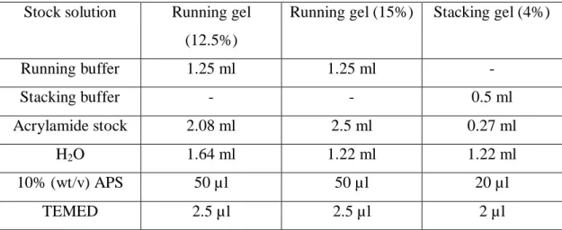
Aliquots of solutions of the purified proteins were analyzed by SDS-PAGE. The composition of the gels prepared is shown in Table 7
Table 6 – Composition of denaturing polyacrylamide gels.
Stock solution Running gel (12.5%)
Running gel (15%) Stacking gel (4%)
Running buffer 1.25 ml 1.25 ml - Stacking buffer - - 0.5 ml Acrylamide stock 2.08 ml 2.5 ml 0.27 ml H2O 1.64 ml 1.22 ml 1.22 ml 10% (wt/v) APS 50 µl 50 µl 20 µl TEMED 2.5 µl 2.5 µl 2 µl
After polymerization, the gels were immersed in running buffer 1X [running buffer 10X contains: 0.25M Tris.base, 1.92M glycin, 1% (wt/s) SDS] and 10µl of denatured protein sample resuspended in gel loading buffer were applied in each well of the gel. Previously, the proteins were denaturated by incubation for 5 minutes at 95ºC. Proteins separation was achieved by applying 160 V, until the dye contained in the loading buffer reached the end of the gel.
The gels were stained by imersion in coomassie blue staining solution [0.2% (wt/v) coomassie blue R-250, 10% (v/v) acetic acid, 47.5% (v/v) ethanol, filtered with Whatman nº1 filter paper] for 25 minutes, after boiling the solution. During incubation, the gel was gently agitated to distribute the dye uniformly over the gel.
After staining, the gel was placed into a previously boiled distaining solution (10% (v/v) acetic acid, 26.25% (v/v) ethanol) for 30 minutes. When the background of the gels were still stained, the gels were moved into fresh distaining solution. The method is sensitive to 0.5 to 20 micrograms of protein.
3.7.
In vitro phosphodiesterase (PDE) activity
In the case of BCAM0580 protein that contains EAL motif, the phosphodiesterase activity was determined by the method described in Lee et al (2010) [56]. The reaction mixtures were prepared in ELISA plates with a total volume of 20 or 40µl, depending of the quantity of enzyme used.
PDE activity was assessed using final concentration of 1mM divalent metal cations MgCl2, MnCl22 or CaCl2, 50mM Tris-HCl (pH 8.5) buffer, and 5mM of bis-p-nitrophenylphosphate (bis-pNPP) as substrate, in the present of 1µM, 2µM and 5µM purified protein. The blank reaction contained all the components except the enzyme. The negative control contained BSA instead of the enzyme. To test the requirement of presence of the divalent cations for PDE activity, another control was prepared with all the components except the cations.
The reaction mixture were added to the wells of an ELISA plate and incubated for 2h at 37ºC. After this time, 80 or 160µl distilled water was added. The amount of p-nitrophenol released was immediately registed using a Low Volume Absorbance Microplate Reader SPECTROstar Nano at of 410 nm using the reaction mixture without enzyme as blank.
3.8.
Determination of C-di-GMP intracellular levels
The diguanylate cyclase enzymatic activity of the proteins with GGDEF domains was determined by the method described by
The c-di-GMP intracellular levels were determined using the colorimetric detection method described by Nakatama et al (2012) [77]. This method is based on the peroxidase activity of the complex formed between c-di-GMP, hemin and proflavine.
For the diguanylate cyclase enzymatic activity of BCAM0580 and BCAM2836, 1, 5 or 10µg of the purified proteins were performed by incubating with 0.5mM GTP, 5 mM MgCl2, 50mM Tris.Hcl (pH 7.9), 50 mM NaCl in a total volume of 3ml. The mix tube was incubated at 30ºC and aliquots of 500 µl were taken after 0, 10, 30, 60 and 120 minutes of incubation. Each reaction was stopped by the addition of with 25µl 0.5 M EDTA (pH 8). After incubation for 5 minutes at 95ºC, samples were centrifuged for 5 minutes to remove protein aggregates. The supernatant was carefully removed and incubated again for 5 minutes at 95ºC, cooled down for 15 minutes at the room
28 The levels of c-di-GMP present in cell lysates of the Burkholderia strains previously grown at 30ºC in 30 ml of S medium with orbital agitation (250rpm) were also quantified. After 24, 48 and 72 cells were harvested by centrifugation, and resuspended to final volume of 2.4 ml of 10 mM Tris-HCL (pH 8.0) containing 100 mM NaCl. Each sample was sonicated (25 seconds, 4 times) and centrifuged one hour at 4ºC, 1200 rpm. The supernatants were filtered using a 0.2 µm filter and 250 µl of each sample were used the c-di-GMP quantification.
In both quantification experiments 50 mM Tris.HCL (pH 7.9), sterilised water were added to obtain a final volume of 500 µl and heated at 65ºC for 5 minutes. The samples were cool down to room temperature, 0.5 μM of Hemin were added and the resulting mixture was incubated 20 minutes at room temperature. After this time was added 30 μMproflavine to the mixture and incubated for 12 hours at 4ºC .
On the following day, 2mM of ABTS was added to all the samples and incubated at 15ºC for 5 minutes. Each sample was divided aliquots and were added on the wells of an ELISA plate. To one of each of the two aliquots 2mM of H2O2 was added and the resulting mixtures were incubated for 5 minutes at 25ºC. The product formated, the radical cation ABTS+, was immediately read in the Low Volume Absorbance Microplate Reader SPECTROstar Nano at a wavelength of 415 nm. Absorbance was normalized by subtracting the absorbance value of the aliquots without H2O2 addition (Figure 7).
Figure 7- Illustration of peroxidase activity of the complex formed between c-di-GMP, hemin and
Negative controls were preformed as described, using 2μg of BSA instead of the purified protein.
An assay with different concentrations of c-di-GMP (BIOLOG) (0, 2, 6, 10, 20, 30, 40 μM) was performed as standard and used as positive control.
3.9.
Expolysaccharide production
The Bcc strains under study were grown overnight in 20ml of LB liquid medium at 30ºC with agitation (250 rmp). These cultures were used to inoculate 30ml S of medium [12.5 g/l Na2HPO4.2H2O, 3 g/l KH2PO4, 1 g/l K2SO4, 1 g/l NaCl, 1 g/l yeast extract, 1 g/l casamino acids, 20 g/l glucose, 0.2 g/l MgSO4.7H2O, 0.01 g/l CaCl.2H2O, 0.001 g/l FeSO4.7H2O] with an initial OD640 of 0.1. Cultures were carried out for 72 hours at 30ºC with orbital agitation (250 rpm).
Samples form cultures were taken after 24, 48 or 72 hours and were mixed with 20ml of 0.9% (wt/t) NaCl. The bacterial cells present in these suspensions were pelleted by centrifugation at 20000xg for 15 minutes. The EPS was precipitated from the cell-free supernatant by the addition of 2.5 volumes of 95% (v/v) cold ethanol. The precipitated EPS was recovered air-dried at room temperature for about three days. The results are the means of at least three independent determinations of EPS dry weight.
3.10. Virulence assays in the nematode Caenorhabditis elegans
The nematode C. elegans DH26 was used as a model of infection by Bcc.
C. elegans was maintained at 20ºC in Nematode Growth Medium I (NGM I) [Per
litre of medium was used: 3 g/l NaCl, 2.5 g/l Tryptone, 17 g/l Agar, 5 ml of Nystatin (10mg/ml in ethanol), 25 ml of 1M KPO4 buffer (pH 6), 1 ml 1M CaCl2, 1 ml 1M MgSO4, 1 ml Uracil (2 mg/ml, sterile filtrated), 0.5 ml Cholesterol (10 mg/ml in ethanol)] petri plates containing a lawn of 100 µL E. coli OP50, previously grown at 37ºC for 24 hours into the surface of agarized (NGM I). NGM I plates can be stored at 4ªC for several weeks, but every 7 days the nematodes were transferred to fresh plates by cutting a piece of agar with the help of a sterile scalpel.
The nematodes were synchronized worms and eggs was rinsed 4 times with 1 ml of sterile bidestilled water and the treatment of the worms and eggs suspension with 500µl of hypochlorite solution [600 µl H2Obidest, 500 µl Sodiumhypochlorite 12%, 400 µl