CLINICAL SCIENCE
Reduced salivary flow and colonization by mutans
streptococci in children with Down syndrome
Cristina Areias,IBenedita Sampaio-Maia,IMaria de Lurdes Pereira,IA´lvaro Azevedo,IPaulo Melo,ICasimiro Andrade,ICrispian ScullyII
IFaculty of Dental Medicine of Porto University, Porto, Portugal.IIUniversity College London, London, UK.
OBJECTIVES: Although individuals with Down syndrome have considerable oral disease, the prevalence of dental caries in this group is low. The present study aimed to compare known risk factors for dental caries development in children with Down syndrome and a matched population (siblings). In both populations, the number of acidogenic microorganisms, such as mutans streptococci, lactobacilli andCandidaspecies, and the paraffin-stimulated pH, flow rate and IgA concentration in whole saliva were evaluated and compared.
METHOD:Saliva was collected, and the caries index was evaluated in 45 sibling pairs aged between 6 and 18 years old. The salivary IgA concentration was determined by immunoturbidimetry. Salivary mutans streptococci, lactobacilli and Candida species were quantified on mitis salivarius agar containing bacitracin and 20% sucrose, rogosa agar supplemented with glacial acetic acid and sabouraud agar supplemented with chloramphenicol, respectively.
RESULTS:Down syndrome children had a higher caries-free rate (p,0.05) and lower salivary mutans streptococci counts (p,0.03) compared to their siblings. Similar numbers of lactobacilli andCandidaspecies were found in both groups. Salivary flow rates were 36% lower in Down syndrome children compared to their siblings (p,0.05). The salivary pH did not differ between Down syndrome children and controls. The Down syndrome children had an IgA secretion rate 29% lower than that of their siblings, but this difference was not statistically significant.
CONCLUSIONS: In conclusion, the lower number of mutans streptococci in the saliva may be one of the factors contributing to the lower caries rate observed in Down syndrome children, despite evidence of hyposalivation.
KEYWORDS: Down Syndrome;Streptococcus Mutans; Immunoglobulin A, Xerostomia.
Areias C, Sampaio-Maia B, Pereira ML, Azevedo A´ , Melo P, Andrade C, et al. Reduced salivary flow and colonization by mutans streptococci in children with Down syndrome. Clinics. 2012;67(9):1007-1011.
Received for publication onDecember 20, 2011;First review completed onApril 20, 2012;Accepted for publication onApril 24, 2012 E-mail: [email protected]
Tel.: 351 917411727
INTRODUCTION
Down syndrome (DS) is a genetic disorder resulting from a trisomy of chromosome 21 that leads to multiple oral abnormalities, including malformations of the small palate and maxilla, delayed tooth eruption and dental agenesis (1-3). DS individuals have a significantly higher prevalence of some oral diseases, including periodontal disease, which develops at early age and is rapidly progressive (4,5), and oral candidiasis (6). However, we (7) and others (8-11) have found a lower prevalence of dental caries in DS populations. Possible causes for the lower prevalence of caries may be dietary and other environmental factors, late tooth eruption, and altered salivary flow and/or composition. Mutans streptococci, including Streptococcus mutansandStreptococcus sobrinus, are the primary
etiological agents of dental caries in most populations (12), and other acid-tolerant oral bacteria, such asLactobacillusspecies, may also be implicated (12). Longitudinal studies have shown that an increase in the numbers of both mutans streptococci and lactobacilli in saliva or plaque over time is associated with caries onset and progression (13,14). Some studies have suggested a link betweenCandida species and dental caries, particularly in children, adolescents and young adults (15), with a possible active role forC. albicansin caries pathogenesis (16). In addition to microbial factors, several salivary components are also related to caries status. Salivary pH and flow play crucial roles in caries development (17). Secretory immunoglobulin A (IgA) in the saliva is also a local defense factor against caries (18).
The aim of the present study was therefore to compare known risk factors for dental caries development in children with DS and a matched population of siblings. In both populations, the number of acidogenic microorganisms, such as mutans streptococci, lactobacilli, and Candida species, and the paraffin-stimulated pH, flow rate, and IgA concentration in whole saliva were evaluated and compared.
Copyrightß2012CLINICS– This is an Open Access article distributed under the terms of the Creative Commons Attribution Non-Commercial License (http:// creativecommons.org/licenses/by-nc/3.0/) which permits unrestricted non-commercial use, distribution, and reproduction in any medium, provided the original work is properly cited.
MATERIALS AND METHODS
All DS children aged between 6 and 18 years old included in the Portuguese national database were invited to participate in the study. For each DS child, the sibling closest in age who was living in the same household was used as the matched control. The exclusion criteria included a lack of siblings, non-Caucasian ethnicity, systemic diseases other than DS, and current medication use. Informed consent was obtained from the participants’ parents, who were provided with detailed information on the study protocol. The ethics committee of Faculty of Dental Medicine of Porto University approved the consent form, and the research protocol was developed in accor-dance with the 1983 revision of the Helsinki Declaration. The present investigation was performed in accordance with European and Portuguese laws.
The final study sample consisted of forty-five Caucasian sibling pairs. Data were gathered using a questionnaire and clinical observations. The present or past history of institu-tionalization and the dietary habits were assessed. The parents answered the questionnaires of both children. Calibrated examiners carried out dental caries examinations using a mirror and explorer in accordance with the World Health Organization criteria and methods. The total number of decayed, missing and filled primary (dmft) and permanent (DMFT) teeth was recorded for each study patient and control to characterize the epidemiological history of caries in both groups.
We elected to study stimulated saliva because the amount obtained without stimulation was insufficient for the planned biochemical and microbiological analyses. Saliva was col-lected in a quiet room over a 5-minute period between 8:00 AM and noon to minimize circadian rhythm effects and at least 2 h after eating, tooth brushing, or mouth washing. Salivary secretion was stimulated with paraffin pellets (Ivoclar Vivadent, NY, USA), and the children were asked to spit into a sterile tube. The total amount collected over a 5-minute period was registered, enabling the calculation of the stimulated salivary flow rate (ml/min). The salivary pH was measured immediately after saliva collection using pH indicator paper (5.0-8.0, Duotest, Germany). Saliva for IgA analysis was frozen directly at -80
˚
C, whereas the saliva collected for microbiological analysis was mixed 1:1 with Brain Heart Infusion broth (Cultimed, Barcelona, Spain) with 15% glycerol and then frozen at -80˚
C until assayed. The saliva samples were rapidly defrosted in a 37˚
C water bath and mixed well. Salivary IgA was determined by immuno-turbidimetry using an automatic analyzer (Pentra C200,Horiba ABX Diagnostics, Switzerland). The IgA secretion rates (mg/min) were calculated by multiplying antibody titers
by the salivary flow rate (19).
For the microbiological analyses, the samples were serially diluted to 10-6with 0.9% sterile NaCl solution and immediately plated in triplicate on the following culture media: mitis salivarius agar containing 0.2 units of bacitracin/ml plus 20% sucrose to detect mutans strepto-cocci; rogosa agar supplemented with 0.13% glacial acetic acid to assess the number of lactobacilli; and sabouraud agar supplemented with chloramphenicol to evaluate the presence ofCandida. Mitis salivarius agar and Rogosa agar were incubated anaerobically for seven days at 37
˚
C, and Sabouraud agar was incubated aerobically for 48 h at 37˚
C. Colonies were counted, and the results were expressed in colony forming units per ml of saliva (CFU/ml). The lower limit of detection was 104CFU/ml for mutans streptococci and 102CFU/ml for lactobacilli andCandida.Statistical analysis
The categorical variables were described as relative frequencies (%), and the continuous variables were described using the mean ¡ standard deviation (SD). When
appro-priate, the chi-square independence test or Fisher’s exact test were used to analyze hypotheses regarding the categorical variables, and Student’st-test was used for the continuous variables. A level of 0.05 was considered significant. The analyses were performed using the statistical analysis soft-ware SPSSHv.17.0 (Statistical Package for Social Sciences).
RESULTS
The mean age of the DS children was 12.7¡4.0 years, and
the mean age of the sibling controls was 12.8¡3.7 years. The
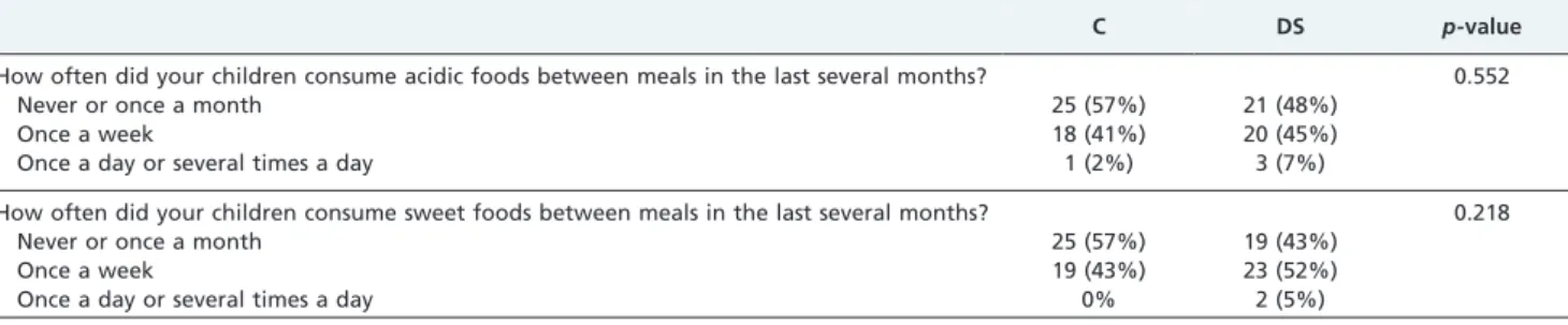
DS group included 49% males, whereas the sibling control group included 60% males. None of the observed children had been institutionalized. The ingestion of a cariogenic diet, i.e., frequency of consuming acidic and sweet foods, was found to be similar between DS children and their siblings (Table 1).
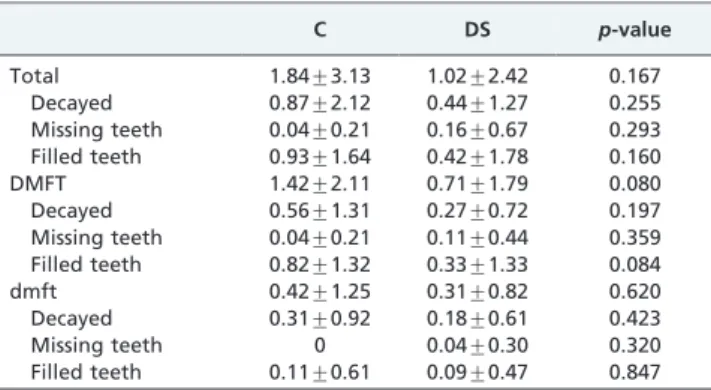
The caries-free rate was significantly higher in the DS group than in the sibling group (Figure 1). The epidemio-logical history of caries in both populations was evaluated based on the dmft and DMFT scores, as shown in Table 2.
Table 3 shows the mutans streptococci, lactobacilli and Candidaspecies relative frequencies in saliva samples from the DS children and the sibling controls. All children in both groups had detectable mutans streptococci in the saliva, but some children did not have detectable lactobacilli orCandida.
Table 1 -Dietary habits in Down Syndrome (DS) and sibling control (C) children, namely regarding the frequency of acidic or sweet food ingestion.
C DS p-value
How often did your children consume acidic foods between meals in the last several months? 0.552
Never or once a month 25 (57%) 21 (48%)
Once a week 18 (41%) 20 (45%)
Once a day or several times a day 1 (2%) 3 (7%)
How often did your children consume sweet foods between meals in the last several months? 0.218
Never or once a month 25 (57%) 19 (43%)
Once a week 19 (43%) 23 (52%)
Once a day or several times a day 0% 2 (5%)
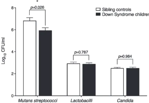
Compared to their siblings, DS children had lower mutans streptococci relative frequencies but similar numbers of lactobacilli andCandidaspecies (Figure 2).
Table 4 shows the pH, salivary flow rates and IgA concen-trations of sibling controls and DS children. The salivary flow rate was 36% lower in DS children than in their siblings, but the pH did not differ between the two groups (Table 4).
Total salivary IgA was similar between the DS and sibling groups (Table 4). Although the DS group presented an IgA secretion rate 29% lower than that of their siblings, the difference was not statistically significant (Table 4).
DISCUSSION
The present study was undertaken to elucidate the factors involved in the lower dental caries prevalence in Down
syndrome (DS) children. Compared to their siblings, DS children showed lower counts of mutans streptococci in parallel with a higher caries-free rate, despite evidence of hyposalivation.
Although DS children had a lower dental caries pre-valence compared with their siblings, the caries history, evidenced by DMFT/dmft scores, was not significantly different between the two groups.
Only a few previous studies have evaluated the number of Streptococcus mutans bacteria in DS children. Some authors have found non-statistically reduced levels (11), while others have found increases (20,21) in comparison to non-DS children. However because oral microbial coloniza-tion is strongly correlated with diet, oral hygiene, and familial pre-disposition, the recommended control group should be a non-DS sibling age-matched group, as used in the present study, rather than unrelated healthy children. In a previous study, we showed that oral hygiene habits were similar between DS children and their siblings (7). Given that the remaining methodology in the present work was similar to that of previous studies, the differences are likely to be related to the different control groups used.
Regarding dietary habits, many epidemiological studies have linked the incidence of caries and food habits, identifying both protective and exacerbating factors. The consistency, viscosity, cooking process, and even the time of ingestion of food containing simple carbohydrates change the cariogenicity of a food. Although the main meals were shared between DS and their siblings, the healthy controls could have different dietary habits because they can more easily buy candy or snacks independently. Interestingly, the consumption of sweet and acidic foods was similar between the DS children and their siblings. Therefore, diet cannot be responsible for the differences found regarding the caries prevalence in these two groups.
Furthermore, higherC. albicanscounts have been reported in DS children in comparison to healthy controls (22), as have candidal infections (6). Again, the differences we found could be related to the control group used. We, like Mathias et al. (21), did not find any differences in Lactobacillus counts between DS children and healthy controls.
There are conflicting reports regarding the salivary pH of individuals with DS in comparison to controls; no difference (8,10,11,23), higher pH (24) and a lower pH (25) have been observed. We believe that the conflicting reports in the literature about the pH levels in DS may be attributed to different measurement methods. The present study, similar to most studies, measured the pH of saliva collected intra-orally and analyzed extra-intra-orally. Some authors believe that this method may be inaccurate because whole saliva does not represent the intra-oral micro environment, the buffer-ing system may change once saliva is removed from the oral cavity, and the pH level of the salivary film covering the soft Figure 1 -The caries-free rates of Down syndrome (DS) children
and sibling controls (C). The bars represent the means, and error bars represent the SDs. p-value was calculated using the chi-square test. *Values are significantly different between DS and sibling controls.
Table 2 -The total numbers of decayed, missing and filled primary (dmft) and permanent (DMFT) teeth in Down syndrome (DS) children and sibling controls (C).
C DS p-value
Total 1.84¡3.13 1.02¡2.42 0.167
Decayed 0.87¡2.12 0.44¡1.27 0.255
Missing teeth 0.04¡0.21 0.16¡0.67 0.293 Filled teeth 0.93¡1.64 0.42¡1.78 0.160
DMFT 1.42¡2.11 0.71¡1.79 0.080
Decayed 0.56¡1.31 0.27¡0.72 0.197
Missing teeth 0.04¡0.21 0.11¡0.44 0.359 Filled teeth 0.82¡1.32 0.33¡1.33 0.084
dmft 0.42¡1.25 0.31¡0.82 0.620
Decayed 0.31¡0.92 0.18¡0.61 0.423
Missing teeth 0 0.04¡0.30 0.320
Filled teeth 0.11¡0.61 0.09¡0.47 0.847
Values are in means¡SD.
Table 3 -The mutans streptococci, lactobacilli orCandida relative frequencies in saliva samples from Down syndrome (DS) children and sibling controls (C).
C DS p-value
Mutans streptococci 100% 100% 1.000
Lactobacilli 73.3% 82.2% 0.310
Candida 77.3% 59.1% 0.067
and hard tissues may not correspond precisely to that of the secreted saliva (8,11,23,26). However, measuring pH intra-orally, especially in cognitively and physically impaired children, such as DS children, is challenging and also raises ethical issues. Further studies evaluating the different pH measurement methods available for saliva are needed.
The stimulated salivary flow of the sibling controls was lower than the common mean value for stimulated salivary flow in adults (,1.2 ml/min) (27). This result could be explained by the wide range of values characteristic of salivary flow and by the fact that children present lower salivary flow rates than adults (28). In DS individuals, most other researchers (19,25), although not all (8), have also reported reductions in salivary flow. The lower salivary flow of DS children could in theory increase their caries susceptibility (12,17), but other factors clearly favor these children in a caries-protective manner. In addition, the lower salivary flow observed in DS children prompted us to study stimulated saliva instead of unstimulated saliva, given that the amount obtained without stimulation would have been insufficient to perform all laboratory analyses.
Salivary IgA may play role in the immune defense against dental caries (29). IgA antibodies may neutralize extracellular enzymes and reduce the initial adherence of bacteria (30) by inhibiting sucrose-independent or sucrose-dependent
streptococcal accumulation on tooth surfaces (31). Chaushu et al. (19) reported deficient secretory immunity in the oral cavity of DS individuals. In the present study, the DS children had less secreted salivary IgA, but the difference was not statistically significant. This result may reflect an early phase of immunodeficiency in young DS individuals. Thus, although DS children present with hyposalivation and a lower IgA concentration, they have a lower rather than a higher caries prevalence, suggesting that the low counts of mutans streptococci may contribute more to reduced caries prevalence than do the blunted salivary flow or lower IgA secretion.
In conclusion, the lower number of mutans streptococci in the saliva may be one of the factors contributing to the lower caries rate observed in DS children, despite evidence of hyposalivation. Further studies are clearly indicated.
ACKNOWLEDGMENTS
The Faculty of Dental Medicine of the University of Porto, Portugal, supported this investigation.
AUTHOR CONTRIBUTIONS
Areias C designed the clinical study and the laboratory approach; performed the clinical oral evaluation and the laboratory procedures; discussed the results and wrote the manuscript. Sampaio-Maia B designed the clinical study and the laboratory approach; performed the laboratory procedures and the statistical analyses; discussed the results and wrote the manuscript. Melo P designed the clinical study and the laboratory approach and performed the clinical oral evaluation. Andrade C designed the clinical study and the laboratory approach and discussed the results. Pereira ML performed the statistical analyses and discussed the results. Azevedo A performed the statistical analyses. Scully C discussed the results and wrote the manuscript.
REFERENCES
1. Desai SS. Down syndrome: a review of the literature. Oral Surg Oral Med Oral Pathol Oral Radiol Endod. 1997;84(3):279-85.
Figure 2 - Mutans streptococci, lactobacilli andCandidasalivary levels in Down syndrome children and sibling controls. The bars represent the means, and error bars represent the SD.p-values were calculated using the Student’s t-test. *Values are significantly different between DS and sibling controls.
Table 4 -The salivary pH, flow, and IgA concentration of Down syndrome (DS) children and sibling controls (C).
C DS p-value
pH 7.33¡0.30 7.40¡0.41 0.282
Salivary flow, ml/min 0.47¡0.29 0.30¡0.24 0.046*
IgA, mg/l 83.2¡36.4 79.5¡42.2 0.677
IgA secretion rate,mg/min 40.1¡39.1 28.5¡24.9 0.125
2. Roper RJ, Reeves RH. Understanding the basis for Down syndrome phenotypes. PLoS Genet. 2006;2(3):e50, http://dx.doi.org/10.1371/ journal.pgen.0020050.
3. Shore S, Lightfoot T, Ansell P. Oral disease in children with Down syndrome: causes and prevention. Community Pract. 2010;83(2):18-21. 4. Reuland-Bosma W, van Dijk J. Periodontal disease in Down’s syndrome:
a review. J Clin Periodontol. 1986;13(1):64-73, http://dx.doi.org/ 10.1111/j.1600-051X.1986.tb01416.x.
5. Shapiro S, Gedalia I, Hofman A, Miller M. Periodontal disease and blood citrate levels in patients with trisomy 21. J Dent Res. 1969;48(6):1231-3. 6. Scully C, van Bruggen W, Diz Dios P, Casal B, Porter S, Davison MF.
Down syndrome: lip lesions (angular stomatitis and fissures) and Candida albicans. Br J Dermatol. 2002;147(1):37-40, http://dx.doi.org/ 10.1046/j.1365-2133.2002.04741.x.
7. Areias CM, Sampaio-Maia B, Guimaraes H, Melo P, Andrade D. Caries in Portuguese Down syndrome children Clinics. 2011;66(7):1183-6. 8. Cogulu D, Sabah E, Uzel A, Ozkinay F. Genotyping of Streptococcus
mutans by using arbitrarily primed polymerase chain reaction in children with Down Syndrome. Arch Oral Biol. 2006;51(3):177-82, http://dx.doi.org/10.1016/j.archoralbio.2005.07.008.
9. Fung K, Allison PJ. A comparison of caries rates in non-institutionalized individuals with and without Down syndrome. Spec Care Dentist. 2005;25(6):302-10, http://dx.doi.org/10.1111/j.1754-4505.2005.tb01405.x. 10. Lee SR, Kwon HK, Song KB, Choi YH. Dental caries and salivary
immunoglobulin A in Down syndrome children. J Paediatr Child Health. 2004;40(9-10):530-3, http://dx.doi.org/10.1111/j.1440-1754.2004.00457.x. 11. Stabholz A, Mann J, Sela M, Schurr D, Steinberg D, Shapira J. Caries
experience, periodontal treatment needs, salivary pH, and Streptococcus mutans counts in a preadolescent Down syndrome population. Spec Care Dentist. 1991;11(5):203-8, http://dx.doi.org/10.1111/j.1754-4505.1991. tb01732.x.
12. Takahashi N, Nyvad B. The role of bacteria in the caries process: ecological perspectives. J Dent Res. 2011;90(3):294-303.
13. Featherstone JD. Prevention and reversal of dental caries: role of low level fluoride. Community Dent Oral Epidemiol. 1999;27(1):31-40, http://dx.doi.org/10.1111/j.1600-0528.1999.tb01989.x.
14. Mandel ID. Relation of saliva and plaque to caries. J Dent Res. 1974;53(2):246-66.
15. Raja M, Hannan A, Ali K. Association of oral candidal carriage with dental caries in children. Caries Res. 2010;44(3):272-6.
16. Klinke T, Guggenheim B, Klimm W, Thurnheer T. Dental Caries in Rats Associated with Candida albicans. Caries Res. 2011;45(2):100-6, http:// dx.doi.org/10.1159/000324809.
17. Tenovuo J. Salivary parameters of relevance for assessing caries activity in individuals and populations. Community Dent Oral Epidemiol. 1997;25(1):82-6, http://dx.doi.org/10.1111/j.1600-0528.1997.tb00903.x. 18. Bratthall D, Serinirach R, Hamberg K, Widerstrom L. Immunoglobulin A
reaction to oral streptococci in saliva of subjects with different combinations
of caries and levels of mutans streptococci. Oral Microbiol Immunol. 1997;12(4):212-8, http://dx.doi.org/10.1111/j.1399-302X.1997.tb00381.x. 19. Chaushu S, Chaushu G, Zigmond M, Yefenof E, Stabholz A, Shapira J,
et al. Age-dependent deficiency in saliva and salivary antibodies secretion in Down’s syndrome. Arch Oral Biol. 2007;52(11):1088-96, http://dx.doi.org/10.1016/j.archoralbio.2007.06.002.
20. Linossier AG, Valenzuela CY, Toledo H. Differences of the oral colonization by Streptococcus of the mutans group in children and adolescents with Down syndrome, mental retardation and normal controls. Med Oral Patol Oral Cir Bucal. 2008;13(9):E536-9.
21. Mathias MF, Simionato MR, Guare RO. Some factors associated with dental caries in the primary dentition of children with Down syndrome. Eur J Paediatr Dent. 2011;12(1):37-42.
22. Linossier A, Vargas A, Villegas R, Chimenos E. Quantitative relationship between salivary level of Streptococcus mutans and Candida albicans in children with Down’s syndrome. Med Oral. 2002;7(4):284-92.
23. Yarat A, Akyuz S, Koc L, Erdem H, Emekli N. Salivary sialic acid, protein, salivary flow rate, pH, buffering capacity and caries indices in subjects with Down’s syndrome. J Dent. 1999;27(2):115-8, http:// dx.doi.org/10.1016/S0300-5712(98)00030-X.
24. Winer RA, Cohen M, Feller RP, Chauncey HH. Composition of Human Saliva, Parotid Gland Secretory Rate, and Electrolyte Concentration in Mentally Subnormal Persons. J Dent Res. 1965;44:632-4, http:// dx.doi.org/10.1177/00220345650440040401.
25. Siqueira WL, de Oliveira E, Mustacchi Z, Nicolau J. Electrolyte concentrations in saliva of children aged 6-10 years with Down syndrome. Oral Surg Oral Med Oral Pathol Oral Radiol Endod. 2004;98(1):76-9.
26. Davidovich E, Aframian DJ, Shapira J, Peretz B. A comparison of the sialochemistry, oral pH, and oral health status of Down syndrome children to healthy children. Int J Paediatr Dent. 2010;20(4):235-41, http://dx.doi.org/10.1111/j.1365-263X.2010.01045.x.
27. Sreebny LM. Salivary flow in health and disease. Compend Suppl. 1989(13):S461-9.
28. Soderling E, Pienihakkinen K, Alanen ML, Hietaoja M, Alanen P. Salivary flow rate, buffer effect, sodium, and amylase in adolescents: a longitudinal study. Scand J Dent Res. 1993;101(2):98-102.
29. Michalek SM, Katz J, Childers NK. A vaccine against dental caries: an overview. BioDrugs. 2001;15(8):501-8, http://dx.doi.org/10.2165/ 00063030-200115080-00002.
30. Hajishengallis G, Nikolova E, Russell MW. Inhibition of Streptococcus mutans adherence to saliva-coated hydroxyapatite by human secretory immunoglobulin A (S-IgA) antibodies to cell surface protein antigen I/II: reversal by IgA1 protease cleavage. Infect Immun. 1992;60(12):5057-64. 31. Takeuchi H, Kanehisa J, Hori Y, Ueda M, Koayakawa H, Kimura M, et al.