Revista da
ASSOCIAÇÃO MÉDICA BRASILEIRA
w w w . r a m b . o r g . b r
Original article
Arthroscopic capsular release for refractory shoulder
stiffness
夽
Marcos Rassi Fernandes
Department of Ortopedics and Traumatology, Medical School, Universidade Federal de Goiás, Goiânia, Go, Brazil
a r t i c l e
i n f o
Article history:
Received 7 August 2012 Accepted 11 February 2013 Available online 10 July 2013
Keywords: Frozen shoulder Capsular release Arthroscopy
Passive range of motion Glenohumeral joint Shoulder pain
a b s t r a c t
Objective:To evaluate the results of the arthroscopic treatment of refractory adhesive capsulitis of the shoulder with two to nine years of follow-up, comparing the pre- and postoperative range of motion.
Methods:This was an observational study (case series) of 18 patients who underwent arthro-scopic capsular release for refractory shoulder stiffness. The mean age was of 53.6 years (range: 39 to 68), with female predominance (77.77%) and nine cases left shoulders. There were 6 primary (33.33%) and 12 secondary cases (66.67%). Arthroscopic capsular release was performed in all patients after a mean of 9.33 months of physical therapy (range: 6 to 20 months) with a minimum follow-up of two years (range: 26 to 110 months).
Results:The mean active and passive forward flexion, external rotation and internal rota-tion increased from 94.4◦/103.3◦, 11.9◦/21.9◦, and S1/L5 vertebral level, respectively, to 151.1◦/153.8◦, 57.2◦/64.4◦, and T12/T10 vertebral level, respectively. There was a significant difference between the pre- and postoperative range of motion (p < 0.001). According to the Constant-Murley functional score (ROM), the value increased from 14 (preoperative mean) to 30 points (postoperative mean). Postoperatively, all patients showed diminished shoulder pain (none or mild/15 or 10 points in the Constant-Murley score).
Conclusion: Arthroscopic treatment is an effective treatment for refractory shoulder stiff-ness.
© 2013 Elsevier Editora Ltda. All rights reserved.
Liberac¸ão capsular artroscópica para a rigidez refratária do ombro
Palavras-chave: Ombro congelado Liberac¸ão capsular Artroscopia
Amplitude de movimento articular Articulac¸ão do ombro
Dor de ombro
r e s u m o
Objetivo:Avaliar os resultados do tratamento artroscópico da capsulite adesiva refratária do ombro com dois a nove anos de seguimento, comparando o arco de movimentos pré e pós-operatório.
Métodos:Foi realizado um estudo observacional (série de casos) em 18 pacientes com ombros rígidos resistentes a tratamento conservador submetidos à capsulotomia artroscópica. A idade média foi de 53,6 anos (39 a 68), com predomínio do sexo feminino (77,77%) e nove ombros esquerdos. Houve seis primários (33,33%) e 12 secundários (66,67%). A liberac¸ão cap-sular artroscópica foi realizada em todos os pacientes, após uma média de 9,33 meses de fisioterapia (6 a 20 meses), com seguimento mínimo de dois anos (26 a 110 meses).
夽
Study conducted at Universidade Federal de Goiás, Goiânia, Go, Brazil. E-mail: [email protected] (M.R. Fernandes).
348
r e v a s s o c m e d b r a s .2 0 1 3;5 9(4):347–353Resultados: A média da elevac¸ão anterior, rotac¸ão lateral e rotac¸ão medial ativa e pas-siva aumentaram de 94,4◦/103,3◦, 11,9◦/21,9◦ e S1/L5 níveis vertebrais para 151,1◦/153,8◦, 57,2◦/64,4◦e T12/T10 níveis vertebrais, respectivamente. Houve uma significativa diferenc¸a entre a amplitude de movimentos pré- e pós-operatório (p < 0,001). De acordo com o escore funcional de Constant-Murley, o valor aumentou de 14 (média pré-operatória) para 30 pontos (média pós-operatória). No pós-operatório, todos os pacientes demonstraram uma diminuic¸ão da dor no ombro (nenhuma ou leve/15 ou 10 pontos no escore de Constant-Murley).
Conclusão: O tratamento artroscópico é eficaz para a rigidez refratária do ombro.
© 2013 Elsevier Editora Ltda. Todos os direitos reservados.
Introduction
Adhesive capsulitis, frozen shoulder, and stiff shoulder are terms used for a common, poorly understood disorder whose hallmark is a restriction of active and passive range of motion associated with pain. Multiple etiologies of this disease have been reported. A primary idiopathic form develops with no specific cause, with a prevalence of 2%, and the secondary form arises after surgery, trauma, or systemic disease, such as diabetes and hypothyroidism1–3.
Some authors have described the disorder as a self-limiting condition with spontaneous tendency to recover after 12 to 24 months3. Others, however, have demonstrated that some patients remain symptomatic and with restricted motion, even after five to seven years of follow-up2.
Regardless of the etiology, the initial treatment of choice is always conservative, such as drugs3,4, intra-articular steroid injections5, manipulation under anesthesia6, nerve block7, and/or physical therapy8,9. The duration of the conservative approach has been discussed, but the authors have recom-mended six months3.
However, it has been shown in multiple studies that some patients have limitation of motion and long-term residual pain, and do not respond adequately to these therapeutical modalities. They consequently require open10or arthroscopic surgical release6,11,12.
Arthroscopic treatment has been proven to be very effec-tive in shoulder stiffness, as a minimally invasive surgical option with reliably good results11,12. The hypothesis of the present study was that arthoscopic release, including the divi-sion of the intra-articular portion of the subscapularis tendon, would lead to a significant improvement in the shoulder range of motion.
The effectiveness of arthroscopic capsular release in refrac-tory shoulder stiffness was evaluated.
Methods
This was a retrospective descriptive (case series) study of 18 patients who underwent arthroscopic capsular release for resistant shoulder stiffness, operated on between August of 2002 and August of 2009. All data were collected as part of a patient database.
A refractory shoulder stiffness patient was defined as an individual who presented constant and severe pain (0 points in the pain category of the Constant-Murley score)13, with no
or minimal improvement with nonoperative management for a six-month period, and limited active and passive shoulder range of motion, such as forward elevation up to 130◦, external
rotation up to 50◦, and internal rotation up to L5 vertebral level.
The inclusion criteria were patients with stiff shoulder diagnosis; in stage II of the disease14; age between 35 and 70 years; operated on by the same surgeon; and who had a follow-up of at least two years. The exclusion criteria were concomitant glenohumeral osteoarthritis, partial or full-thickness cuff tear, humerus fracture or dislocation, infection, and incomplete follow-up. The abovementioned conditions were excluded by X-ray, MRI, or joint inspection.
Preoperatively, all patients underwent a supervised reha-bilitation for a six-month period; 11 of them also presented suprascapular nerve blocks (mean of three), with failure to regain a functional motion with minimal or no pain; no patient received intra-articular corticosteroid injections or manip-ulation under anesthesia. The radiographic evaluation was performed with true anteroposterior, scapular-Y, and axillary views.
The range of motion was measured pre- and postop-eratively, in accordance with the American Academy of Orthopaedics Surgeons criteria15. The Constant-Murley score was used to evaluate the shoulder function13 and the crite-ria by Zuckerman et al. was used to classify the stiffness and its clinical severity16. The primary endpoint was the shoulder range of motion.
This study was approved by the Research Ethics Committee of the HGG General Hospital under Protocol No. 477- 2009.
Statistical analysis
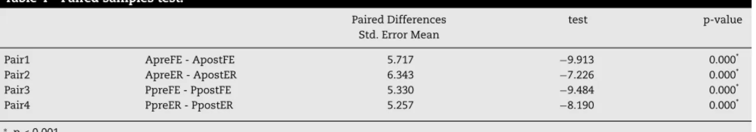
Data analysis was performed using the Statistical Package for Social Sciences (SPSS) version 19.0. The Kolmogorov-Smirnov test was initially used to verify the normality of the values. Then, the active and passive forward elevation and external rotation pre- and postoperative values were com-pared by Student’s parametric pairedt-test. Internal rotation was analysed by Friedman’s test, with the risk assumed by the researcher of 5% and probability of rejecting the null hypoth-esis < 0.05.
Surgical technique
patient in the operating room, passive range of motion was documented while the patient was under anesthesia.
The entire upper extremity was then prepared in a ster-ile fashion, and the glenohumeral joint was approached from the posterior arthroscopic portal. After an articular inventory of the synovium, biceps tendon, humeral head, capsule, and rotator cuff, the anterosuperior portal was made and a can-nula was inserted directly underneath the long head biceps (inside-out) and above the subscapularis tendon.
First, a synovectomy was performed. The next key step in all cases was to release the rotator interval region, which was represented as contracted capsule between the anterior edge of the supraspinatus tendon and the superior border of the subscapularis tendon, with subsequent release of the coraco-humeral ligament, which was identified by probe palpation of the coracoid process. This release allowed the humeral head to move laterally away from the glenoid, and the stiff anterior capsule could then be released.
Then, a subscapularis tenotomy was performed, which is carefully separated from the middle glenohumeral ligament using a radiofrequency device. A partial tenotomy was per-formed in some cases (1, 2, 3, 5, 14, 15, 16); the remaining cases were submitted to total tenotomy.
The next step was the anterior capsule division. The release was made from the two o’clock positionto the five o’clock posi-tion along the glenoid rim, and continued down to the six o’clock position.
Afterwards, the arthroscope was first placed through the anterior cannula with inflow switched to that cannula, and the radiofrequency device was changed to the posterior por-tal to proceed with the posterior capsule release for persistent loss of internal rotation. This was perfomed along the glenoid rim, from directly behind the biceps tendon down to approx-imately the eight o’clock position on the glenoid. Finally, the
inferior capsule was released for flexion and abduction restric-tions.
There were no concomitant diseases, as verified by pre-operative MRI and joint inspection. After the arthroscopic release, no manipulation was performed.
Aftercare
All patients with interscalene catheter received a bolus of 15 mL to 20 mL of 0.5% bupivacaine 30 minutes before each ses-sion of rehabilitation. The patients were admitted to the hospi-tal for 72 hours to undergo immediate physical therapy, which consisted of passive range of motion performed twice daily.
Patients were discharged without a shoulder sling and were instructed to start supervised rehabilitation five days per week and to use their shoulder for activities of daily living.
Results
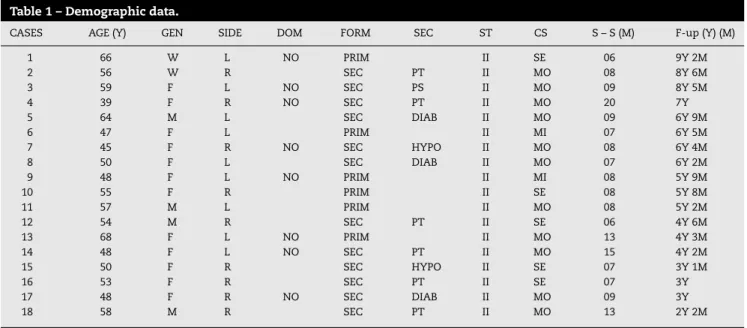
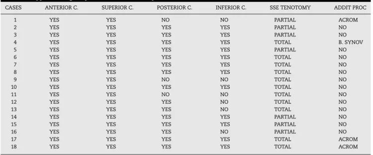
Demographic data are exhibited in Table 1. Table 2 demon-strates additional procedures and and the steps performed in each case.
The mean active and passive forward flexion, external rotation, and internal rotation increased from 94.4◦/103.3◦,
11.9◦/21.9◦, and S1/L5 vertebral level, respectively, to
151.1◦/153.8◦, 57.2◦/64.4◦, and T12/T10 vertebral level,
respec-tively (Table 3). The mean increase was 56.7◦/50.5◦ in the
forward elevation, 45.3◦/42.5◦ in the external rotation, and
06/07 levels. The values had normal distribution.
Regarding the range of motion assessment (40 points) with the Constant–Murley functional score, it increased 16 points; from 14 (preoperative mean) to 30 points (postop-erative mean). There were no intraop(postop-erative complications or
Table 1 – Demographic data.
CASES AGE (Y) GEN SIDE DOM FORM SEC ST CS S – S (M) F-up (Y) (M)
1 66 W L NO PRIM II SE 06 9Y 2M
2 56 W R SEC PT II MO 08 8Y 6M
3 59 F L NO SEC PS II MO 09 8Y 5M
4 39 F R NO SEC PT II MO 20 7Y
5 64 M L SEC DIAB II MO 09 6Y 9M
6 47 F L PRIM II MI 07 6Y 5M
7 45 F R NO SEC HYPO II MO 08 6Y 4M
8 50 F L SEC DIAB II MO 07 6Y 2M
9 48 F L NO PRIM II MI 08 5Y 9M
10 55 F R PRIM II SE 08 5Y 8M
11 57 M L PRIM II MO 08 5Y 2M
12 54 M R SEC PT II SE 06 4Y 6M
13 68 F L NO PRIM II MO 13 4Y 3M
14 48 F L NO SEC PT II MO 15 4Y 2M
15 50 F R SEC HYPO II SE 07 3Y 1M
16 53 F R SEC PT II SE 07 3Y
17 48 F R NO SEC DIAB II MO 09 3Y
18 58 M R SEC PT II MO 13 2Y 2M
350
r e v a s s o c m e d b r a s .2 0 1 3;5 9(4):347–353Table 2 – Types of surgical procedures in the sample.
CASES ANTERIOR C. SUPERIOR C. POSTERIOR C. INFERIOR C. SSE TENOTOMY ADDIT PROC
1 YES YES NO NO PARTIAL ACROM
2 YES YES YES YES PARTIAL NO
3 YES YES YES YES PARTIAL NO
4 YES YES YES YES TOTAL B. SYNOV
5 YES YES YES YES PARTIAL NO
6 YES YES YES YES TOTAL NO
7 YES YES YES YES TOTAL NO
8 YES YES YES YES TOTAL NO
9 YES YES NO NO TOTAL NO
10 YES YES YES YES TOTAL NO
11 YES YES NO NO TOTAL NO
12 YES YES YES NO TOTAL NO
13 YES YES YES NO TOTAL NO
14 YES YES YES YES PARTIAL NO
15 YES YES YES YES PARTIAL NO
16 YES YES YES NO PARTIAL NO
17 YES YES YES YES TOTAL ACROM
18 YES YES YES YES TOTAL ACROM
C, capsulotomy; SSE, subscapular; ADDIT PROC, additional procedure; ACROM, acromioplasty; B. SYNOV, bursal synovectomy.
instability, and no postoperative neurological injury. When the patients’ range of motions means were compared, there was difference between the pre- and postoperative values (p < 0.001) (Table 4).
All patients showed substantial gains in shoulder range of motion, as well as diminished shoulder pain (none to mild/15 to 10 points in the Constant-Murley pain score) (p < 0.001).
Discussion
Adhesive capsulitis is a common disease and remains an enigmatic condition despite many attempts to elucidate the underlying pathologic process17. Inflammatory and fibrous modifications of the joint capsule and synovial sheath of the shoulder are responsible for the obliteration of the axillary recess and capsule adhesions to the humeral head. These changes cause capsular retraction, with reduced volumetric capacity and shoulder stiffness1,2,18.
Ozaki et al. reported that the contracted coracohumeral lig-ament and rotator interval appear to be the main lesions in the stiff shoulder. The pathological findings of these structures are extremely important when treating such patients19.
The disease occurs more predominantly between the ages of 40 and 60 years, in females, in their non-dominant side, without any racial preference2,3,9. The present study showed a mean age at the time of capsular release of 53.6 years, and 77.77% of the patients were female, which coincides with the literature data, although ten of the 18 patients presented the disease on the dominant side.
Stiff shoulder responds well to nonsurgical treatment in 70% to 90% of patients4,9,20. A therapeutic option is the supras-capular nerve block, which is an efficient method when compared to placebo and intra-articular injections21–23. The procedure is justified, since this nerve is responsible for 70% of the shoulder capsule sensitivity, which is retractable and has its volume decreased in the case of shoulder stiffness3,7,22,23. However, 11 patients of the sample underwent this method
associated with rehabilitation, without any efficacy. Some authors reported good results with manipulation under anesthesia, but this does not allow for a controlled release of the pathological tissue, with increased risk of humerus fractures3,5,6,24,25.
Surgical capsular release should be performed in patients unresponsive to conservative treatment for at least six months3, which is in agreement with this study with regard to the same minimum time from onset of symptoms to the proposed surgery, after unsuccessful conservative measures.
The coracohumeral ligament exploration demonstrates that this is the thickest and abnormal part of the stiff capsule10. As an extra-articular anatomical structure, its arthroscopic release is only possible after opening of the rotator interval and exposure of the lateral and inferior coracoid surface. The main objective of the procedure is to restore external rotation19. In this study, all patients had the coracohumeral ligament released, and the mean active increase of external rotation was 45.3◦, unlike the study by
Beaufils et al., who performed this procedure in only one of 26 patients, and concluded that the capsular release was of little benefit in primary frozen shoulder with long recovery time, without pain relief26.
Subacromial fibrosis with hypertrophic synovium was observed in several studies, and both debridement and acromioplasty were performed27,28. Chen et al. reported that 86% of the patients underwent subacromial decompression, and that the procedure contributed to the pain relief of the shoulder29.
Capsular release was performed in the present study, with two additional procedures (cases 1, 4, 17, 18), and with a sub-stantial improvement of pain in all cases. However, it is not possible to conclude that acromioplasty has contributed to the improvement of the variable pain, since the present study was not of association.
rev
assoc
med
bras.
2013
;
5
9(4)
:347–353
351
CASES A. pre FE A. pre ER A. pre IR/level P. pre FE P. pre ER P. pre IR/level A. post FE A. post ER A. post IR/level P. post FE P. post ER P. post IR/level
1 70◦ 5◦ S1 80◦ 10◦ L5 180◦ 40◦ T12 180◦ 50◦ T12
2 90◦ 30◦ L5 100◦ 40◦ L5 120◦ 40◦ L3 130◦ 50◦ L1
3 90◦ 10◦ S1 90◦ 20◦ L5 180◦ 50◦ T12 180◦ 60◦ T12
4 90◦ 0◦ GT 100◦ 10◦ GT 170◦ 40◦ T12 170◦ 50◦ T12
5 100◦ 10◦ S1 110◦ 20◦ S1 120◦ 20◦ L3 130◦ 30◦ L2
6 120◦ 40◦ L5 120◦ 40◦ L5 170◦ 50◦ L1 170◦ 70◦ T12
7 100◦ 0◦ S1 110◦ 10◦ L5 170◦ 40◦ T12 170◦ 50◦ T10
8 90◦ 10◦ S1 100◦ 20◦ S1 120◦ 50◦ L3 120◦ 50◦ L1
9 110◦ 0◦ L5 120◦ 10◦ L4 170◦ 40◦ T12 170◦ 50◦ T11
10 60◦ 0◦ L5 80◦ 10◦ L5 90◦ 30◦ L3 110◦ 50◦ L3
11 100◦ -10◦ S1 110◦ 0◦ S1 150◦ 90◦ T10 150◦ 90◦ T10
12 90◦ 10◦ S1 90◦ 20◦ L5 130◦ 70◦ T11 140◦ 80◦ T10
13 110◦ 30◦ L4 120◦ 45◦ L3 150◦ 70◦ T11 150◦ 80◦ T10
14 110◦ 20◦ S1 120◦ 40◦ L5 160◦ 90◦ T10 160◦ 90◦ T10
15 80◦ 0◦ S1 90◦ 10◦ S1 160◦ 80◦ L1 160◦ 80◦ T12
16 80◦ -10◦ GT 90◦ 0◦ GT 160◦ 90◦ T10 160◦ 90◦ T10
17 100◦ 30◦ S1 110◦ 40◦ L5 160◦ 70◦ T12 160◦ 70◦ T11
18 110◦ 30◦ L5 120◦ 40◦ L5 160◦ 70◦ T11 160◦ 70◦ T10
Mean 94.4◦ 11.9◦ S1 103.3◦ 21.9◦ L5 151.1◦ 57.2◦ T12 153.8◦ 64.4◦ T10
352
r e v a s s o c m e d b r a s .2 0 1 3;5 9(4):347–353Table 4 – Paired samples test.
Paired Differences test p-value
Std. Error Mean
Pair1 ApreFE - ApostFE 5.717 −9.913 0.000*
Pair2 ApreER - ApostER 6.343 −7.226 0.000*
Pair3 PpreFE - PpostFE 5.330 −9.484 0.000*
Pair4 PpreER - PpostER 5.257 −8.190 0.000*
∗ p < 0.001.
be performed in the inferior, but not in the posterior capsule30. Jerosch described his technique performing both releases27. Chen et al., in 74 randomized patients, where the first group received only the anterior capsulotomy, while in the second group the release extended to the inferior and posterior cap-sule, concluded that shoulder function and range of motion were similar in six months29. Snow et al. also observed no differences when adding posterior capsulotomy into the procedure11. The patients of the present study increased their range of motion with the posterior (except cases 1, 9, 11) and inferior release (except cases 1, 9, 11, 12, 13, 16), regardless of whether the adhesive capsulitis was primary or secondary.
There is also concern of the axillary nerve injury in the infe-rior capsulotomy performance. As it is closer to the humeral insertion of the capsule, the release should be close to the glenoidal edge27. None of our 12 of 18 patients presented axillary nerve palsy, as in the study by Jerosch27; however, Harryman et al. had one case, with spontaneous resolution31. Pearsall et al.28 and Ogilvie-Harris et al.30 reported the release of the intra-articular portion of the subscapu-laris tendon, lateral to the musculotendinous junction, although most studies showed excellent results without this procedure27,29,32. This portion represents only 25% of the cephalocaudal length of the subscapularis muscle28. For this reason, and because it is an external rotation restrictor, this tenotomy was added to the presented technique with good results. It was performed partially in some cases (1, 2, 3, 5, 14, 15, 16) and totally in the others. Increased range of motion and decreased pain was observed in all patients, regardless of which tenotomy was performed (p < 0.001).
The performance of the tenotomy allowed for the avoidance of any type of joint manipulation in the post-operative period, which can be an advantage of the pre-sented technique. It is important to note that no recur-rence occurred postoperatively.
Did the subscapularis tenotomy contribute to this absense? Since this was not a randomized clinical trial, this ques-tion remains unanswered. It is important to understand whether this section of the subscapularis undermines the anterior shoulder stability. Pearsall et al. observed 97% of patients with minimal or no sign of instability28. Comparing the results of this study with partial or total subscapularis tenotomy, there were no cases of anterior instability after arthroscopic surgery.
Berghs et al.33presented their results on the arthroscopic treatment of shoulder stiffness, and the mean of the forward elevation improved from 73.7◦to 163◦(89.3◦); the external
rota-tion improved from 10.6◦to 46.8◦(36.2◦), and internal rotation
improved nine levels. Elhassan et al.34, in the analysis of the
averages in the three directions, observed that they increased by 38◦, 24◦, and six levels, respectively, a finding similar to that
of the present study, which showed improvement in forward elevation of 56.7◦, 45.3◦in external rotation, and six levels in
internal rotation (p < 0.001).
The limitations of this study include its retrospective design, with a small sample size without comparison group, since frozen shoulder is a disease with predominantly non-surgical treatment, with few cases progressing to surgery. This study, however, has importance in that the same surgical technique was performed in all patients, regardless of the eti-ology of the shoulder stiffness, but the insufficient number of patients does not allow for conclusions in this regard. The other issue that needs to be highlighted is that internal rota-tion strength was not measured. It can be a subject of further research.
Conclusion
Arthroscopic treatment is effective in shoulder stiffness unre-sponsive to conservative treatment.
Conflicts of interest
The author declares no conflicts of interest.
r e f e r e n c e s
1. Zuckerman JD, Rokito A. Frozen shoulder: a consensus definition. J Shoulder Elbow Surg. 2011;20:322–5.
2. Hand C, Clipsham K, Rees JL, Carr AJ. Long-term outcome of frozen shoulder. J Shoulder Elbow Surg. 2008;17:231–6. 3. Tasto JP, Elias DW. Adhesive capsulitis. Sports Med Arthrosc
Rev. 2007;15:216–21.
4. Levine W, Kashyap CP, Bak SF, Ahmad C, Blaine T, Bigliani L. Nonoperative management of idiopathic adhesive capsulitis. J Shoulder Elbow Surg. 2007;16:569–73.
5. Jacobs LG, Smith MG, Khan SA, Smith K, Joshi M.
Manipulation or intra-articular steroids in the management of adhesive capsulitis of the shoulder? A prospective randomized trial. J Shoulder Elbow Surg. 2009;18:348–53. 6. Massoud SN, Pearse EO, Levy O, Copeland SA. Operative
management of the frozen shoulder in patients with diabetes. J Shoulder Elbow Surg. 2002;11:609–13.
8. Alvado A, Pelissier J, Benaim C, Petiot S, Herisson C. Physical therapy of frozen shoulder: literature review. Ann Readapt Med Phys. 2001;44:59–71.
9. Jewell DV, Riddle DL, Thacker LR. Interventions associated with an increased or decreased likelihood of pain reduction and improved function in patients with adhesive capsulitis: a retrospective cohort study. Phys Ther. 2009;89:419–29. 10. Omari A, Bunker TD. Open surgical release for frozen
shoulder: surgical findings and results of the release. J Shoulder Elbow Surg. 2001;10:353–7.
11. Snow M, Boutros I, Funk L. Posterior arthroscopic capsular release in frozen shoulder. Arthroscopy. 2009;25:19–23. 12. Baums MH, Spahn G, Nozaki M, Steckel H, Schultz W, Klinger
HM. Functional outcome and general health status in patients after arthroscopic release in adhesive capsulitis. Knee Surg Sports Traumatol Arthrosc. 2007;15:638–44. 13. Constant CR, Murley AHG. A clinical method of functional
assessment of the shoulder. Clin Orthop Relat Res. 1987;214:160–4.
14. Reeves B. The natural history of the frozen shoulder syndrome. Scand J Rheumatol. 1975;4:193–6.
15. American Academy of Orthopaedics, Surgeons., Joint motion: method of measuring and, recording. Chicago: American Academy of Orthopaedics Surgeons. 1965.
16. Zuckerman JD, Cuomo F, Rokito S. Definition and classification of frozen shoulder: a consensus approach. J Shoulder Elbow Surg. 1994;3(1):S72.
17. Vastamaki H, Kettunen J, Vastamaki M. The natural history of idiopathic frozen shoulder. A 2- to 27-year follow up study. Clin Orthop Relat Res. 2012;470:1133–43.
18. De Carli A, Vadalà A, Perugia D, Frate L, Iorio C, Fabbri M, et al. Shoulder adhesive capsulitis: manipulation and arthroscopic arthrolysis or intra-articular steroid injections? Int Orthop. 2012;36:101–6.
19. Ozaki J, Nakagawa Y, Sakurai G, Tamai S. Recalcitrant chronic adhesive capsulitis of the shoulder. Role of contracture of the coracohumeral ligament and rotator interval in pathogenesis and treatment. J Bone Joint Surg Am. 1989;71:1511–5. 20. Lorbach O, Anagnostakos K, Scherf C, Seil R, Kohn D, Pape D.
Nonoperative management of adhesive capsulitis of the shoulder: Oral cortisone application versus intra-articular cortisone injections. J Shoulder Elbow Surg. 2010;19:172–9. 21. Favejee MM, Huisstede BMA, Koes BW. Frozen shoulder:
the effectiveness of conservative and surgical interventions-systematic review. Br J Sports Med. 2011;45:49–56.
22. Fernandes MR, Barbosa MA, Souza ALL, Ramos GC.
Suprascapular nerve block: an important procedure in clinical practice. Part II. Rev Bras Reumatol. 2012;52:616–22.
23. Checchia SL, Fregoneze M, Miyazaki NA, Santos PD, Silva LA, Ossada A, et al. Tratamento da capsulite adesiva com bloqueios seriados do nervo supraescapular. Rev Bras Ortop. 2006;41:245–52.
24. Dodenhoff RM, Levy D, Wilson A, Copeland SA. Manipulation under anesthesia for primary frozen shoulder. Effect on early recovery and return to activity. J Shoulder Elbow Surg. 2000;9:23–6.
25. Fox A, Board T, Srinivasan MS. Improvement in shoulder function following manipulation for adhesive capsulitis: how long does it last? J Bone Joint Surg Br. 2005;88:138–9. 26. Beaufils P, Prévot N, Boyer T, Allard M, Dorfmann H, Frank A,
et al. Arthroscopic release of the glenohumeral joint in shoulder stiffness: a review of 26 cases. French Society for Arthroscopy. Arthroscopy. 1999;15:49–55.
27. Jerosch J. 360◦arthroscopic capsular release in patients with adhesive capsulitis of the glenohumeral joint – indication, surgical technique, results. Knee Surg Sports Traumatol Arthrosc. 2001;9:178–86.
28. Pearsall IV AW, Osbahr DC, Speer KP. An arthroscopic technique for treating patients with frozen shoulder. Arthroscopy. 1999;15:2–11.
29. Chen J, Chen S, Li Y, Hua Y, Li H. Is the extended release of the inferior glenohumeral ligament necessary for frozen
shoulder? Arthroscopy. 2010;26:529–35.
30. Ogilive-Harris DJ, Biggs DJ, Fitsialos DP, MacKay M. The resistant frozen shoulder: manipulation versus arthroscopic release. Clin Orthop Relat Res. 1995;(319):238–48.
31. Harryman II DT, Sidles J, Matsen III F. Arthroscopic management of refractory shoulder stiffness. Arthroscopy. 1997;13:133–47.
32. Cinar M, Akpinar S, Derincek A, Circi E, Uysal M. Comparison of arthroscopic capsular release in diabetic and idiopathic frozen shoulder patients. Arch Orthop Trauma Surg. 2010;130:401–6.
33. Berghs BM, Sole-Molins X, Bunker TD. Arthroscopic release of adhesive capsulitis. J Shoulder Elbow Surg. 2004;13: 180–5.