589
NOVEL ACUTE STRESSOR EFFECTS ON INTERSCAPULAR BROWN ADIPOSE TISSUE SYMPATHETIC INERVATION AND UCP-1 CONTENT IN CHRONICALLY ISOLATED
AND SPONTANEOUSLY HYPERTENSIVE RATS
IVA LAKIC1, TAMARA DRENCA1, JELENA DJORDJEVIC1, P. VUJOVIC1, N. JASNIC1,
S. DJURASEVIC1, SLADJANA DRONJAK-CUCAKOVIC2 and GORDANA CVIJIC1
1 Institute of Physiology and Biochemistry, University of Belgrade, Faculty of Biology, 11000 Belgrade, Serbia 2 Laboratory of Molecular Biology and Endocrinology, Institute of Nuclear Sciences “Vinča”, 11001 Belgrade, Serbia
Abstract - Interscapular brown adipose tissue (IBAT) is an energy storing organ involved in the maintenance of homeosta-sis in stress conditions when the balance of energy supplies is disturbed. he major regulator of IBAT activity is the sym-pathetic nervous system (SNS). Since genetic background is responsible for the individual diferences in neuroendocrine stress responsivity, spontaneously hypertensive rats (SHR) that have a genetically increased general sympathetic output are a useful model for studying adaptive processes in stress conditions. Our aim was to test the efect of acute and/or chronic exposure to various stressors (thermal-cold, psychophysical-immobilization and psychosocial-isolation) on IBAT SNS and the metabolic activity in SHR, by measuring the number of monoamine-containing nerve endings and uncoupling protein-1 (UCP-1) content. he obtained results show that the IBAT SNS activity of unstressed SHR was stimulated by the administration of a single acute or chronic stressor and was independent of the duration or type of stressor, while chronic pre-stress of isolation suppressed further the SNS reaction to novel acute stress exposure. he IBAT UCP-1 content fol-lowed SNS changes, suggesting that this system is dominant in the regulation of IBAT metabolic rate in SHR.
Key words:Hypertension, SHR, sympathetic innervations, IBAT, stress.
UDC 577.1:591.4
INTRODUCTION
Early studies of Cannon (Cannon, 1929) proposed a concept according to which the sympathetic nerv-ous system (SNS) and adrenal medulla (AM) prepare an animal for the “ight or light” reaction to stress by raising blood glucose, mobilizing fatty acids from adipose tissue, increasing the heart rate, redistrib-uting blood low and elevating the metabolic rate. Cannon suggested that the AM is more important than the SNS. However, it has been shown that sym-pathetic denervation leads to an increase in adipose tissue weight. Nerve stimulation results in fatty acids release and sympathetic or ganglionic blockades in-hibits the mobilization of lipid (Gilgen et al., 1962;
Rebufe-Scrive, 1991). On the other hand, adrenal demedullation is not efective, which suggests that the SNS might be more important than the AM (Rayner, 2001) at least, in the metabolic response to stressors.
Seydoux, 1986) by stimulating uncoupling protein-1 (UCP-1) synthesis (Cassard-Doulcier et al., 1993). UCP-1 transforms electrochemical energy into heat (Nicholls and Locke, 1984), enabling small mammals to tolerate cold (Nedergaard et al., 1999) or other conditions when energy homeostasis is disturbed. Our results with 6-hydroxydopamine administration indicate that the intact sympathetic activity is nec-essary for the maintaining basal level of brown fat UCP-1 (Davidovic et al., 2004). Bearing in mind that sympathetic stimulation also leads to an increase in fatty acid release from white adipose tissue (Rebufe-Scrive, 1991), we can assume that the SNS is the ma-jor regulator of both white and brown fat mobiliza-tion to provide homeostasis of energy supplies under conditions of stress.
As far as the inluence of genetic factors on adaptive processes is concerned, some diferences in individual responses can occur in diferent animal strains or social paradigms in humans. he genetic background partly contributes to the emergence of individual diferences in stress reactivity (Blanchard et al., 1995). he psychoneuroendocrine responses to social stress can also have a genetic origin and therefore the use of SHR can provide an important model for studying adaptive processes (Berton et al., 1998).
SNS has been considered to play an important role in the development of human essential hyper-tension (Esler et al., 1977, Julius, 1996). hus the SHR is a widely used model of hypertension in despite the fact that the etiology of this physiological state is not well understood.
Bearing in mind all the above-mentioned, our aim was to test whether animals with a genetically-induced increased general sympathetic output, such as SHR, also have an enhanced local IBAT SNS out-put and if so, how does it inluence the metabolic re-sponse of tissue to stress. Special emphasis was on the type (environmental-thermal-cold, psychophys-ical-immobilization and psychosocial-isolation) and duration (acute and chronic exposure) of stressor and on stressor combinations.
MATERIALS AND METHODS
Experiments were performed on 15 week-old spontaneously hypertensive male rats (SHR). Age-matched normotensive Wistar rats (n=6) were used as the control group for determining blood pressure and the catecholaminergic nerve proile density in IBAT. he rats were acclimatized to 22±1 ºC, kept at a 12:12 h light-dark cycle, with dark onset at 6 p.m. he animals were given commercial rat food and tap water ad libitum and housed two per cage for 15 days before the experiment. he blood pressure of Wis-tar and SHR was monitored one week prior to the experiment, using tail cuf plethysmography of re-strained conscious animals. he mean arterial pres-sure for Wistar rats was 100±5 mmHg and for SHR 180±5 mmHg.
he SHR animals were divided into six groups each containing six animals. he irst SHR group con-sisted of intact controls. he second SHR group was subjected to social isolation for 21-days and killed on the 22nd day. Social isolation was performed by
plac-ing one rat in a cage. Visual, acoustic and olfactory communication between isolated rats was reduced to the minimum. he third and fourth groups were chronically stressed in the same way as the second group (21 days of the isolation) and then on the 22nd
day subjected to acute exposure to a novel stressors: cold (6 ºC for 2 h) or immobilization (2 h). Immo-bilization stress was performed according to Kvetn-ansky and Mikulaj (1970) by ixing all four limbs to a board with adhesive tape. he head was also ixed by a metal loop over the neck area, thus limiting its motion. hese animals were killed at the end of ex-posure to acute stressors. he SHR from the ith and sixth groups were exposed to acute stressors, cold (6 ºC for 2 h) and immobilization (2 h) respectively, and decapitated ater stress termination. Acute stress ex-posure was exerted inlicted between 8:00 a.m. and 11:00 a.m. to avoid efects of circadian rhythms. All animals were decapitated with a guillotine (Harvard-Apparatus, Holliston, MA, USA).
Protection Law and proposed guidelines and proto-cols that have been approved by the Ethical Commit-tee of Faculty of Biology, University of Belgrade.
Blood was collected from the trunk and IBATs were rapidly excised (4ºC) and stored at -70ºC. Be-fore freezing, the same part of IBAT was always im-mediately dipped into frozen-section medium (OKT, Galen-Focus) and later used for determination of the intensity of luorescent staining of monoamine-con-taining IBAT nerve proiles according to the method of De la Tore (1980). Five and 10 µm cryostat sections of IBAT were melted onto uncoated glass slides and dipped into a solution containing 2% glyoxylic acid, 10% sucrose, 0.1 M PBS pH 7.4, incubated at room temperature for 10 min and dried under cold airlow. he dried sections were then covered with a drop of glycine-glycerol bufer, heated at 95ºC for 2.5 min-utes, allowed to cool and cover-slipped. he sections were examined using a BH2 luorescence
photomi-croscope (Olympus, Tokyo, Japan) equipped with ex-citation ilter BP-405 and barrier ilter Y-475. Image-J sotware was used for quantifying the number of noradrenaline-containing nerve ibers per total area.
IBAT UCP-1 levels were estimated by Western blot analysis. Samples of the solubilized mitochon-drial fraction (containing 5 µg of the IBAT mito-chondrial proteins) were added to an equal volume of bufer (0.125 M Tris-HCl, 0,14 M SDS, 20% glyc-erol, 0.2 mM dithiothreitol, 0.03 mM bromophe-nol blue; pH = 6.8). Ater denaturation by heating to 100ºC for 5 min, the samples were separated on a 12.5% polyacrylamide gel and electro-transferred to a PVDF membrane. Ater removal of nonspeciic binding, the membrane was incubated with a solu-tion of rabbit antibody raised against rat UCP-1 (Sig-ma, U 6382), followed by secondary goat antibodies raised against rabbit immunoglobulin and coupled with horseradish peroxidase (Santa Cruz, goat anti-rabbit IgG-HRP SC-2004). he UCP-1 content was visualized by ECL Western Blotting Detection Rea-gents (Amersham), by exposure of an X-ray ilm for 5 min. he intensity of signals was evaluated by the Image Quant program (Molecular Dynamics, Hab-ersham Biosciences). he number of pixels obtained
for the control specimen that had the lowest expo-sure density represented one arbitrary unit.
Sera were frozen and later used for determina-tion of free fatty acids (FFA) concentradetermina-tions by the method of Duncombe (1964). he blood glucose level was determined by a glucose analyzer Exac-Tech (MediSense, Cambridge, MA, USA) using Dex-trostix reagent strips. Proteins were quantiied by the method of Lowry (1951).
he results were expressed as means ± S.E.M. For comparison of diferences between groups, one way ANOVA, followed by the Holm-Sidak posterior comparison test were employed with a level of sig-niicance set at p<0.05.
RESULTS
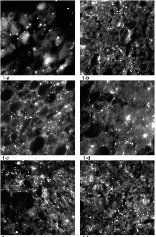
he number of luorescent stains representing the monoamine-containing IBAT sympathetic nerve ib-ers (40 spots) revealed that in SHRs (Fig. 1-a), SNS activity was less pronounced when compared to nor-motensive Wistar rats (110 spots) (Fig. 2). It was also observed that all of the applied stressors provoked changes in the number of IBAT noradrenergic (NA) nerve endings when compared to non-stressed SHR. Figure 1-b shows that chronic isolation stress leads to an increase of luorescent spots (167), represent-ing increased numbers of monoaminergic nerve proiles. Under the inluence of both acute stressors cold (1-c) and immobilization (1-e), the density of nerve endings also increased. he increased density of monoaminergic nerves was signiicant in chroni-cally stressed SHR that were exposed to acute novel stressors cold (1-d) and immobilization (1-f), only when compared to non-stressed SHR controls (1-a), but not when compared to isolated SHR (1-b) or acutely stressed rats (1-c; 1-e).
of the IBAT UCP-1 content that was statistically sig-niicant only ater the latter stress (p<0,001) in com-parison to unstressed SHR controls. he chronic stress of 21 day isolation also increased the IBAT 1 content (p<0.05). However, the IBAT UCP-1 content signiicantly decreased (p<0,05) when chronically isolated rats were exposed to the acute stress of immobilization, while it remained statisti-cally unchanged when chronistatisti-cally isolated rats were exposed to the acute stress of low environmental temperature.
As a peripheral metabolic marker, blood FFA (Fig. 4) and glucose (Fig. 5) concentrations were determined. he only signiicant changes in these parameters were observed in SHR exposed to im-mobilization stress with or without the previous 21-day isolation (Fig. 4: p<0.05, p<0.001; Fig. 5: p<0.01, p<0.001).
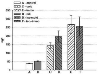
As judged by the change in blood ACTH concen-tration (Fig. 6), all applied stressors, whether chronic
(isolation-p<0.01) or acute (cold- p<0.01; immobili-zation p<0.05), or combined (p<0.05), induced acti-vation of the HPA axis.
Fig. 2. he density of monoamine containing sympathetic nerve proiles in IBAT of normotensive Wistar rats (110 spots), visua-lised by sucrose–phosphate–glyoxylic acid (SPG) histoluores-cence method (De la Torre, 1980)
Fig. 3. IBAT UCP-1 content (AU – arbitrary units) in SHR: A – control non-stressed rats; B – chronic isolation (21 days); C – acute cold (2 h); D – chronic isolation (21 days) + acute cold (2 h); E – acute immobilization (2 h); F – chronic isolation (21 days) + acute immobilization (2 h). he values are means of six animals ±S.E.M. Statistical signiicance: A:B - p<0.05; C:D - p<0.05; A:E - p<0.001; B:F - p<0.05.
DISCUSSION
Both white and brown adipose tissues are regarded as organs for energy storage, with the sympathetic nervous system being the major regulator of their activity, which is to maintain homeostasis of the en-ergy supply (Rayner, 2001). In small mammals, the changes in SNS activity are accompanied by changes in IBAT thermogenic capacity induced by NA. he noradrenaline which is secreted from sympathet-ic nerve endings is the important trophsympathet-ic factor of this tissue that stimulates thermogenesis (Himms-Hagen, 1990; Puigserver et al., 1992; Waldbillig and Desautels, 1992). It was shown that the decrease in the IBAT sympathetic NA turnover is associated with fasting, while an increase occurs in cold expo-sure or overfeeding (Rayner, 2001). Since SHR ex-hibits enhanced generalized SNS activity, we hypoth-esized that these rats might also have an enhanced IBAT sympathetic outlow and consequently could respond diferently to applied cold, as well as other stressors of diferent type and duration. It was shown that these rats react to some environmental stimuli with exaggerated cardiovascular and sympathetic
re-sponses (Chambers et al., 2000). However, our results showed that IBAT SNS activity was less pronounced in unstressed SHR in comparison to normotensive Wistar rats. Ater exposure to cold (2h) SHR IBAT UCP-1, the major metabolic marker of its activity, insigniicantly increased. his result difers from the results we obtained in normotensive rats (Cvijic et al., 2004). he photomicrographs demonstrating the number of monoamine-containing nerve proiles in the IBAT of cold exposed SHR showed that there is increased density of SNS nerve endings surrounding the cells. he peripheral metabolic response, judged by the glucose and FFA blood levels, was not sig-niicantly changed. Exposure to mobilization (2 h), characterized as a strong psychophysical stressor, also inluenced all of these parameters. he density of NA-containing nerve endings, the levels of IBAT UCP-1 and circulating energents glucose and FFA in immobilized SHRs were signiicantly increased. It is interesting to speculate why immobilization pro-voked these changes in brown fat since it is not con-sidered a thermal stressor. Intense muscle activity occurred in SHR as they tried to free the ixed limbs and head. It is clear that these processes provoke the Fig. 6. Plasma ACTH level (ng/l) in SHR. A – control non-stressed rats; B – chronic isolation (21 days); C – acute cold (2 h); D – chronic isolation (21 days) + acute cold (2 h); E – acute immobilization (2 h); F – chronic isolation (21 days) + acute im-mobilization (2 h). he values are means of six animals ±S.E.M. Statistical signiicance: A : B - p<0.01.; A : C - p<0.01; A : E - p<0.05; B : D - p<0.05; B : F - p<0.05.
disturbance of energy homeostasis. It was also shown that muscle tissue NA sympathetic innervation is more pronounced in SHR than in normotensive rats (Cabassi et al., 1998).
Judging by the level of circulating ACTH, all of the applied stressors, in addition to activating SNS, also activated the HPA axis. he pronounced quan-titative diferences depended on the nature of the stress and duration of exposure. Quantitatively, the most pronounced hormonal response was observed in immobilization stress, additionally proving that this stressor is the strongest. Bearing in mind that ACTH secretion is stimulated by hypothalamic CRH, it can be expected that secretion of this hor-mone is also increased. It was shown that CRH in-jection into rat brain produces hyperglycemia and that neither hypophysectomy nor adrenalectomy prevent this efect. However, pretreatment with the ganglionic blocker chlorisondamine completely pre-vents the CRH-induced increase in plasma glucose (Brown et al., 1988). his result suggests that CRH acts in the brain to stimulate sympathetic outlow which results in the development of hyperglycemia. In SHR, the adrenomedullary response to CRH ad-ministration is more intense than in normotensive rats (Brown et al., 1988). herefore, we can assume that in these animals the adrenal medulla is equally involved in the stress response. Our results are con-sistent with the inding of Dronjak et al. (2004) that ater 21 days of isolation, SHR exhibit a signiicant increase in NA and even a greater increase in circu-lating adrenaline (A) in response to additional im-mobilization. hese results are in agreement with those of Sohn et al. (2002) who showed that SHR are hyperactive in a novel environment and hyper-responsive to environmental stimuli, exhibiting a pronounced behavioral, sympathetic and cardio-vascular responsiveness.
In conclusion, while being less pronounced in SHR than in normotensive Wistar rats, IBAT SNS ac-tivity was signiicantly stimulated by acute cold and immobilization stress, as well as by chronic isolation in comparison to unstressed controls. he responses did not depend on the type or duration of stressor.
Exposure to chronic pre-stress suppressed the addi-tional SNS reaction to the novel acute stressor. Since the IBAT UCP-1 contents followed the SNS changes, it can be assumed that this system also assumes a dominant role in the regulation of SHR IBAT meta-bolic rate.
Acknowledgments - his work was supported by the Ministry
of Education and Science of the Republic of Serbia, Grant No 173023.
REFERENCES
Berton, O., Aguerre, S., Sarrieau, A., Mormede, P., and F. Chaou-lof (1998). Diferential efects of social stress on central serotonergic activity and emotional reactivity in Lewis and spontaneously hypertensive rats. Neuroscience, 82, 147-159.
Blanchard, D. C., Spencer, R. L., Weiss, S. M., Blanchard, R. J., McEwen, B. S., and R. R. Sakai (1995). Visible burrows system as a model of chronic social stress: behavioral and neuroendocrine correlates. Psychoneuroendocrinology,20, 117-134.
Brown, M .F., Hauger, R., and L. A. Fisher (1988). Autonomic and cardiovascular efects of corticotrophin-releasing factor in the spontaneously hypertensive rat. Brain Res.441, 33-40.
Cabassi, A., Vinci, S., Calzolari, M., Bruschi, G., and A. Borghetti
(1998). Regional sympathetic in pre-hypertensive phase of spontaneously hypertensive rats. Life Sciences, 62, 1111-1118.
Cannon, W.B. (1929). Organization for physiological homeosta-sis. Phys. Rev. 9, 399-431.
Cassard-Doulcier, A. M., Gelly, C., Fox, N., Schrementi, J., Raim-bault, S., Klaus, S., Forest, C., Bouillaud, F., and D. Ricquier
(1993). Tissue-speciic and beta-adrenergic regulation of the mitochondrial uncoupling protein gene: control by cis-acting elements in the 5’ lanking region. Mol. Endo-crinol. 7, 497-506.
Chambers, J. B., Williams, T. D., Nakamura, A., Henderson, R. P., Overton, J.M., and M. E. Rashotte (2000). Cardiovascu-lar and metabolic responses of hypertensive and normo-tensive rats to one week of cold exposure. Am. J. Physiol. Regulatory Comp. Physiol. 279, 1486-1494.
Cvijic, G., Petrovic, N., Djordjevic, J., Davidovic, V., and V. M. Petrovic (2004). Efect of cold exposure on serum DBH and interscapular brown adipose tissue MAO activity in hypothyroid and hyperthyroid T3- and T4-treated rats.
Davidovic, V., Petrovic, N., Djordjevic, J., Djurasevic, S., and G. Cvijic (2004). Acute efect of cold on the antioxidant en-zymes activities and uncoupling protein-1 content in the brown fat of 6-hydroxydopamine-treated rats. Journal of hermal Biology,29, 825-830.
De la Torre, J. C. (1980). Standardization of the sucrose-potas-sium phosphate-glyoxylic acid histoluorescence method for tissue monoamines. Neurosci. Lett.17, 339-340.
Dronjak, S., Gavrilovic, Lj., Filipovic, D., and B. M. Radojcic
(2004). Immobilization and cold stress afect sympatho-adrenomedullary system and pituitary-adrenocortical axis of rats exposed to long-term isolation and crowding.
Physiology & Behavior,81, 409-415.
Duncombe, W. G. (1964). he colorimetric micro-determination of non-esterifed fatty acids in plasma. Clin. Chim. Acta,9, 122-125.
Esler, M., Julius, S., Zweiler, A., Randall, O., Harburg, E., Gardin-er, H., and V. Dequattro (1977). Mild high-renin essential hypertension. Neurogenic human hypertension? N. Engl. J. Med.296, 405-411.
Gilgen, A., Maickel, R. P., Nikodijevic, O., and B.B. Brodie (1962). Essential role of catecholamines in the mobilization of free fatty acids and glucose ater exposure to cold. Life Sciences,
1, 709-715.
Girardier, L., Seydoux, J. (1986). Neural control of brown adipose tissue. In: Brown adipose tissue. P hajhurn, DG Nicholas (eds),Edvard Arnold, London.
Himms-Hagen, J. (1990). Brown adipose tissue thermogenesis: interdisciplinary studies. Faseb J. 4, 2890-2898.
Julius, S. (1996). he evidence for a pathophysiologic signiicance of the sympathetic overactivity in hypertension. Clin. Exp. Hypertens. 18, 305-321.
Kvetnansky, R., and L. Mikulaj (1970). Adrenal and urinary cat-echolamines in rats during adaptation to repeated immo-bilization stress. Endocrinology, 87, 738-743.
Lowry, O. H., Rosenbrough. N. J., Farr, A. L., and R. J. Randall
(1951). Protein measurement with the Folin phenol re-agent. J. Biol. Chem. 193, 265-275.
Nedergaard, J., Matthias, A., Golozoubova, V., Jacobsson, A., and
B. Cannon (1999). UCP1: the original uncoupling protein – and perhaps the only one? New perspectives on UCP1, UCP2 and UCP3 in the light of the bioenergetics of the UCP1-ablated mice. J. Bioenerg. Biomembr. 31, 475-491.
Nicholls, D. G., and R. M. Locke (1984). hermogenic mecha-nisms in brown fat. Physiol. Rev. 64, 1-64.
Puigserver, P., Herron, D., Gianotti, M., Palou, A., Cannon, B.,
and J. Nedergaard (1992). Induction and degradation of the uncoupling protein thermogenin in brown adipocytes in vitro and in vivo. Evidence for a rapidly degradable pool. Biochem. J. 284, 393-8.
Rayner, D. V. (2001). he sympathetic nervous system in white adipose tissue regulation. Proceedings of the Nutrition So-ciety, 60, 357-364.
Rebufe-Scrive, M. (1991). Neuroregulation of adipose tissue: molecular and hormonal mechanisms. International Jour-nal of Obesity, 15, 83-86.
Sohn, H. S., Park, Y. N., and S. R. Lee (2001). Efect of immobi-lization stress on brain polyamine levels in spontaneously hypertensive and Wistar-Kyoto rats. Brain Research Bul-letin, 57, 575-579.