einstein. 2012;10(3):377-9
CASE REPORT
Hemoperitoneum secondary to gastric GIST negative
for c-KIT and positive for antibody anti-DOG1
Hemoperitôneo secundário a GIST gástrico c-KIT negativo e anticorpo anti-DOG1 positivo
Marina Gabrielle Epstein1, Aline Fioravanti Pasquetti1, Sara Venoso Costa1, Murillo de Lima Favaro2, Orlando Contrucci Filho3, Marcelo Augusto Fontenelle Ribeiro Junior4
1 Department of General Surgery, Universidade de Santo Amaro – UNISA, São Paulo (SP), Brazil. 2 Department of Surgical Technique, Universidade de Santo Amaro – UNISA, São Paulo (SP), Brazil.
3 Department of General Surgery and Department of Proctology, Universidade de Santo Amaro – UNISA, São Paulo (SP), Brazil. 4 Department of Surgical Clinic, Universidade de Santo Amaro – UNISA, São Paulo (SP), Brazil
Corresponding author: Marina Gabrielle Epstein – Rua Professor Carlos de Carvalho, 88, apto. 111 – Itaim Bibi – Zip code: 04531080 – São Paulo (SP), Brazil – Phone: (55 11) 3079-1831 – E-mail: [email protected] Received on: Dec 19, 2011 – Accepted on: Jan 1, 2012
ABSTRACT
Although relatively rare, the gastrointestinal stromal tumors comprise most mesenchymal tumors of the digestive tract and account for 5% of all sarcomas. The most common symptoms are pain, gastrointestinal bleeding and palpable mass. This study reported the case of a young patient who developed hemoperitoneum due to gastric neoplasm rupture and required urgent surgical treatment. Pathology and immunohistochemistry analysis showed an epidemiologically rare case: epithelioid and c-KIT negative tumor.
Keywords: Gastrointestinal stromal tumors; Gastrointestinal
hemorrhage/diagnosis; Gastrectomy; Gastrointestinal neoplasms; Endoscopy; Case reports
RESUMO
Os tumores estromais do trato gastrintestinal, embora relativamente raros, compreendem a maioria dos tumores mesenquimais do trato digestivo e constituem 5% de todos os sarcomas. Quanto à apresentação clínica, os sintomas mais comuns são dor, hemorragia digestiva e massa palpável. Este trabalho relatou o caso de um paciente jovem que desenvolveu hemoperitôneo por ruptura de neoplasia gástrica e necessitou de tratamento cirúrgico de urgência. A análise patológica e imunoistoquímica revelou tratar-se de um caso raro epidemiologicamente: tipo celular epitelioide e c-KIT negativo.
Descritores: Tumores do estroma gastrintestinal; Hemorragia
gastrintestinal/diagnóstico; Gastrectomia; Neoplasias gastrintestinais; Endoscopia; Relatos de casos
INTRODUCTION
The gastrointestinal stromal tumors (GIST) occur mainly in individuals aged 40- 80 years, and the
incidence is similar in both sexes(1). They may
originate anywhere in the gastrointestinal tract, and 50-60% of the lesions arise from the stomach, 20-30% from the small bowel, 10% from the colon, 5% from the esophagus and 5% from other sites in
the abdominal cavity(2). These tumors derive from
interstitial cells of Cajal, located at the level of the myoenteric plexus, between the longitudinal and circular muscle layers of the gastrointestinal tract(3).
They have immunophenotypical and ultrastructural characteristics, both of smooth muscle and of neural differentiation, and express the KIT receptor (CD117), similar to the GIST(4).
The clinical presentation of GIST patients varies and primarily correlates with the size of the lesion(5).
einstein. 2012;10(3):377-9
378 Epstein MG, Pasquetti AF, Costa SV, Favaro ML, Contrucci Filho O, Ribeiro Junior MA
CASE REPORT
Male patient LGS, 28-year-old, born in the state of Ceara, was admitted to the Emergency Department of the Hospital Municipal Dr. Moysés Deutsch complaining of epigastralgia and hyporexia that started the day before. The patient did not report nausea, vomiting or fever. As to past history, he reported a gastric ulcer diagnosed two years before and treated with a proton pump inhibitor. Upon examination, he was in good general conditions, hemodinamically stable, normal colored mucosae and afebrile. The abdomen was flat, flacid, painful upon palpation in the epigastrium and hypogastrium, with no signs of peritoneal irritation.
He was submitted to esophagogastroduodenoscopy on the day of admission, which showed submucosal lesion in the greater curvature and on the posterior wall of the distal gastric body, with erosion in its apex, and measuring approximately 2.5cm in diameter. On the 5th day of hospitalization, the patient presented
progressive drop in hemoglobin levels (from 14.0 to 8.3), with no hemodynamic repercussion. The abdomen was flacid, painful upon diffuse palpation with sudden positive decompression. Based on this clinical picture, an abdominal computed tomography was ordered, and demonstrated free fluid in the abdominal cavity and a tumor in the greater curvature of the stomach (Figure 1). The presumptive diagnosis was hemoperitoneum and exploratory laparotomy was indicated. During surgery hemoperitoneum was confirmed, with a semi-pediculated tumor lesion attached to the gastric body and fundus, measuring roughly 8cm, and signs of rupture. No other lesions or bleeding in other peritoneal structures or carcinomatosis were observed. Partial wedge gastrectomy was the surgery chosen, with resected margins of 6cm and suture of the gastric
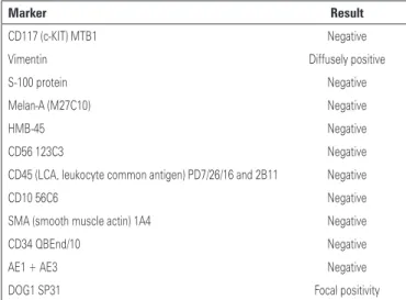
wall in two planes. The pathological examination demonstrated GIST, of epithelioid architectural pattern, mitotic index of 1 per 50 high-magnification fields and moderate cell pleomorphism. The lesion had no signs of necrosis or of gastric mucosa infiltration. The immunohistochemistry was negative for DC117 and the results are demonstrated on chart 1. Despite being a severe case, the patient progressed with no intercurrent events and was discharged from hospital on the 5th postoperative day. The patient remained on
outpatient monitoring with no signs of disease relapse.
Figure 1. Computed tomography revealed a tumor in the greater curvature of the stomach, with heterogeneous contrast uptake, measuring 9.2x7.7cm
DISCUSSION
GIST account for only 1% of all primary gastric tumors. In immunohistochemistry they are positive for c-KIT (CD117). It is known that approximately 95% of all GIST are positive for this receptor(5). Histologically
the GIST derive from smooth muscle cells. In the 1990´s, some inconsistencies in broad classification of tumors as GIST were reported. An important discovery was the identification of CD117 receptor expression for almost all GIST, unlike leiomyomas, leiomyosarcomas and other spindle cell tumors of the tract gastrointestinal that are often CD117 negative(5,6).
As described in the literature, the first acute symptom of this patient was gastrointestinal hemorrhage. Bleeding is present in approximately 40% of cases(6,7). DOG1 is
a membrane protein discovered through analysis of the gene expression patterns of GIST, which showed
to be a useful immunohistochemical marker(8). The
immunohistochemical results of the concomitant use of DOG1 and KIT in GIST demonstrated that 85% of the cases were positive for both markers (DOG+/KIT+), 6% were negative for DOG1 and KIT, 6% were DOG+/ Chart 1. Immunohistochemical results of the surgical specimen
Marker Result
CD117 (c-KIT) MTB1 Negative
Vimentin Diffusely positive
S-100 protein Negative
Melan-A (M27C10) Negative
HMB-45 Negative
CD56 123C3 Negative
CD45 (LCA, leukocyte common antigen) PD7/26/16 and 2B11 Negative
CD10 56C6 Negative
SMA (smooth muscle actin) 1A4 Negative
CD34 QBEnd/10 Negative
AE1 + AE3 Negative
379
Hemoperitoneum secondary to gastric GIST
einstein. 2012;10(3):377-9
KIT- and 3% were DOG-/KIT+. DOG1 specificity was very high for few types of non-GIST tumors that are
immunoreactive for DOG1(8). The CD117- DOG1+
patients can be treated with tyrosine kinase inhibitors like the C-KIT positive individuals. As to surgical approach, exploratory laparotomy was chosen, but
some authors(9) described a case of hemoperitoneum
secondary to gastric GIST treated by laparoscopy and vertical gastrectomy, with no intercurrent events, and surgeries that started by laparoscopy and were converted to laparotomy, due to intense bleeding and hemodynamic instability(10).
REFERENCES
1. Demetri GD, Morgan J, Raut CP. Epidemiology, classification, clinical presentation, prognostic features, and diagnostic work-up of gastrointestinal mesenchymal neopslasms, including GIST [Internet]. UpToDate Feb 2010 [cited 2010 Jun 24]. Available from: http://www.uptodate.com/contents/ epidemiology-classification-clinical-presentation-prognostic-features- and-diagnostic-work-up-of-gastrointestinal-mesenchymal-neoplasms-including-gist
2. Miettinen M, Lasota J. Gastrontestinal stromal tumors: pathology and prognosis at different sites. Semin Diagn Pathol. 2006;23(2):70-83.
3. Kang YN, Jung HR, Hwang I. Clinicopathological and immunohistochemical features of gastrointestinal stromal tumors. Cancer Res Treat. 2010;42(3): 135-43.
4. Medeiros F, Corless CL, Duensing A, Hornick JL, Oliveira AM, Heinrich MC, et al. KIT-negative gastrointestinal stromal tumors: proof of concept and therapeutic implications. Am J Surg Pathol. 2004;28(7):889-94.
5. Huang HY, Li CF, Huang WW, Hu TH, Lin CN, Uen YH, et al. A modification of NIH consensus criteria to better distinguish the highly lethal subset of primary localized gastrointestinal stromal tumors: a subdivision of the original high-risk group on the basis of outcome. Surgery. 2007;141(6):748-56. 6. Kawanowa K, Sakuma Y, Sakurai S, Hishima T, Iwasaki Y, Saito K, et al. High
incidence of microscopic gastrointestinal stromal tumors in the stomach. Hum Pathol. 2006;37(12):1527-35.
7. Bachet JB, Hostein I, Le Cesne A, Brahimi S, Beauchet A, Tabone-Eglinger S, et al. Prognosis and predictive value of KIT exon 11 deletion in GISTs. Br J Cancer. 2009;101(1):7-11.
8. Lee CH, Liang CW, Espinosa I. The utility of discovered on gastrointestinal stromal tumor 1 (DOG1) antibody in surgical pathology-the GIST of it. Adv Anat Pathol. 2010;17(3):222-32.
9. Costi R, Le Bian A, Creuze N, Prevot S, Cauchy F, Violi V, et al. Hemoperitoneum caused by a ruptured GIST located in the posterior gastric wall managed by endoscopic diagnosis and laparoscopic treatment: case report and literature review. Surg Laparosc Endosc Percutan Tech. 2011;21(6):e316-8.