ORIGIN
AL RESEAR
CH
Corresponding author: Éder Kröef Cardoso – Universidade Federal do Pará – Av. Gastão Mazeron, 265, ap 301, Medianeira – Porto Alegre (RS), Brazil – CEP 90880-370 E-mail: [email protected] – Financing source: Nothing to declare – Conlict of interest: Nothing to declare – Presentation: Dec. 2015 – Accepted for publication: Oct. 2016. Approved by the Research Ethics Committee of the Federal University of Health Sciences of Porto Alegre – UFCSPA under number 1492/11.
1Graduate program in Rehabilitation Sciences, Universidade Federal de Ciências da Saúde de Porto Alegre (UFCSPA) –
Porto Alegre (RS), Brazil.
2Department of Physical Therapy, Universidade Federal de Ciências da Saúde de Porto Alegre (UFCSPA) – Porto Alegre (RS), Brazil. 3Scholarship holder of DTI – Research Support Foundation of the State of Rio Grande do Sul (FAPERGS).
ABSTRACT | The model of brace developed consists
of a neoprene kneepad that has an inlatable cuf positioned over the popliteal region of the knee. It provides stimuli on joint structures as well as a better biomechanical alignment during the stance phase of the gait. The aim of this study was to quantify the changes and adaptations induced by gait training with the use of orthotic device in hemiparetic patients. A quasi-experimental before-after study was held with twelve adult patients with hemiparesis due to cerebrovascular accident. The peaks of plantar pressure were the markers used to compare the three moments of the study: the baseline, when they were using the brace, and post-gait training. After the sessions of gait retraining with the orthosis, the redistribution of plantar pressures showed increased symmetry during the stance phase, mainly by reducing the pressure on the paretic forefoot (p=0.024) and by the increase in the rearfoot in the paretic side (p=0.010). In addition, these changes were associated with a decrease in pressure on all regions of the foot not afected, especially in the rearfoot after training. The results of the study suggest a change in the gait pattern of participants after using the brace. There has been greater symmetry of the values of the plantar pressure peaks when the afected side was compared with the nonafected side. Training with the brace helps in the rehabilitation process, since it provides baropodometric
372
values approaching the normal pattern of plantar distribution.
Keywords | Stroke; Gait; Orthotic Devices.
RESUMO |O modelo de brace desenvolvido consiste em uma joelheira de neoprene que apresenta um balonete inlável posicionado sobre a região poplítea do joelho. Ele proporciona estímulos sobre estruturas articulares, bem como um melhor alinhamento biomecânico durante a fase de apoio da marcha. O objetivo do estudo foi quantiicar as alterações e adaptações induzidas pelo treinamento da marcha com a utilização do dispositivo ortótico em pacientes hemiparéticos. Realizou-se um estudo quase-experimental do tipo antes e depois com doze pacientes adultos com hemiparesia decorrente de acidente vascular encefálico. Os picos de pressão plantares foram os marcadores utilizados para comparar os três momentos do estudo: na linha de base, quando estivessem utilizando o brace e após o treinamento da marcha. Após as sessões de reeducação da marcha com auxílio da órtese, a redistribuição das pressões plantares evidenciou aumento na simetria durante a fase de apoio, principalmente pela diminuição da pressão sobre o antepé parético (p = 0,024) e pelo aumento no retropé no lado parético (p = 0,010). Além disso, estas alterações foram associadas a uma diminuição da pressão sobre todas as regiões do pé não afetado, especialmente no retropé no
A diferent brace model to retrain hemiparetic gait
with
genu recurvatum
: efects on plantar pressure
distribution
Um modelo diferente de
brace
para a reeducação da marcha hemiparética com
genu
recurvatum
: efeitos sobre a distribuição das pressões plantares
Un modelo distinto de rodillera en la rehabilitación de la marcha hemiparética con
genu
recurvatum
: efectos sobre la distribución de las presiones plantares
momento de pós-treinamento. Os resultados do estudo sugerem uma mudança no padrão de marcha dos participantes após a utilização do brace. Houve maior simetria dos valores dos picos de pressão plantares quando se comparou o lado afetado com o não afetado. O treino com o brace contribui no processo de reabilitação, uma vez que forneceu valores baropodométricos que se aproximaram ao padrão normal de distribuição plantar.
Descritores | Acidente Vascular Cerebral; Marcha; Aparatos Ortopédicos.
RESUMEN | En una rodillera de neoprene se desarrolló un tipo de rodillera ortopédica, que lleva un manguito inlable, puesto en la región poplítea de la rodilla, y que les proporciona estímulos a las estructuras articulares, así como mejora la alienación biomecánica durante la fase de apoyo de la marcha. El propósito del estudio es cuantiicar las alteraciones y adaptaciones producidas por el entrenamiento con este dispositivo ortótico en pacientes hemiparéticos. Se trata de un estudio casi experimental de tipo antes y después, del cual participaron doce adultos hemiparéticos debido al accidente cerebrovascular. Se utilizaron
como marcadores los picos de presión plantar para comparar tres momentos del estudio: el inicio del estudio; el momento en que utilizaban la rodillera y tras entrenar la marcha. Después de las sesiones de rehabilitación de la marcha con ayuda de la rodillera, en la redistribución de las presiones plantares ocurrió un aumento en la simetría durante la fase de apoyo, principalmente disminución de la presión sobre el antepié parético (p=0,024) y aumento en el retropié en el lado parético (p=0,010). Además, estas alteraciones se las asociaron a la disminución de la presión sobre todas las regiones del pie no alterado, especialmente en el retropié en el momento posterior al tratamiento. Los resultados del estudio muestran un cambio en el patrón de marcha de los participantes después de utilizar este tipo de rodillera. Hubo una mayor simetría de los valores de los picos de presión plantar cuando se comparó el lado alterado con el no alterado. El entrenamiento con este tipo de rodillera ayudó en el proceso de rehabilitación, puesto que presentó valores baropodométricos cerca del patrón prestablecido de distribución plantar.
Palabras clave | Accidente Cerebrovascular; Marcha; Aparatos Ortopédicos.
INTRODUCTION
Retraining the gait after a stroke represents one of the greatest challenges of the rehabilitation process, once the locomotion allows greater autonomy and has direct impact on the levels of functionality1. About half of the stroke survivors exhibit motor deiciencies, such as muscle weakness, abnormal muscle tone, and sensory disabilities, often combined with spasticity or contractures of muscles of the paretic lower limbs2. he hemiparetic gait is characterized by changes in the kinematic and spatial-temporal parameters. During the stance phase, the hyperextension of the knee often occurs, which is known as genu recurvatum2.
From the biomechanical point of view, the genu recurvatum is characterized by a ground reaction force vector that passes exactly in front of the knee. In patients with quadriceps weakness, this phenomenon raises a knee extensor moment, avoiding the fall in lexion during the midstance, as a strategy for greater stability in the lower limb. Besides being a walking aesthetic problem, it can cause pain and therefore limit the autonomy of the patient in daily life activities3. It can be caused due to various phenomena, such as weakness or spasticity of the quadriceps muscle, spasticity and/
or contracture of the plantarlexors, and proprioceptive deicits2. he proile of plantar pressure distribution of patients with stroke proves to be very diferent from what it is observed in healthy individuals4. In this sense, the measurement of plantar pressures quantiies the degree of commitment of the hemiparetic patient and assesses objectively the efectiveness of the intervention with an orthotic device in the rehabilitation process2.
the aim of this study was to assess the distribution of plantar pressures during the walking of patients with hemiparesis after using a diferent brace to train the gait of hemiparetic people with genu recurvatum.
METHODOLOGY
Quasi-experimental before-after study. Twelve (12) adult patients who have had a stroke participated, they received physical therapy follow-up in a clinic. Inclusion criteria were the presence of hemiparesis after stroke with evolution between six and 24 months, ability to walk without auxiliary devices, and presence of genu recurvatum during the gait midstance phase. Exclusion criteria were the conditions that obstructed the assessment and training with the orthosis, as well as the presence of ixed deformities or contractures in lower limbs and moderate-to-severe compromises of cognitive, perceptual, of attention or language nature. his irst screening was performed by one of the physical therapists responsible for the study through physical and functional assessment and by data from patient records.
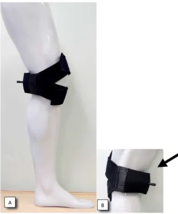
he researchers developed the orthotic device used. It has four neoprene strips ixed with hook and loop tape above and below the patella, gathered in the posterior region through neoprene and leather fabrics (Figure 1A). It also has, on the popliteal fossa, a rubber inlatable cuf wrapped with neoprene, which must be positioned in a way that ensures more pressure on that local (Figure 1B).
he pressure of the cuf was established at 0.35 kg/cm2 with a calibration sensor so that it could ofer a moderate degree of pressure on the popliteal region. he hook and loop tapes on the anterior surface of the thigh and leg were adjusted according to the patient’s response, they should be comfortable and securely ixed.
After receiving explanations about the study and signing an informed consent form, the irst gait assessment was held. he lowchart of assessments and training was divided into three moments. At irst, we held an assessment of plantar pressure measurement. he records of plantar pressures were collected through dynamic baropodometry. To obtain the baropodometry data, we used a digital baropodometer of the pressure platform type of the brand Novel, Emed-X model (690mmx403mmx190mm) resolution of 4 sensor/cm2, maximum sampling rate of 100Hz.
he platform was positioned in the center of a runway of Ethylene-vinyl acetate (EVA) of approximately 7 meters in length. We asked the patient to walk naturally and barefoot in self-selected speed on the runway. he patient started the gait with parallel feet from the mark previously delineated, allowing the implementation of the third step on the platform, according to the three-step protocol6. Each individual walked ive times the path to become familiar with the procedures and, after this period, the software was enabled to carry out the acquisitions. he Peak Plantar Pressure in kilopascals (kPa) –maximum pressure recorded in speciic plantar areas – of four regions of the foot (rearfoot, midfoot, forefoot, and toes) was established through the Novel Scientifc 12.3.30 software.
Figure 1: Orthosis for gait rehabilitation. (A) lateral view of the neoprene strips above and below the patella; (B) greater detail of the position of the inlatable cuf on the popliteal region (arrow)
he day after that assessment, patients began four weeks of gait training with the orthosis under the supervision and observation of a physical therapist who belongs to the group of authors of this study. Twice a week, for ive weeks, before the sessions in the physical therapy clinic, the patients who participated in the study were asked to walk with self-selected speed on level ground, a hall with 50 meters in length, wearing the orthosis to retrain the gait in the afected side during ifteen minutes. he amount of interventions was based on a study that showed signiicant and lasting efects on the control of recurvatum in the gait after 10 electrogoniometric biofeedback sessions7. On the irst day following the tenth session of training, we held the moment 3 to assess the plantar pressures without the orthosis.
Data processing and analysis
For the statistical analysis, we calculated the average and the standard deviation of both feet of the variables of Peak Plantar Pressure (maximum pressure recorded in the plantar speciic areas: rearfoot, midfoot, forefoot, and toes). hus, eight independent analyses were performed, aiming to compare the situations without brace, with brace, and post-training, one for each limb and regions of the foot. he values of peak pressure in these areas of both feet were compared with the afected and nonafected sides in diferent moments of the gait assessment. hese comparisons were made using parametric tests (ANOVA for repeated measures) or non-parametric tests (Friedman), according to the presence or absence of normal distribution of the data. Shapiro-Wilk test was used to verify the normality of the data. he version 17 of the program SPSS® (Statistical Package for the Social Sciences, Inc., Chicago, USA) was used to analyze the data. he signiicance level adopted was p≤0.05.
RESULTS
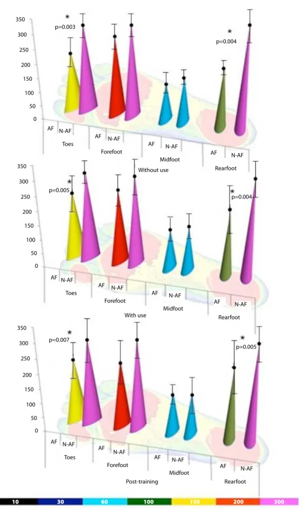
From a total of twelve hemiparetic patients, the average age was 61.5±10.7 years, 7 of them were male. he average time post-stroke was 12.9±5.6 months. he graphical representations of the variables of peak pressure in the four regions of the feet on the sides afected (AF) and nonafected (N-AF) with and
without the orthosis in the paretic knee are exposed in Figures 2, 3, and 4.
Moment 1 (baseline)
In the rearfoot, the plantar pressure behavior showed signiicantly diferent average values on the afected side compared with the nonafected side with 209.65 (SD 51.38) and 349.16 (SD 120.68), respectively, (p=0.003). As well as on the region of the toes, the average values were 229.51 (SD 107.87) and 335.32 (SD 180.69), (p=0.004), with the largest distributed pressures on the nonafected side.
Moment 2 (wearing the brace)
Comparing the afected side with the nonafected side, the orthosis did not show changes in the pattern identiied in the baseline. Diferences in the pressures of the rearfoot remained with 215.55 (SD 50.52) and 322.62 (SD 109.16), (p=0.005), as well as in the toes with 260.44 (SD 118.15) and 327.92 (SD 133.06), (p=0.004) being the biggest pressures presented on the healthy side.
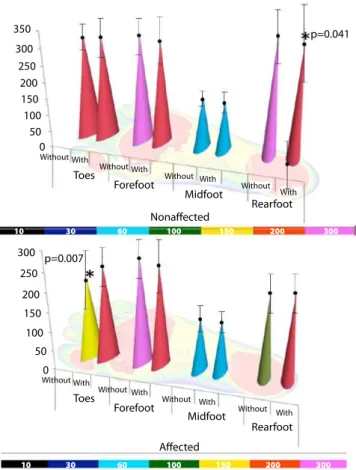
When comparing the AF wearing the orthosis with its baseline values, we identiied increase in peak plantar pressure in the region of the toes, going from 229.51 (SD 107.87) to 260.44 (SD 118.15), (p=0.007) and a decreasing trend in the pressure on the forefoot on the paretic side, going from 287.53 kPa to 265.41 kPa. he healthy side showed decreased values in all regions, in particular on the rearfoot, 349.16 (SD 120.68) to 322.62 (SD 109.16), (p=0.041).
Moment 3 (gait training)
350
p=0.003
AF Toes N-AF
AF Forefoot
N-AF
AF Midfoot Without use
N-AF
AF Rearfoot
N-AF p=0.004
p=0.005
AF Toes N-AF
AF Forefoot
N-AF
AF
Midfoot With use
Post-training N-AF
AF Rearfoot
N-AF p=0.004
p=0.007
AF Toes
10 30 60 100 150 200 300
N-AF
AF
Forefoot N-AF
AF
Midfoot N-AF
AF Rearfoot
N-AF p=0.005 300
250 200
150 100
50 0
350 300
250 200
150 100
50 0
350 300
250 200
150 100
50 0
350 300 250
200 150 100
50 0
300 250 200 150 100 50
0
Without
Toes
With Without
Forefoot
Affected Nonaffected
With Without
Midfoot
With
Without
Rearfoot
With
Without
Toes
With Without
Forefoot
With Without
Midfoot
With
Without
Rearfoot
With
p=0.041
p=0.007
10 30 60 100 150 200 300
10 30 60 100 150 200 300
Figure 3: Graphical representations of the variables of peak pressure compared without and with the use of orthosis on the side afected and nonafected in diferent regions of the foot at moments 1 and 2
350 300 250 200 150 100 50
0
300 250 200
150 100
50 0
No use
Toes
Post Tr No
usePostTr No
usePostTr No use PostTr
Forefoot
Affected Nonaffected
Midfoot
Rearfoot
Toes
Forefoot
Midfoot
Rearfoot p=0.010
p=0.024
10 30 60 100 150 200 300
10 30 60 100 150 200 300
No use
Post Tr No
use Post
Tr No
use PostTr No use PostTr
DISCUSSION
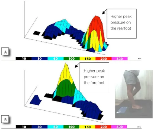
he pattern of contact with the ground of the patients assessed in our study showed a predominance of higher peak pressure on the forefoot paretic region. his aspect can be noted when comparing the pressure areas in three-dimensional format of the initial contact of a hemiparetic patient with those of a healthy individual. Figure 5 shows an example of comparison between a paretic foot with that of a healthy individual. Insuicient dorsilexion and spasticity of plantilexors can explain this form of “heel strike” in hemiparetic patients4,8.
When comparing the values of pressure peaks between the afected and the nonafected side, smaller values were observed on the region of the rearfoot and toes on the afected side without the orthosis, constituting the basal pattern of the patients. he distribution of the peak plantar pressure, during the gait without the orthosis for gait rehabilitation, showed patterns similar to other studies involving baropodometry in hemiparetic people8,9-12. he dynamic plantar pressure of hemiplegic patients shows, on the afected side, a change of initial contact, from the rearfoot to the forefoot area, an increase in the lateral plantar stance, a limited bearing, and a reduced or absent heel-of movement in terminal stance14.
On the afected side, the pressures are mainly displaced on the forefoot, on the basis of spasticity of triceps surae, on the N-AF side, however, there is a greater transfer of stance on the foot not afected in patients with severe motor impairment15. he peak of reduced pressure at the base of the third metatarsal, according to studies, denotes a higher transverse arch of the forefoot: that would be connected to the greater spasticity of the intrinsic foot muscles12. he comparison of peak pressure between the nonafected and afected side of nine hemiparetic patients showed that the nonafected side had the highest peaks of pressure, while on the afected side, the forefoot and the lateral plantar arch showed greater values8. his can occur due to equinism and spasticity, which commonly occur in hemiparetic patients, as well as due to coordination disturbance, sensory alteration, and insuicient transfer of weight on the paretic side11. he analysis of distribution in peak plantar pressure of 25 hemiparetic patients and of 31 healthy patients showed reduction of peak pressure on the sides afected, mainly in the medial metatarsal area of hemiparetic patients13. Other studies have also observed that the distribution of plantar pressures of hemiparetic patients showed low-pressure peak around the foot of the afected side12.
10 30 60 100 150 200 300
10 30 60 100 150 200 300
Higher peak pressure on the rearfoot
Higher peak pressure on the forefoot
In the second part of the study, the asymmetries found in the baseline remained, from a comparison of the afected side with the orthosis versus the nonafected side. Comparing the peaks of baseline pressure with the ones of paretic knee use, some changes were observed: on the afected side, the region of the toes showed more pressure against the ground, a tendency to decrease the pressure on the forefoot and increase on the rearfoot. All nonafected regions of the foot showed decreased peak pressure, mainly in the region of the rearfoot. Studies with analysis of normal patterns of plant distribution consider that an ordered sequence of events occurs, during the stance, starting by the heel, heel and forefoot strike, only the forefoot and, inally, forefoot and toes. In this inal moment of contact with the ground, the weight is being transferred to the other foot with heel strike14. he reduction of the pressure on the rearfoot of the N-AF side after the training can be attributed to a better motor control, conferred by a better angle of the calcaneus entry and subsequent softening in the heel strike, observed in better distribution of plantar pressures in the stance phase on the AF side.
he veriication of the efects of the orthosis on the gait, at moment 3, showed positive functional results. In the forefoot region of the afected side, where usually the highest peak pressures occur in the hemiparetic foot12, there was a signiicant reduction of the values after the training. On the nonafected side, the peak plantar pressure decreases in all regions, in particular in the rearfoot. he reduction of the pressures on the foot on that side, both when using the orthosis, and post-training, suggest that the body weight is being distributed more to the paretic side. Hemiparetic individuals transfer approximately 70% of the total weight on the nonafected lower limb16. he postural alignment and symmetry in transfers of weight, in patients with stroke, and its relationship with functional skills, of balance and gait are essential for a successful rehabilitation17.
Irregularities and asymmetries in the distribution patterns of plantar pressures between the limbs relect a disturbance in the articular movement9,10,18. Possibly the decreased dorsal lexion by spasticity of the triceps surae contributes with the highest peaks of pressure on the forefoot on the afected side. he diiculties in the transfer of weight from the rearfoot to the forefoot can be due to genu recurvatum, tilted uterus and insuicient hip extension on the paretic side18.
he results of the study revealed that the orthosis developed must necessarily be accompanied by a period
of adjustment and gait training to obtain better results at the level of the forefoot. We found that only the use itself does not cause signiicant changes in the pattern of plantar pressures. After ten sessions, there was more adequate distribution of plantar pressure to the rearfoot on the afected side and greater symmetry in relation to the N-AF side. On the nonafected side, the pressure, which was greater in the rearfoot, showed a decreasing trend in the pressure peaks in this region after the training. We can therefore infer that training with the orthosis contributes to the rehabilitation process, since it provides favorable biomechanical alignment in the joint, and thus a best performance of knee lexors that are weaker in the genu recurvatum. In addition, it can provide proprioceptive input, giving the patient a feedback about the knee hyperextension, informing the patient of the need to correct this unwanted movement.
Among the limitations of the study, in addition to the reduced sample, we highlight the lack of a control group to document normal developments with the conventional physical therapy. In an upcoming assessment, in addition to the incorporation of placebo group, it would be important to obtain baropodometry data associated with electromyography and/or kinematic analysis to quantify the degree of genu recurvatum at diferent speeds, correlating the muscular behavior with changes in plantar pressure and in kinematic parameters. When considering the mechanical characteristics of the brace, we understand that it proves to be a diferentiated management of the gait with genu recurvatum, compared with the other existing resources. his device does not restrict the movement of the knee in a ixed angle, but “reports” to the patient by means of tactile and proprioceptive stimuli the occurrence of hyperextension during the midstance. his study suggests a better gait pattern in the participants by presenting greater symmetry in plantar pressure peak values, i.e., a contact of the foot with the ground closer to normal. We can thus infer that the training with the orthosis can help hemiparetic individuals to obtain better motor control and biomechanical alignment of the knee during the gait.
REFERENCES
2. Boudarham J, Zory R, Genet F, Vigné G, Bensmail D, Roche N, et al. Efects of a knee-ankle-foot orthosis on gait biomechanical characteristics of paretic and non-paretic limbs in hemiplegic patients with genu recurvatum. Clin Biomech (Bristol, Avon). 2013;28(1):73-8. doi: 10.1016/j. clinbiomech.2012.09.007.
3. Bleyenheuft C, Bleyenheuft Y, Hanson P, Deltombe T. Treatment of genu recurvatum in hemiparetic adult patients: a systematic literature review. Ann Phys Rehabil Med. 2010;53:189-99. doi: 10.1016/j.rehab.2010.01.001.
4. Femery V, Moretto P, Renaut H, Thévenon A. Spasticité et distribution des pressions plantaires chez des enfants atteints d’hémiplégie cérébrale infantile. Ann Readapt Med Phys. 2001;44(1):26-34. doi: http://dx.doi.org/10.1016/ S0168-6054(00)00060-X.
5. Isakov E, Mizrahi J, Onna I, Susak Z. The control of genu recurvatum by combining the Swedish knee-cage and an ankle-foot brace. Disabil Rehabil. 1992;14(4):187-91.
6. Robinson CC, Detânico RC, Zaro MA, Andrade MC. Comparação entre dois protocolos de baropodometria dinâmica utilizando plataforma de pressão. Tecnicouro. 2010;249:70-4.
7. Basaglia N, Mazzini N, Boldrini P, Bacciglieri P, Contenti E, Ferraresi G. Biofeedback treatment of genu-recurvatum using an electrogoniometric device with an acoustic signal. One-year follow-up. Scand J Rehabil Med. 1989;21(3):125-30. 8. Wong AM, Pei YC, Hong WH, Chung CY, Lau YC, Chen CP.
Foot contact pattern analysis in hemiplegic stroke patients: an implication for neurologic status determination. Arch Phys Med Rehabil. 2004;85(10):1625-30.
9. Valentini FA, Granger B, Hennebelle DS, Eythrib N, Robain G. Repeatability and variability of baropodometric and spatio-temporal gait parameters – results in healthy subjects and in stroke patients. Neurophysiol Clin. 2011;41(4):181-9. doi: 10.1016/j.neucli.2011.08.004.
10. Boza R, Duarte E, Belmonte R, Marco E, Muniesa JM, Tejero M, et al. Estudio baropodométrico en el hemipléjico vascular:
relación con la discapacidad, equilibrio y capacidad de marcha. Rehabilitación (Madr. Internet). 2007;41(1):3-9. doi: http://dx.doi.org/10.1016/S0048-7120(07)75350-1.
11. Schuster RC, Zadra K, Luciano M, Polese JC, Mazzola D, Sander I, et al. Análise da pressão plantar em pacientes com acidente vascular encefálico. Rev Neurociênc. 2008;16(3):179-83.
12. Meyring S, Diehl RR, Milani TL, Hennig EM, Berlit P. Dynamic plantar pressure distribution measurements in hemiparetic patients. Clin Biomech (Bristol, Avon). 1997;12(1):60-5. 13. Titianova EB, Pitkänen K, Pääkkönen A, Sivenius J,
Tarkka IM. Gait characteristics and functional ambulation proile in patients with chronic unilateral stroke. Am J Phys Med Rehabil. 2003;82(10):778-86. doi: 10.1097/01. PHM.0000087490.74582.E0.
14. Perry J. Gait analysis: normal and pathological function. New Jersey: SLACK Incorporated; 1992.
15. Gaviria M, D’Angeli M, Chavet P, Pelissier J, Peruchon E, Rabischong P. Plantar dynamics of hemiplegic gait: a methodological approach. Gait Posture. 1996;4(4):297-305. doi: http://dx.doi.org/10.1016/0966-6362(95)01055-6. 16. Shumwaycook A, Anson D, Haller S. Postural sway biofeedback:
its efect on reestablishing stance stability in hemiplegic patients. Arch Phys Med Rehab. 1988;69(6):395-400.
17. Trindade APNT, Barboza MA, Oliveira FB, Borges APO. Inluência da simetria e transferência de peso nos aspectos motores após acidente vascular cerebral. Rev Neurociênc. 2011;19(1):61-7.
18. Robain G, Valentini F, Renard-Deniel S, Chennevelle JM, Piera JB. A baropodometric parameter to analyze the gait of hemiparetic patients: the path of center of pressure. Ann Readapt Med Phys. 2006;49(8):609-13. doi: 10.1016/j. annrmp.2006.05.002.