Neurology Service, Hospital de Clínicas, Federal University of Paraná (UFPR), Curitiba PR, Brazil: 1Associate Professor of Neurology,
Medical Clinic Department.
Received 1 September 2000, received in final form 22 June 2001. Accepted 1 July 2001.
Dr. Ehrenfried Wittig - Hospital de Clínicas - Rua General Carneiro 181 / 12º andar - 80069-900 Curitiba PR - Brasil.
OCULAR CYSTICERCOSIS
An epidemiological study
Ehrenfried O. Wittig
1ABSTRACT The objetive of the research was to verify the incidence of the ocular form of cysticercosis among the patients who consulted professionals of the field, during the first statistic inventory accomplished in the State of Paraná, Brazil. The 1996 research was performed through a questionnaire sent to all 220 members of Paraná Ophthalmology Association and to the 17 members of Pathology and Cytology Department of the Paraná Medical Association. A pre-stamped envelop for the return of the answers was included in the package. We received 99 answers from ophthalmologists and 10 answers from pathologists. Retrospective and epidemiological research about the incidences of the ocular cysticercosis in the State of Paraná, added to national related published reports, rendered 95 cases probably adquired in Paraná, 71 of the cases were referred in this research and 24 in prior publication. The most frequent localization was vitreous and subretinal, as literature had already pointed out.
KEY WORDS: ocular cysticercosis, Paraná State Brazil, incidence.
Cisticercose ocular: um estudo epidemiológico
RESUMO O objetivo da pesquisa foi o de verificar a incidência da forma ocular de cisticercose entre os pacientes que consultam profissionais da área, no primeiro levantamento estatístico realizado no Estado do Paraná. A pesquisa retrospectiva e epidemiológica foi realizada em 1996 através de encaminhamento por correio, de um questionário com envelope com porte pago para resposta aos 220 membros da Associação Paranaense de Oftalmologia e aos 17 membros do Departamento de Patologia e Citologia da Associação Médica do Paraná, dos quais obtivemos 99 cartas-respostas dos oftalmologistas e 10 dos patologistas. A pesquisa retrospectiva, associada aos relatos de outros casos já referidos na literatura nacional, permitiu-nos reunir 95 casos de cisticercose ocular, provavelmente adquiridos no Paraná, sendo 71 casos referidos nesta pesquisa e 24 em publicação anterior. A localização ocular mais frequente foi a vítrea e subretiniana, fato já salientado na literatura.
PALAVRAS-CHAVE: cisticercose ocular, Estado do Paraná, incidência.
Man is the only certain natural host for Taenia
so-lium although other animals apparently can be
in-fected. By chance, man can be the intermediate host for Taenia solium cysticercotic larva. In these circum-stances, he may suffer important consequences cau-sed by its presence. Man only shelters Taenia solium
cysticercus. As a guest of foreign body, the
Cysticer-cus cellulosae, or the metacystoid form of Taenia
so-lium as the parasitologists prefer to call, can pro-duce immunological reactions with fairly intense in-flammatory signs and symptoms in addition to caus-ing compressive and obstructive phenomena in sur-rounding structures and cavities .
The clinical features of cysticercosis may be a re-sult of just one or hundreds of cysts, which may be
distributed in one or several organs and tissues. The combination of the localization within the body, the amount of cysts, the hosts immunological reaction, the viability duration of the cyst and the intensity of the inflammatory reaction, may produce fairly exu-berant manifestations, especially when it is acting within the meninges, central nervous system or eye-ball. Cysts may have diverse localization in the hu-man body and their frequency in tissues and organs may vary. Brazilian1-6 and American7 studies in
gene-ral report the encephalic localization as the predomi-nant one. Some European statistics from the be-ginning of the century8-12, when cysticercosis was
the encephalic predominance be a result of larval adaptation modified after its introduction in America? Human and animal cysticercosis are frequently encountered in Paraná State, although there has been a progressive decline13. The neurologic form is more
predominant than the ocular or cutaneous-muscu-lar ones. National literature1-4 describes more than
2500 cases of the neurologic form in Paraná State. This epidemiological research sought to verify the prevalence of this ocular form of larval infection in Paraná, a State with 9 258 813 inhabitants (1998).
METHOD
The 1996 research was performed through a question-naire sent to all 220 members of Paraná Ophthalmology Association and to the 17 members of Pathology and Cy-tology Department of the Paraná Medical Association. A pre-stamped envelop for the return of the answers was included in the package. We received 99 answers from ophthalmologists and 10 answers from pathologists, (Table 2). Most of the professionals of these two specializations in the State are affiliated to both associations. The inten-tion of this research was to verify statistical changes in this last century, when at least, in its beginning, the scientific studies used to show the ocular form predominance.
RESULTS
Ninety letters were received from the ophthal-mologists (40.9%) and 10 from the
anatomopatho-Table 1. Cysticercosis distribution in France.
Vosgien (1911)9 Volovatz (1902)8
cysts - 807 cases cysts - 414 cases
Eyes & appendage 372 (46.0%) Eyes & appendage 248 (59%)
Nervous system 330 (41%) Nervous system 149 (35%)
Skin & subcut. cel. 51 (6%) Others 1 (4.1%)
Muscles 28 (3%)
Other organs 26 (3%)
Table 2. Questionnaire model.
I - Have you already diagnosed any case of ocular cysticercosis? Yes ______ No ______
II - If yes, how many cases? _______________ III - Which was the local, side and number:
A) Extra Ocular Right site Left site 1 palpebral ________ ________ 2 sub-conjuntural ________ ________ 3 orbital ________ ________ 4 lacrimal g. ________ ________ B) Intra Ocular Right site Left site
1 Anterior Chamber ________ ________ a) cornea ________ ________ b) aquoso humor ________ ________
c) iris ________ ________
d) crystalline ________ ________ 2 Posterior Chamber
a) vítreos humor ________ ________ b) subhyaloidal ________ ________ c) subretinal ________ ________ d) subchoroidal ________ ________ IV - The diagnostic was obtained through:
Physical examination ________ surgery ________ Anatomopathologic examination ________
serologic test ________
V - Was there associated cysticercosis in other local or organs ? _____________________
VI - Specify which were the most important ocular manifestations?
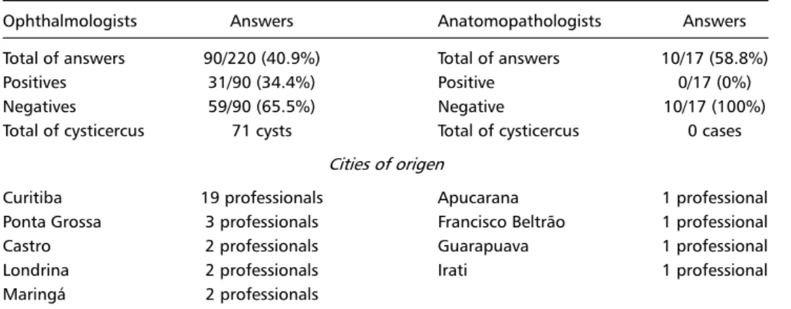
Table 3. Analysis of information received.
Ophthalmologists Answers Anatomopathologists Answers Total of answers 90/220 (40.9%) Total of answers 10/17 (58.8%)
Positives 31/90 (34.4%) Positive 0/17 (0%)
Negatives 59/90 (65.5%) Negative 10/17 (100%)
Total of cysticercus 71 cysts Total of cysticercus 0 cases
Cities of origen
Curitiba 19 professionals Apucarana 1 professional
Ponta Grossa 3 professionals Francisco Beltrão 1 professional
Castro 2 professionals Guarapuava 1 professional
Londrina 2 professionals Irati 1 professional
logists (58,8%). We noticed that most of the profes-sionals who answered the questionnaire worked in University Hospitals and in the biggest private clin-ics around the State, and that most of the reported cases were observed in the last 80 years (1918-1998). After analyzing the information received, we gathered the data on Table 3.
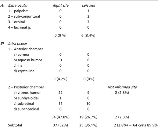
The classification of ocular cysts found in this re-search was made according to the system proposed by Fontan14 (Table 4).
DISCUSSION
The data attained in our research can be com-pared with those presented by Almeida & Oliveira15
in São Paulo State (Brazil), in 1971. They did an ex-cellent review in the Instituto Penido Burnier Archives, in Campinas (São Paulo). Their material has already been described by Lech16 and Queiroz17, who
re-ported the findings of Table 5.
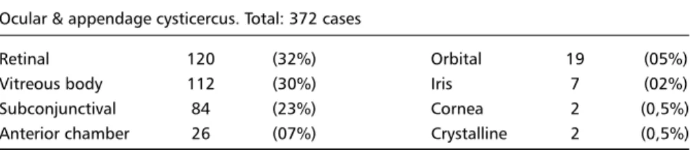
The data gathered in our inquiry concerning the ocular topography may also be compared with those reported in 1911 by Vosgien in his thesis (Table 6)9,11,12.
Among European statistics from the beginning of the century that were consulted, the works of A. Von Graefe, Vosgien and Volovatz stand out because
of the number of cysticercosis cases and because of their significance concerning the ocular form in that period (Table 7).
Statistical distribution of cysts in their ocular form, in that time, represents the findings of a period, of a country and of a limited group of researchers from specialized clinics and coincides with the ophthal-mologic surgical therapy emergence. The studies cited, one from Germany and two from France, which are neighboring countries, showed a rising number of cysticercosis cases by the end of last century. To-day there are no autochthonous cases in these coun-tries.
Vosgien and Volovatz showed the ocular form prevalence over neurological or another forms (Table 1). This is an interesting fact to be observed since in the American statistics the central nervous system localization was always predominant, although the data came from more recent research. Since in those countries there is hardly any autochthonous human cysticercosis, it is not possible to compare the distri-bution by periods.
In 1829, in Germany, the first case of ocular cysti-cercosis was reported by Schott and Soemmering18.
In 1864, in Brazil, Dr. Pedraglia described the first case of human Cysticercus cellulosae in the eyeball
Table 4. Number and topographic distribution of the cases (1916-1996).
A) Extra ocular Right site Left site
1 palpebral 0 1
2 sub-conjuntural 0 2
3 orbital 0 3
4 lacrimal g. 0 0
0 (0 %) 6 (8.4%)
B) Intra ocular
1 Anterior chamber
a) cornea 0 0
b) aquoso humor 3 0
c) iris 0 0
d) crystalline 0 0
3 (4.2%) 0 (0%)
2 Posterior chamber Not informed site
a) vítreos humor 22 9 2 (2.8%)
b) subhyaloidal 1 0
c) subretinal 11 10
d) subchoroidal 0 0
34 (47.8%) 19 (26.7%) 2 (2.8%)
Table 5. Cases collected between 1916-1970: 299.
Cases studied: 283
Number of affected eyes
RE 151 eyes LE 137 eyes Obs. Binocular (both eyes) = 4 cases
Ocular cysticercus topographic localization
a) Vitreous 147 eyes e) Anterior chamber
Clinical Diagnosis 126 eyes Clinical Diagnosis 3 eyes Anat.path. Diagnosis 21 eyes f) Subconjunctival
b) Subretinal 90 eyes Clinical Diagnosis 6 eyes
Clinical Diagnosis 83 eyes g) Subcutaneous
Anat. Pathologic 7 eyes Clinical Diagnosis 1 eye
c) Subhyaloidal h) Imprecise local. 13 eyes
Clinical Diagnosis 23 eyes Binocular 4 cases
d) Intraretinal Vitreous + subhyaloidal 1 case
Clinical Diagnosis 2 eyes Vitreous + subretinal 2 cases
Subretinal 1 case
Cysticercus racemous 1 case (6 vitreous vesicles)
Table 6. Ocular cysticercus topographic distribution.
Ocular & appendage cysticercus. Total: 372 cases
Retinal 120 (32%) Orbital 19 (05%)
Vitreous body 112 (30%) Iris 7 (02%)
Subconjunctival 84 (23%) Cornea 2 (0,5%)
Anterior chamber 26 (07%) Crystalline 2 (0,5%)
to the Academia Nacional de Medicina. This study was published in Annaes Brasileiros de Medicina Vol. XVI, p.42.18,19 Dr.Rubens de Lacerda Manna, a
neurologist, reported the first case of neurocysti-cercosis in Paraná State in 1945, to the Paraná Medi-cal Association. In 1923, Nercolino Machado thesis18
in São Paulo referred to the first 25 cases of ocular cysticercosis, collected from various papers published in national literature. He found 9 subretinal, 8 vitre-ous, 5 conjunctival and 3 located in the anterior chamber.
If we consult the works of Lech16, Queiroz17 and
Al-meida & Oliveira15 (and other ophthalmologists) about
the ocular or ophthalmologic cysticercosis form in Bra-zil, we will notice that their statistics were similar to the best statistical works concerning the neurologi-cal form described at that time. But they were also based on aberrant ophthalmological statistics. Because of the surgical therapy and the good reputation of the hospital, there was a great national flow of pa-tients to the Ophthalmologic Institute (Penido Burnier), where the studies of the three authors originated.
Some old research can illustrate the comparison between the statistical frequency of ocular and neu-rological forms. Andrade20, in his dissertation for the
Rio de Janeiro Medical School (1908), reports 2 cys-ticercosis cases with neurological manifestations. In one, the necropsy was incomplete. Tretiakoff & Pache-co e Silva, working in Juquery Hospital (São Paulo), a great psychiatric hospital in 1924, found in 250 necropsies, 9 brain cysticercosis cases6. Toledo
Gal-vão19, in his thesis from 1928 (São Paulo), found in
1248 necropsies, perfomed in the São Paulo Medi-cal School and Juquery Hospital, 26 cases of brain lodging. In Rio de Janeiros Hospital dos Alienados (1934), Hélion Póvoa observed only 11 cases of brain cysticercosis among 1163 necropsies21. Pinheiro e
Mel-lo21 (1941) working in the same hospital, after
Therefore, a simple statistical presentation may not represent the reality of the situation. Some coun-tries situated in the same meridian and parallel have different disease incidence rates. This phenomenon has been explained by climate, cultural, religious or economic factors. In the USA and Europe taeniasis and human cysticercosis were eliminated in the last fifty years, mainly by educational modifications in the hygienic habits of the population. No more au-tochthonous cases are found in these places. In 1911, Gomes Faria referred, through coprologic tests, to the infrequency of Taenias (especially solium) in the population of Rio de Janeiro16. Concerning this, Lech
Júnior16 (1949) stated, while there is Taenia solium,
cysticercosis will be prevalent, although both pro-phylaxis are easily executed.
In the ocular form the cysts are lodged in the orbital structures. In Tables 6 and 7 and in the statis-tics presented by Almeida & Oliveira15, the site
varia-tion was shown with retinal and vitreous predomi-nance. Nevertheless, in India 63% of the ocular cases described were subconjunctival22. We do not have
an explanation for these findings. According to most authors, in the topographic distribution of head cys-ticercosis the central nervous system and eyeball lo-calization prevails because of the disposition of the cervical arteries and the amount of cephalic blood flow. The left side predominance, mentioned by sev-eral authors, is due to a more direct disposition of blood on the left side. This argumentation may not be true since the investigation about the cutaneous-muscular form has not been so systematically un-dertaken.
It is possible that the cutaneous-muscular form has a higher incidence than that reported in pub-lished studies, but because of its small clinical mani-festation it has not been routinely examined neither clinically nor radiologically. Corroborating these facts, Malik et al.23 and Dixon & Lipscomb24 studying the
disease more systematically found the cutaneous-muscular form predominant.
It is understood that the statistical prevalence of the ocular form in the beginning of the century was due more to the emergence of surgical therapy and the possibility of an easy diagnosis than to the scarce-ness of other forms. Because the ocular cases deter-mine acute and important ophthalmic or painful clinical manifestations, it was possible to diagnose the processes early and rapidly25. Probably, the
con-centration of well equipped ophthalmologic clinics (the ophthalmoscope had already been invented, im-proving the accuracy of the diagnosis) in big cities had determined the predominance of the ocular form. Cysticercosis in the brain and other organs and tissues normally does not show clinical features and it is only verified through necropsies or image stud-ies such as radiography, tomography and magnetic resonance.
Analyzing the data collected in our research, a difficulty to evaluate the information was clearly observed. Because some information was incom-plete, and some were given from memory, it was diffi-cult to consult or recover the data. Duplicity of info-rmation about the same case and loss of records about other cases are possible. Items IV, V and VI of the questionnaire were not analyzed because the
Table 7. Ocular cysticercosis topographic distribution according to statistics of several authors.
Ocular cysticercosis Cases Vitreous Sub- Vit./sub- Sub- Ant- Orbital retinal retinal conjunt. chamber
Von Graefe10 Germany 1866 179 90 80 - 5 3 1
Volovatz8 France 1902 248 - - - - -
-Vosgien9,13 France 1911 372 112 120 - 84 26 19
Galvão19 Brazil 1928 21 - - 21 - -
-Laigner-Terrasse22 France 1932 35 - - 26 7 1
-Michaeil25 Romania 1935 11 - 10 - - 1
-Queiroz17 Brazil 1945 92 42 41 - 7 2
-Lech16 Brazil 1949 116 51 44 - 7 2 1
Tolkovskey22 Russia 1951 1216 - - 73% - -
-Fontan14 Mexico 1964 70 29 31 - 7 2 1
Malik23 India 1968 110 11 1 - 10 -
-Almeida15 Brazil 1971 283 147 90 - 6 3
data was insufficient for a statistic analysis. In the ana-tomopathologists records this fact may be well ob-served because none of them informed the record or analysis of an ocular cysticercosis case. This prob-ably was due to a flaw in the nosologic filling of the material. Through personal contact with some oph-thalmologists and the applied questionnaire, we found out that they frequently did not send the surgical material for laboratory testing because there were no doubts about the etiology of the problem. Only three ophthalmologists mentioned sending the sur-gical piece to antomopatholosur-gical analysis. None-theless, the survey represents the reality quite well and allows us to have a broad view of the problem. In our research in Paraná State (Brazil) we obser-ved 71 ocular cysticercosis cases probably acquired in our State, in the period 1916-1996. Almeida & Oliveira15 examined and referred 24 other cases of
people resident in Paraná state in his studies at Pe-nido Burnier Institute (São Paulo State, 1971), mak-ing a total of 95 cases. This collection of ocular cases is significantly smaller than the already reported neu-rological cysticercosis cases in Paraná State1-4.
In Brazil, except for the research done by Almeida & Oliveira that reported a great number of cases, we were able to verify only 1 to 8 cases26-29. A
com-parison among studies from several countries does not show significant statistical or topographical mo-difications.
Ocular cysticercosis is, in most cases, diagnosed through anamnesis, physical examination entailing ophthalmoscopic evaluation, biomicroscopy, transil-lumination, direct and indirect ophthalmoscopy. Thus, in many cases it is possible to identify a suspected alteration and in others to see the cystic larva alive and moving in the aqueous or vitreous humor.
Nowadays, computerized tomography, magnetic resonance and sonography give us more elements for diagnosis. The final etiologic confirmation is given by macro and/or microscopic anatomopathological analysis. Although infrequent, the association be-tween the ocular form with other localization has not been sought most of the time, especially the muscular one, which is ordinarily asymptomatic23,24.
The main therapeutic procedure for ocular cys-ticercosis is surgical removal of intact or degenera-ted cyst. Countless techniques were used according to the localization, time and surgical resources. There is great controversy concerning the use of drugs. The more recommended cysticides have been prazi-quantel and albendazole, isolated or in association. There has been some advantage for the use of
al-bendazole because of its lower price and apparent higher effectiveness29,30. Dexamethasone and
dexa-chlorfeniramine (antihistaminic) should be used as supporting treatment. Cysticides have little use in in-traocular cysts because the penetration of therapeu-tic substances there is difficult.
REFERENCES
1. Narata AP, Arruda WO, Uemura E, et al. Neurocisticercose: diagnóstico tomográfico em pacientes neurológicos. Arq Neuropsiquiatr 1998; 56:245-249.
2. Bruck I, Antoniuk S, Figimura S, Mogami K, Miyaki M. Neurocysti-cercosis: analysis of 114 patients (abstr ) Pediatric Neurol 1994;11:149. 3. Arruda WO, Menezes MS, Antoniuk AS. Neurocisticercose: diagnóstico
e tratamento. Rev Bras Neurol 1996;32:15-20.
4. Almeida SM, Torres LFB, Stegoni MM, Vicente L, Werneck LC. Neurocisticercose: estudo retrospectivo de laudos de necrópsia (1977-1994) (abstr). Arq Neuropsiquiatr 1994;52(Suppl):P-009.
5. Spina-França A. Cisticercose do sistema nervoso central. Rev Paulista Med 1956;48:59-70.
6. Canelas HM. Neurocisticercose: incidência, diagnóstico e formas clínicas. Arq Neuropsiquiatr 1962;20:1-16.
7. Sotelo J, Guerrero V, Rubio F. Neurocysticercosis: a new classification based on active and inactive form: a study of 753 casos. Arch Intern Med 1985;145:442-445.
8. Volovatz E. Ladrerie ou cysticercose chez l’homme. Thèse, Faculté de Medecine de Paris. Paris, 1902 (citado por Brumpt).
9. Vosgien Y. Le Cysticercus cellulosae chez l’homme et chez les animaux. Thèse, Faculté de Medecine de Paris. Paris, 1911.Nº 373 (cit por Rey e por Brumpt).
10. Von Graefe A. Bemerkungen über Cysticercus. Archv fur Ophtalmol Zwölfeter Jahrgang 1866;12:174 (citado por Queiroz).
11. Rey L. Cisticercose humana. Revista Roche 1958; nº 7 a 11:182-284. 12. Brumpt E. Précis de parasitologie. Ed.3. Paris: Masson, 1923:408-434. 13. Giovannoni M, Fernandes BF, Buseti ET, França DC, Wittig EO.
Considerações sobre a hidatidose e cisticercose em suínos nos Estados do Paraná e Santa Catarina.Arq Biol Tecnol Curitiba 1972;15:73-85. 14. Sanchez Fontan R. Cisticercosis ocular. These, Faculdad de Medicina
de la Universidad Nacional Autonoma de Mexico. Mexico, 1964. 15. Almeida AA, Oliveira JEB. Cisticercose ocular. Rev Inst Med Trop São
Paulo 1971;13:1-8.
16. Lech J. Cisticercose ocular. Arq Inst Penido Burnier 1949;8:13-64. 17. Queiroz LS. Cisticercose ocular. Arq Inst Penido Burnier 1945;7:129-150. 18. Machado NR. Contribuição ao estudo da cysticercose ocular no Brasil. These, Faculdade de Medicina e Cirurgia de São Paulo. São Paulo, 1923:1-118.
19. Galvão ST. Incidencia e prophylaxia da cysticercose e do kysto hydatico em São Paulo. These, Faculdade de Medicina de São Paulo. São Paulo, 1928;1-52.
20. Andrade AD. Pequena contribuição ao estudo da cysticercose humana. These, Faculdade de Medicina do Rio de Janeiro. Rio de Janeiro, 1908;1-80.
21. Pinheiro J, Mello AR. Considerações sobre a cisticercose cerebral. Arch Bras Med 1941;31:192-216.
22. Kapoor S. Ocular cysticercosis in India. Trop Geogr Med 1978; 30:253-256. 23. Malik SRK, AK Gupta AK, Choudhry S. Ocular cysticercosis. Am J
Ophthalmol 1968;66:1168-1171.
24. Dixon HBF, Lipscomb FM. Cysticercosis: an analysis and follow-up of 450 cases. Special Report Series. Medical Research Council 1961; 299:1-58.
25. Michäil D. La cysticercose oculaire en Roumanie. Ann d’Oc 1935;172:385-402.
26. Fialho A. Caso de cisticercose ocular sub-conjuntival. A Patologia Geral. Maio-1918 (cit Machado NR).
27. Adam A Neto, Setter V. Cisticercose ocular: análise de oito casos. Rev Bras Oftalmol 1983;42:205-215.
28. Almeida W. Contribuição ao estudo clínico da cysticercose cerebral. Arq Bras Psiquiat Neurol Med Legal 1915;11:229-264.
29. Nóbrega JPS. Neurocisticercose: tratamento clínico. In Machado LR et al. Neuroinfecção 1996. São Paulo: Clínica Neurológica HC/FMUSP, 1996:219-226.