Short Report
S
Printed in Brazil - ©2013 Sociedade Brasileira de Química 0103 - 5053 $6.00+0.00*e-mail: [email protected]
Talaroxanthone, a Novel Xanthone Dimer from the Endophytic Fungus
Talaromyces
sp.
Associated with
Duguetia stelechantha
(Diels) R. E. Fries
Hector H. F. Koolen,*,a,b Laís S. Menezes,a Mayane P. Souza,a Felipe M. A. Silva,a
Fabiana G. O. Almeida,a Antonia Q. L. de Souza,c Angelita Nepel,d Andersson Barison,d
Flávio Henrique da Silva,e Danilo Elton Evangelistae and Afonso D. L. de Souzaa
aDepartamento de Química, Universidade Federal do Amazonas, 69077-000 Manaus-AM, Brazil
bInstituto de Química, Universidade Estadual de Campinas, 13084-971 Campinas-SP, Brazil
cEscola Superior de Ciências da Saúde, Universidade do Estado do Amazonas, 69065-001 Manaus-AM, Brazil
dDepartamento de Química, Universidade Federal do Paraná, 81531-990 Curitiba-PR, Brazil
eDepartamento de Genética e Evolução, Universidade Federal de São Carlos, 13565-905 São Carlos-SP, Brazil
DgCr22.1b, um isolado endofítico de Talaromyces sp., foi coletado na Floresta Amazônica a partir da planta medicinal Duguetia stelechantha. Do fracionamento do extrato metanólico de sua massa micelial, foi isolada uma nova xantona dimérica talaroxantona. A estrutura deste novo composto foi elucidada com base em análises espectroscópicas incluindo experimentos de ressonância magnética nuclear (RMN) 2D.
DgCr22.1b, an endophytic isolate of Talaromyces sp., was collected in the Amazonian Rainforest from the medicinal plant Duguetia stelechantha. From the fractionation of the methanolic mycelial extract, a new xanthone dimer talaroxanthone was isolated. The structure of this new compound was elucidated based on spectroscopic analyses including 2D nuclear magnetic resonance (NMR) experiments.
Keywords: Duguetia stelechantha, endophytic fungi, Talaromyes sp., xanthone
Introduction
Fungi from the genus Talaromyces are known to be a rich and reliable source of biologically active and/ or chemically novel compounds.1 So far, almost all
secondary metabolites from this telemorphic state of the genus Penicillium appear to be of polyketide origin.2-4
Endophytic organisms have been fructuous producers of new xanthones,5,6 as illustrated by the antibacterial and antifungal
microsphaeropsones from Microsphaeropsis spp.,7 the
antimalarial phomoxanthones A and B from Phomopsis sp.,8
the cytotoxic tajixanthone from Emericella variecolor9and
the antibacterial blennolides from Blennoria sp.10 Xanthones
found in fungi are always found as tetrahydroxanthone group of dimers and heterodimers.5 Many of these xanthone
dimers from fungi displayed several biological activities such as antibacterial, antifungal and anticancer.7 Therefore,
endophytic fungi are considered to be a promising resource for new lead compounds in drug development. Our research group focuses on the discovery of new structural and bioactive secondary metabolites from endophytic fungi that have been isolated from Amazonian endemic plants.11 This
work describes the chemical investigation of Talaromyces
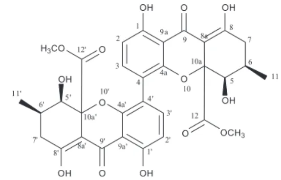
sp., an endophytic fungus strain from the Amazonian Rainforest plant Duguetia stelechantha (Annonaceae), which resulted in the identification of a new xanthone dimer, talaroxanthone (Figure 1).
Results and Discussion
Talaroxanthone (Figure 1) was isolated as a yellow powder from the extract of mycelia from flask cultures of Talaromyces sp. DgCr22.1b. The molecular formula C32H30O14 was established by high-resolution mass spectrometry (HRMS). The 13C nuclear magnetic resonance
Koolen et al. 881 Vol. 24, No. 5, 2013
symmetric homodimer structure of this metabolite. Both the presence of a conjugated carbonyl (dC 187.0) bound to
two adjacent hydroxyl groups at dH 11.41 and 13.66 ppm
(characteristic shifts of a keto-enol structure), as well as the analysis of the long-range (HMBC (Heteronuclear Multiple-Bond Correlation Spectroscopy)) 1H-13C NMR
correlation experiments, suggested the presence of a tetrahydroxanthone ring as shown in Figure 2. The orientation of the methylene hydrogen at dH 2.27 (H-7α)
(dd, J 10.4, 19.4 Hz) was suggested by the vicinal coupling constant at J6,7 10.4 Hz, indicating a pseudoaxial
orientation, and consequently the pseudoequatorial orientation of the hydrogens at dH 2.71 (H-7β) (dd, J 6.4,
19.4 Hz) by the absence of this coupling constant. A trans
diaxial conformation (Figure 2) between the carbinolic hydrogen at H-5 (dH 3.75) along with the methine hydrogen
at H-6 (dH 2.42) was observed by the coupling constant
J5,6 12 Hz previously reported for the eumitrin A2.12 The
comparison of the 13C NMR resonances for the C-5
(dC 76.4) with other xanthones presenting the hydroxyl
group at C-5 with pseudoequatorial orientation such as blennolide B (dC 77.0, Dd = +0.4)10 and archexanthone
A (dC 75.9, Dd = –0.5)
13 presented close chemical shifts,
whereas xanthones with pseudoaxial hydroxyl groups at C-5 have lower chemical shifts, secalonic acid B (dC 71.4,
Dd = – 5.0)10 and blennolide A (d
C 71.3, Dd = –5.1).10 These
comparisons led to the establishment of the orientations of the hydroxyl group at C-5 and the methyl connected to C-6 as pseudoequatorial (Figure 2).
The aromatic hydrogens at dH 6.60 (H-2) and dH 8.04 ppm
(H-3) were assigned as being ortho to each other due to their coupling constants (J2,3 8.7 Hz) and COSY (correlation
spectroscopy) data. The observed chemical shift for H-3 is different than the observed for phomoxanthone A,8 but any
other connection between the aromatic rings was dismissed by the observed data. The symmetry observed by 1H and 13C NMR and confirmed by HRMS/MS indicates that this
molecule is a homodimer connected at C4-C4’, which is rare in fungal metabolites.8 This connectivity was also
confirmed by HMBC correlations of the hydroxyl group at
C-1. Talaroxanthone differs from the phomoxanthones at positions C-10a, C-10a’ and C-5, presenting as substituents methylacetate at C-10a and C-10a’ and a hydroxyl at C-5. The Phomopsis xanthones, on the other hand, contain –CH2OCCH3 groups at C-10a and C-10a’ and an
acetoxy group at C-5a. Talaroxanthone is related to the secalonic acids, also called ergochromes,5 which are also
tetrahydroxanthones, but with a C2-C2’ linkage pattern,14
usually found in compounds from the Claviceps,15
Penicillium,16Pyrenochaeta and Aspergillus genera.17
Conclusions
The potential of endophytic fungi as sources of novel substances was demonstrated by the isolation of a new xanthone dimer named talaroxanthone from the mycelial mass of the endophytic fungus Talaromyces DgCr22.1b. This compound is responsible for the yellow pigmentation observed for this strain. To the best of our knowledge, compound 1 is the first example of a xanthone homodimer related to the secalonic acids with C4-C4’ linkage along with the presence of methylacetate groups at the positions C10a and C10a’ differently from the previous isolated phomoxanthones.
Experimental
General procedures
1D and 2D NMR experiments were recorded on a Bruker AVANCE 400 spectrometer operating at 9.4 T, equipped with a 5 mm multinuclear direct detection probe with z-gradient operating at 400 MHz for 1H and 100 MHz
for 13C. The chemical shifts were referenced to the solvent
peak CDCl3 at d 7.27 and 77.0 ppm, respectively. High
resolution electrospray ionization mass spectrometry (HRESIMS) measurements were recorded on a Waters Synapt HDMS instrument with quadrupole time-of-flight (QTOF) geometry. Optical rotations were recorded on a Jasco P-1020 polarimeter. Fourier transform infrared
Figure 1. Structure of talaroxanthone.
(FTIR) spectra were recorded on a Bomem MB102 spectrophotometer. A Shimadzu LC-6AD pump equipped with a Shimadzu SPD-10AV UV detector and a Rheodyne injector was used for high pressure liquid chromatography (HPLC). Silica gel 60 (70-230 mesh) was used for column chromatography, while silica gel 60 F254 was used for
analytical (0.25 mm) thin layer chromatography (TLC). A Phenomenex Phenyl Hexyl 5 µm preparative column (10 mm × 250 mm) was used for the HPLC separations. All solvents used for chromatography and mass spectrometry were from Tedia (HPLC grade), and H2O was ultrapure
(Milli-Q, Millipore). All microbiological culture media were purchased from Biosystems.
Fungal material
The roots of Duguetia stelechantha (Diels) R. E. Fries were collected in the experimental farm of the Universidade Federal do Amazonas, Amazonas State, Brazil. The plant material was washed with detergent and sterile water for external cleaning, then fragments were immersed in 70% alcohol, later in 3% hypochlorite solution and finally in sterile water.18 After this process, plant fragments were
inoculated in Petri dishes containing ISP2-agar medium and incubated for five days. The fragments containing hyphae of fungi were subsequently transferred to test tubes with the same medium and incubated at 26 °C for 30 days. After this period, the mitosporic strain was purified by the Tween technique.19 The isolated strain was identified according to
traditional morphological criteria and sequencing of the fungus ITS-1 to ITS-2 rDNA and compared with sequences from the GenBank.18 A voucher was deposited in the fungal
collection of the GEMMA group of the Universidade Federal do Amazonas under the code DgCr22.1b.
Production and isolation
An isolated culture of the strain DgCr22.1b was grown on ISP2 broth medium (International Streptomyces Project 2)
at 25 °C for 19 days, into 39 × 1 L Erlenmeyer flasks each containing 300 mL of medium. The mycelia were separated from the broth and extracted with EtOAc 1:1 MeOH (1 L, 2 days), the solvent was evaporated under reduced pressure and provided an red solid gum (30.2 g). The crude extract was subjected to column chromatography (silica gel, 4 × 20 cm) with EtOAc as eluent. A yellow pigment was eluted after ca. 500 mL elution with EtOAc. This process was repeated twice to yield a yellow gum (2.1 g). This yellow gum was subjected to another column chromatography (silica gel, 2.5 × 30 cm) eluted with a gradient of hexane and EtOAc (9:1, 0:1) to give 17 main fractions. The fractions 11-13 (1:9,
104 mg) were subjected to semi-preparative HPLC using isocratic MeOH as the eluent at a flow rate of 8.7 mL min-1
to obtain 1 (70.6 mg, tR 5 min).
Talaroxanthone (1): yellow needles; mp 214-216 °C; [α]D
25 +98 (CHCl
3; c 0.0027); UV λmax/nm (log ε) 222
(4.45), 255 (4.26) and 334 (4.60); IR (KBr) vmax/cm-1
1744, 1615, 1586, 1470, 1366, 1223, 1044, 897, 830; HRESIMS m/z 639.1636 [M + H]+; calculated
639.1636; product ions (CID, 15 eV) 621.1507 [M + H−H2O]+, 589.1324 [M + H−MeOH]+, 579.1443
[M + H−COOCH3]
+, 561.1329 [M + H−COOCH 3−H2O]
+,
543.1218 [M + H−COOCH3−2H2O]+, 288.2895
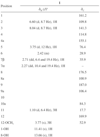
[M + H monomer – CO]+; 1H and 13C NMR data are given
in Table 1.
Supplementary Information
Supplementary information (1H, 13C, COSY, HMBC,
HSQC, NOESY spectra and MS data) are available free of charge at http://jbcs.sbq.org.br as PDF file.
Table 1.1H and 13C NMR spectral dataa for compound 1
Position 1
dH (J)b dC
1 161.2
2 6.60 (d, 8.7 Hz), 1H 109.8
3 8.04 (d, 8.7 Hz), 1H 141.3
4 114.8
4a 155.1
5 3.75 (d, 12 Hz), 1H 76.4
6 2.42 (m) 28.9
7β 2.71 (dd, 6.4 and 19.4 Hz), 1H 35.9
7α 2.27 (dd, 10.4 and 19.4 Hz), 1H –
8 176.5 8a 100.9 9 187.0 9a 106.4 10 10a 84.3
11 1.10 (d, 6.4 Hz), 3H 17.7
12 169.9
12-OCH3 3.77 (s), 3H 52.9
1-OH 11.41 (s), 1H
8-OH 13.66 (s), 1H
aExperiments were carried out at 400 MHz for 1H and 100 MHz for 13C in CDCl
Koolen et al. 883 Vol. 24, No. 5, 2013
Acknowledgments
This work was funded by grants from the Conselho Nacional de Desenvolvimento Científico e Tecnológico (CNPq), the Coordenação de Aperfeiçoamento de Pessoal de Nível Superior (CAPES) and the Fundação de Amparo a Pesquisa do Estado do Amazonas (FAPEAM).
References
1. Liu, F.; Cai, X. L.; Yang, H.; Xia, X. K.; Guo, Z. Y.; Yuan, J.; Li, M. F.; She, Z. G.; Lin, Y. C.; Planta Med. 2010, 76, 185. 2. Dong, Y.; Yang, J.; Zhang, H.; Lin, J.; Ren, X.; Liu, M.; Lu, X.;
He, J.; J. Nat. Prod.2006, 69, 128.
3. Dethoup, T.; Manoch, L.; Kijjoa, A.; Pinto, M.; Gales, L.; Damas, A. M.; Silva, A. M. S.; Eaton, G.; Herz, W.; J. Nat. Prod.2007, 70, 1200.
4. Li, L. Q.; Yang, Y. G.; Zeng, Y.; Zou, C.; Zhao, P. J.; Molecules
2010, 15, 3993.
5. Masters, K. S.; Brase, S.; Chem. Rev.2012, 112, 3712. 6. Cao, S.; McMillin, D. W.; Tamayo, G.; Delmore, J.; Mitsiades,
C. S.; Clardy, J.; J. Nat. Prod.2012, 75, 793.
7. Krohn, K.; Kouam, S. F.; Kuigoua, G.; Hussain, H.; Brandt, S. C.; Florke, U.; Kurtán, T.; Pescitelli, G.; Di Bari, L.; Draeger, S.; Schulz, B.; Chem. Eur. J.2009, 15, 12121. 8. Isaka, M.; Jaturapat, A.; Rukseree, K.; Danwisetkanjana, K.;
Tanticharoen, M.; Thebtaranonth, Y.; J. Nat. Prod.2001, 64, 1015.
9. Pornpakakul, S.; Liangsakul, J.; Ngamrojanavanich, N.; Roengsumran, S.; Sihanonth, P.; Piapukiew, J.; Sangvichien, E.; Puthong, S.; Petsom, A.; Arch. Pharmacal Res.2006, 29, 140.
10. Zhang, W.; Krohn, K.; Ullah, Z.; Flçrke, U.; Pescitelli, G.; Di Bari, L.; Antus, S.; Kurtán, T.; Rheinheimer, J.; Draeger, S.; Schulz, B.; Chem. Eur. J. 2008, 14, 4913.
11. Koolen, H. H. F.; Soares, E. R.; Silva, F. M. A.; Almeida, R. A.; Souza, A. D. L.; Medeiros, L. S.; Rodrigues Filho, E.; Souza, A. Q. L.; Quim. Nova2012, 35, 771.
12. Yang, D. M.; Takeda, N.; Iitaka, Y.; Sankawa, U.; Shibata, S.;
Tetrahedron1973, 29, 519.
13. Isaka, M.; Palasarn, S.; Kocharin, K.; Saenboonrueng, S.;
J. Nat. Prod. 2005, 68, 945.
14. Franck, B.; The biosynthesis of the Ergochromes – A Study in Secondary Metabolism, vol. 1, 1st ed.; Academic Press: London,
UK, 1980.
15. Andersen, R.; Buchi, G.; Kobbe, B.; Demain, A. L.; J. Org. Chem. 1977, 42, 352.
16. Kurobane, I.; Vining, L. C. McInnes, A. G.; J. Antibiot. 1979,
32, 1256.
17. Steyn, P. S.; Tetrahedron 1970, 26, 51.
18. Souza, A. D. L.; Rodrigues Filho, E.; Souza, A. Q. L.; Pereira, J. O.; Calgarotto, K.; Maso, V.; Marangoni, S.; Silva, S. L.;
Toxicon2008, 51, 240.
19. Souza, A. Q. L.; Souza, A. D. L.; Astolfi Filho, S.; Pinheiro, M. L. B.; Sarquis, M. I. M.; Pereira, J. O.; Acta Amazon.2004,
34, 185.
Submitted: June 26, 2012
Published online: April 30, 2013
Supplementary Information
Printed in Brazil - ©2013 Sociedade Brasileira de Química0103 - 5053 $6.00+0.00S
I
*e-mail: [email protected]
Talaroxanthone, a Novel Xanthone Dimer from the Endophytic Fungus
Talaromyces
sp.
Associated with
Duguetia stelechantha
(Diels) R. E. Fries
Hector H. F. Koolen,*,a,b Laís S. Menezes,a Mayane P. Souza,a Felipe M. A. Silva,a
Fabiana G. O. Almeida,a Antonia Q. L. de Souza,c Angelita Nepel,d Andersson Barison,d
Flávio Henrique da Silva,e Danilo Elton Evangelistae and Afonso D. L. de Souzaa
aDepartamento de Química, Universidade Federal do Amazonas, 69077-000 Manaus-AM, Brazil
bInstituto de Química, Universidade Estadual de Campinas, 13084-971 Campinas-SP, Brazil
cEscola Superior de Ciências da Saúde, Universidade do Estado do Amazonas, 69065-001 Manaus-AM, Brazil
dDepartamento de Química, Universidade Federal do Paraná, 81531-990 Curitiba-PR, Brazil
eDepartamento de Genética e Evolução, Universidade Federal de São Carlos, 13565-905 São Carlos-SP, Brazil
Figure S1.1H NMR (400 MHz, CDCl
Talaroxanthone, a Novel Xanthone Dimer from the Endophytic Fungus Talaromyces sp. J. Braz. Chem. Soc.
S2
Figure S3.13C NMR (100 MHz, CDCl
3) spectrum of talaroxanthone.
Figure S2. Amplification of the 1H NMR (400 MHz, CDCl
Figure S6. HSQC (1H NMR 400 MHz, 13C NMR 100 MHz, CDCl
3) map of talaroxanthone.
Figura S5. COSY (1H NMR, 400 MHz) map of talaroxanthone.
Figure S4. HMBC (1H NMR 400 MHz, 13C NMR 100 MHz, CDCl
Talaroxanthone, a Novel Xanthone Dimer from the Endophytic Fungus Talaromyces sp. J. Braz. Chem. Soc.
S4
Figure S7.NOESY (1H NMR 400 MHz, CDCl
Figure S9. HRESIMS/MS spectrum of talaroxanthone.