Metal Ions and Protein Folding
Conformational and Functional Interplay
Hugo Miguel Raposo Correia Botelho
Dissertation presented to obtain a PhD degree in Biochemistry
at Instituto de Tecnologia Química e Biológica
Universidade Nova de Lisboa
Supervisor
Cláudio Emanuel Moreira Gomes
Opponents
Teresa J. T. Pinheiro & Peter Faller
Instituto de Tecnologia Química e Biológica
Universidade Nova de Lisboa
Second edition, December 2010
Protein Biochemistry, Folding and Stability Laboratory
Instituto de Tecnologia Química e Biológica, Universidade Nova de Lisboa Av. da República (EAN), 2785-572 Oeiras, PORTUGAL
http://www.itqb.unl.pt/pbfs
This dissertation describes the work performed under the supervision of Cláudio M. Gomes in the Protein Biochemistry Folding and Stability Laboratory at the Instituto de Tecnologia Química e Biológica, from September 2006 to October 2010.
The studies presented herein aim at understanding the role of metal ions in modulating the conformation and stability of proteins. Two types of model systems have been used: iron-sulfur (FeS) proteins having permanently bound FeS cofactors, and the S100 proteins, which bind calcium, zinc and copper ions reversibly.
There were many people who have helped and supported me in different ways during my PhD, to whom I am grateful and want to express my gratitude:
First and foremost, I want to thank my supervisor, Cláudio M. Gomes, for allowing me to pursue exciting research during the last four years, for his joy, foresight, advice and for always trusting in me. By learning from his dynamic and inspiring way of doing science I have grasped how an inquisitive mind can make the difference, broaden my view of science and grown as a scientist.
To Günter Fritz, for the collaboration on the S100 proteins, for great discussions and for always being available to undergo new research projects.
To Ludmilla Morozova-Roche, for sharing her knowledge and for so wonderfully welcoming me in Umeå. Also, to her group, specially Kiran Yanamandra for assisting me in experiments and being such a friendly partner.
To Arnulf Kletzin, for providing Rieske ferredoxin and for discussions on this project.
To my colleague friends Ana Paula Batista, Patrícia Refojo, João Vicente, Sandra Santos, Ana Filipa Pinto and Gabriel Martins for the wonderful shared times, support and the unending good mood. Even though we belonged to different groups, no one who saw us together would say that.
To my family, especially my parents, Victor and Eduarda, for all the love, which does not fade with distance.
To Rita, for her love, support, understanding and so much more.
Fundação para a Ciência e a Tecnologia is acknowledged for financial support, by awarding PhD grant SFRH⁄BD⁄31126⁄2006.
Botelho HM, Koch M, Fritz G & Gomes CM (2009) Metal ions modulate
the folding and stability of the tumor suppressor protein S100A2. FEBS J
276, 1776-1786
Botelho HM, Leal SS, Veith A, Prosinecki V, Bauer C, Frohlich R, Kletzin A
& Gomes CM (2010) Role of a novel disulfide bridge within the all-beta
fold of soluble Rieske proteins. J Biol Inorg Chem 15, 271-281
Fritz G, Botelho HM, Morozova-Roche LA, Gomes CM (2010) Natural and
amyloid self-assembly of S100 proteins: structural basis of functional
Metal ions are cofactors in about 30% of all proteins, where they fulfill catalytical and structural roles. Due to their unique chemistry and coordination properties they effectively expand the intrinsic polypeptide properties (by participating in catalysis or electron transfer reactions), stabilize protein conformations (like in zinc fingers) and mediate signal transduction (by promoting functionally relevant protein conformational changes). However, metal ions can also exert have deleterious effects in living systems by incorporating in non-native binding sites, promoting aberrant protein aggregation or mediating redox cycling with generation of reactive oxygen and nitrogen species. For this reason, the characterization of the roles of metal ions as modulators of protein conformation and stability provides fundamental knowledge on protein folding properties and is instrumental in establishing the molecular basis of disease. In this thesis we have analyzed protein folding processes using model protein systems incorporating covalently bound metal cofactors – iron-sulfur (FeS) proteins – or where metal ion binding is reversible and associated conformational readjustments – the S100 proteins.
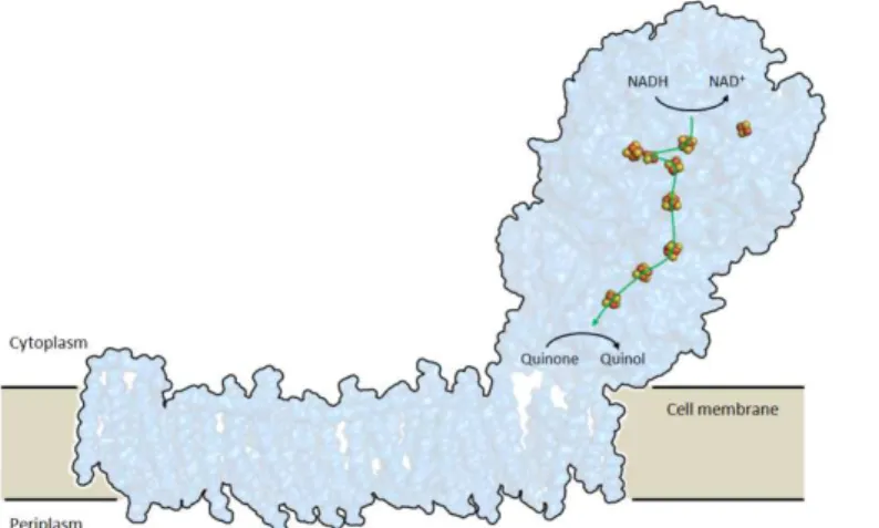
The Rieske [2Fe-2S] domain occurs either as part of electron transfer
system in respiratory (bc1) or photosynthetic (b6f) complexes or as a
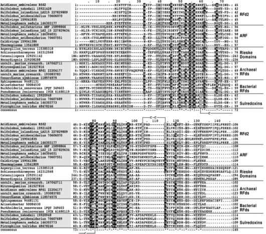
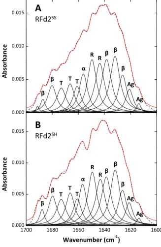
component in soluble dioxygenase systems. Despite the functional plasticity, the all-β Rieske fold is highly conserved, being modified by extensions and mutations modulating the cluster redox or pH properties or introducing features like disulfide bridges. In this respect, we have identified a Rieske
ferredoxin featuring a cysteine pair present in only a subset of Thermoprotei
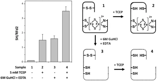
Acidianus ambivalens protein the cysteines form a disulfide bridge in the
as-isolated protein and established the conditions where the disulfide could be selectively reduced (5 mM TCEP) without affecting the overall protein conformation or the redox state of the FeS cluster. Disulfide reduction was
found to regulate protein stability (ΔTm = 9°C), FeS cluster reduction
potential (ΔE0 = +29 mV) and affect the cluster’s pH-dependent properties,
being a putative regulatory element in this protein.
The folding properties of the hyperthermostable zinc-containing seven
iron ([3Fe-4S][4Fe-4S]) ferredoxin from A. ambivalens (Tm = 122°C in water
at pH 7) have been thoroughly studied. Recently, this has revealed the formation of a molten globule conformation upon thermal unfolding at pH 2.5. This conformation was proposed as a candidate for the template upon which the FeS clusters are assembled during protein biosynthesis. Molten globule formation has been shown to include, zinc and FeS dissociation and tertiary structure changes but a detailed description of the conformational changes occurring is missing. By monitoring the thermal denaturation of ferredoxin at pD 2.5 and 12 using singular value decomposition (SVD) of second derivative FT-IR spectra we determined the thermal denaturation profiles of each secondary structure type of ferredoxin. Further, we identified a spectral component describing ferredoxin’s unfolding at acidic and basic conditions. This component was associated with the formation of the molten globule and not of the amorphous unfolded state at basic conditions. The structural state represented by this component undergoes cold unfolding at experimentally accessible temperatures. This is the first description of such event for this protein and constitutes the opportunity of studying cold unfolding of an FeS protein in the absence of external perturbations like co-solvents.
We have built on the knowledge of the folding and stability of rubredoxins
around ~30% of the FeS clusters disintegrating at 95°C, in an EDTA-insensitive process. By monitoring thermal unfolding using FT-IR coupled with spectral band fitting we have described the partial unfolding occurring at 95°C in terms of the secondary structure changes taking place. We have identified two thermal unfolding regimes. In the 25-60°C range α-helices and β-sheets unfold. Starting at 60°C, turns accumulate and β-sheet content increases, forming around 30 and 50% of the protein’s secondary structure at 95°C, respectively. We hypothesize that the highly structured conformation forming at high temperature is correlated with rubredoxin high thermostability.
The S100 proteins are major components in the vertebrate Ca2+ signal
buffer/transducer network regulating cell cycle, cell growth, differentiation and mobility. S100 proteins are small (10-12 kDa) homo- or heterodimers
which bind Ca2+ in EF-hand domains. Zn2+ and/or Cu2+ can also bind
elsewhere in some proteins. Ca2+ binding induces a conformational change
which exposes a protein docking site.
Human S100A2 is a unique family member because it binds Ca2+ and Zn2+,
accumulates in the nucleus, interacts with p53 in a metal-dependent manner and has been assigned a tumor suppression role. The multiplicity of metal
binding sites (2 Ca2+ and 2 Zn2+ sites per monomer), metal-dependent
activation, and S100A2’s role in human pathology makes this protein a model to study metal-dependent conformational changes and the eventual tuning of
conformational stability. We have examined the role of the Zn2+ sites in
modulating protein conformation and stability by using S100A2 variants
with Zn2+ binding site mutations. Circular dichroism analysis has shown that
the protein conformation is prone to subtle readjustments upon Ca2+ and
Zn2+ binding, keeping the same α-helical-rich fold. The conformational
urea and thermal unfolding in different metallation states (apo, Ca2+, Zn2+).
Thermal denaturation experiments indicated that Zn2+ destabilizes and Ca2+
stabilizes the protein conformation. These results suggest an opposite role
for Ca2+ and Zn2+ according to which Ca2+ activates and stabilizes the protein,
and Zn2+ inhibits and destabilizes S100A2, a mechanism with possible
implications in cancer progression.
Recently, S100 proteins have been shown for the first time to form amyloid fibrils in proteinaceous inclusions associated with pre-carcinogenic
inflammatory foci in the prostate named corpora amylacea. These
S100A8/A9 amyloid fibrils could be reconstituted in vitro in the presence of
Ca2+ or Zn2+ but not in metal-free conditions. Since S100 family members
share sequence and structural homology, we hypothesized that other S100 proteins could also be amyloidogenic. By using the Zyggregator and WALTZ algorithms, equivalent amyloid prone regions were detected in most human S100 proteins. Following this observation, we incubated several S100 family members (S100A2, S100A3, S100A4, S100A6, S100A12 and S100B) in amyloid formation prone conditions (pH 2.5, 57°C) while monitoring amyloid formation by extrinsic thioflavin T (ThT) fluorescence. With the exception of S100A12, all other proteins formed amyloid species. The AFM morphological characterization of the S100 amyloid species revealed that S100A2, S100A6 and S100B formed amyloid oligomers and S100A3 formed amyloid fibrils. For S100B, FT-IR monitored amyloid formation assays revealed that native α-helices and coiled regions convert to β-sheets and turns.
S100 proteins exhibit rich metal binding properties. In addition, S100A6,
S100A12, S100B accumulate in corpora amylacea in the brain and are
overexpressed in neurodegenerative diseases like Alzheimer’s, Parkinson’s and Amyotrophic Lateral Sclerosis. By using a combination of ThT, FT-IR and AFM monitored amyloidogenesis assays we showed that the S100 amyloid
the central nervous system. Cu2+ promoted S100A12 amyloid formation and
had the opposite effect towards S100B. For S100A6, Ca2+ completely
inhibited amyloidogenesis, a process which could be reverted by adding
EDTA. Additionally, Ca2+ reverted or alleviated the cytotoxicity of apo S100
amyloids.
Os iões metálicos são cofactores em cerca de 30% de todas as proteínas, desempenhando funções catalíticas e estruturais. Devido às suas propriedades químicas e de coordenação ímpares, alargam as propriedades intrínsecas dos polipéptidos a que se ligam (participando em catálise e reacções de transferência electrónica), estabilizam certas conformações proteicas (como nos dedos de zinco) e servem de mediadores em processos de transdução de sinal (através da promoção de alterações conformacionais funcionalmente relevantes). No entanto, os iões metálicos podem também ter efeitos prejudiciais nos sistemas vivos, ligando-se a locais não nativos, promovendo agregação proteica ou mediando ciclos redox que geram espécies reactivas de oxigénio e azoto. Assim, a caracterização das funções de iões metálicos enquanto modeladores da conformação e estabilidade proteica permite obter conhecimento sobre as propriedades fundamentais do enrolamento e é instrumental na determinação da base molecular de certas doenças. Nesta tese analizámos processos de enrolamento proteico utilizando sistemas modelo que incorporam cofactores metálicos covalentemente ligados – proteínas de ferro-enxofre (FeS) – ou em que a ligação de iões metálicos é reversível e está associada a alterações conformacionais – proteínas S100.
O domínio Rieske [2Fe-2S] ocorre quer como integrante de sistemas de
transferência electrónica em complexos respiratórios (bc1) ou fotossínteticos
(b6f) quer como componente de sistemas dioxigenase solúveis. Apesar da sua
plasticidade funcional, a estrutura Rieske β é altamente conservada, sendo modificada por extensões e mutações que modelam as propriedades redox ou dependentes do pH do centro FeS ou que introduzem características como ligações perssulfureto. Neste sentido, identificámos uma ferredoxina Rieske que inclui um par de cisteínas presente apenas num subgrupo de arquea
uma ligação persulfureto próxima dos centros FeS respiratórios e fotossintéticos. Utilizando uma combinação de quantificação colorimétrica de tióis e os espectros de absorção no visível e infravermelho característicos,
determinámos que as cisteínas na proteína de Acidianus ambivalens formam
um perssulfureto e estabelecemos as condiçõs em que o perssulfureto pode ser reduzido selectivamente (5 mM TCEP) sem afectar a conformação proteica ou o estado redox do centro FeS. A redução do perssulfureto regula
a estabilidade proteica (ΔTm = 9°C), potencial redox do centro FeS (ΔE0 = +29
mV) e afecta as propriedades dependentes do pH, sendo um possível elemento regulatório nesta proteína.
As propriedades de enrolamento da ferredoxina hipertermostável de sete
ferros ([3Fe-4S][4Fe-4S]) com um centro de zinco de A. ambivalens (Tm =
122°C em água a pH 7) foram estudadas em detalhe no passado. Mais
recentemente, identificou-se a formação de um molten globule após
desnaturação térmica a pH 2.5. Esta conformação foi sugerida como candidata à estrutura em que os centros FeS são incorporados durante a biossíntese da proteína. Mostrou-se igualmente que a sua formação compreende a dissociação do zinco e do centro FeS bem como alterações da estrutura secundária mas a descrição das alterações conformacionais envolvidas está em falta. Através da monitorização da desnaturação térmica da ferredoxina a pD 2.5 e 12 utilizando a decomposição em valores singulares (SVD) da segunda derivada dos espectros de FT-IR determinámos os perfis de desnaturação térmica para cada tipo de estrutura secundária. Além disso, identificámos um componente espectral que descreve a desnaturação da ferredoxina em condições ácidas e alcalinas. Este
componente foi associado com a formação do molten globule e não do estado
desnaturação por frio de uma proteína FeS na ausência de perturbações externas como co-solventes.
Contribuímos para o conhecimento do enrolamento e estabilidade de
rubredoxinas estudando a desnaturação térmica da proteína de Desulfovibrio
gigas. Apesar da sua origem mesofílica, a proteína é altamente estável, com
apenas 30% dos centros FeS a desintegrarem-se a 95°C, um processo insensível ao EDTA. Monitorizando a desnaturação térmica através de FT-IR juntamente com o ajuste de bandas espectrais, descrevemos a desnaturação parcial que ocorre a 95°C em termos de alterações na estrutura secundária. Identificámos dois regimes de desnaturação. Na gama 25-60°C ocorre a desnaturação de hélices α e folhas β. A partir de 60°C, acumulam-se voltas e folhas β, que constituem aproximadamente 30 e 50% da estrutura secundária da proteína a 95°C, respectivamente. Assim, colocamos a hipótese de que a conformação altamente estruturada que se forma a alta temperatura está correlacionada com a estabilidade térmica da rubredoxina.
As proteínas S100 são componentes principais na rede de tamponização e
transdução de sinais de Ca2+ em vertebrados, regulando o ciclo, crescimento,
diferenciação e mobilidade celulares. As proteínas S100 são pequenos (10-12
kDa) homo ou heterodímeros que ligam Ca2+ em domínios EF-hand. Zn2+
e/ou Cu2+ também se podem ligar em locais distintos em algumas proteínas.
A ligação de Ca2+ induz uma alteração conformacional que expõe uma zona
de ancoragem de proteínas.
A proteína S100A2 humana é um membro ímpar desta família porque liga
Ca2+ e Zn2+, acumula-se no núcleo, interage com o p53 numa forma
dependente de metais e tem uma função supressora de tumores. A
multiplicidade de locais de ligação a metais (2 locais de Ca2+ e Zn2+ por
alterações conformacionais dependente de metais e o consequente
ajustamento da estabilidade. Examinámos o papel dos locais de Zn2+ na
modelação da conformação e estabilidade proteica utilizando variantes da
S100A2 com mutações ao nível dos locais de Zn2+. Uma análise por dicroismo
circular mostrou que a conformação desta proteína é propensa a ligeiros
reajustamentos em resposta à ligação de Ca2+ e Zn2+, mantendo a mesma
estrutura rica em hélices α. A estabilidade conformacional das variantes de
S100A2 em diferentes estados de metalação (apo, Ca2+, Zn2+) foi quantificada
através de desnaturação por temperatura e ureia monitorizada através de CD
e FT-IR. As experiências de desnaturação térmica indicaram que o Zn2+
desestabiliza e o Ca2+ estabiliza a conformação da proteína. Estes resultados
sugerem um papel antagónico para o Ca2+ e o Zn2+ segundo o qual o Ca2+
activa e estabiliza a proteína e o Zn2+ inibe e desestabiliza a S100A2, um
mecanismo com possíveis implicações na progressão do câncro.
Recentemente, foi mostrado pela primeira vez que as proteínas S100 formam fibras amilóides em inclusões proteicas associadas com focos
pré-inflamatórios na próstata denominadas corpora amylacea. Estas fibras
amilóides de S100A8/A9 podem ser reconstituidas in vitro na presença de
Ca2+ ou Zn2+ mas não em condições livres de metais. Dado que as proteínas
amilóides e que a S100A3 formou fibras amilóides. A formação de amilóides de S100B monitorizada por FT-IR revelou que hélices α e regiões desestruturadas nativas se convertem em folhas β e voltas.
As proteínas S100 possuem ricas propriedades de ligação a metais. Além
disso, a S100A6, S100A12 e S100B acumulam-se em corpora amylacea no
cérebro e são sobre-expressas em doenças neurodegenerativas como Alzheimer, Parkinson e Esclerose Lateral Amiotrófica (ALS). Através da utilização da combinação de ensaios de amiloidogénese monitorizados por fluorescência de ThT, FT-IR e AFM, mostrámos que a cinética de formação de
amilóides S100 e a respectiva estrutura é sensível à presença de Ca2+, Zn2+ e
Cu2+, elementos principais da química biológica da sinapse glutamatérgica no
sistema nervoso central. O Cu2+ promoveu a formação de amilóide S100A12 e
teve o efeito oposto relativamente à S100B. Relativamente à S100A6, o Ca2+
inibiu completamente a amiloidogénese, um processo que pôde ser revertido
pela adição de EDTA. Para além disto, o Ca2+ reverteu ou diminuiu a
citotoxicidade dos amilóides S100 apo.
A citotoxicidade dos amilóides S100 e o papel modelador dos iões metálicos é relevante no âmbito do anteriormente reportado envolvimento das proteínas S100 em processos neurodegenerativos. Devido à co-acumulação de S100B e Aβ na doença de Alzheimer, analizámos a possibilidade de inter-relação entre os processos amiloidogénicos de ambas.
Através de experiências de cross-seeding descobrimos que fibras Aβ
AD Alzheimer’s disease
AFM Atomic force microscopy
ALS Amyotrophic lateral sclerosis
ANS 1-anilino-8-naphthalene-sulfonic
acid
APP Amyloid precursor protein
ATR Attenuated total reflectance
Aβ Amyloid β
CAPS
N-cyclohexyl-3-aminopropanesulfonic acid
CD Circular dichroism
Cm Midpoint denaturant
concentration
DLS Dynamic light scattering
DNA Deoxyribonucleic acid
DTT Dithiothreitol
E0 Standard reduction potential
EDTA Ethylenediaminetetraacetic acid ELISA Enzyme-linked immunosorbent
assay
EPR Electron paramagnetic resonance
ESI-MS Electrospray ionization mass spectrometry
Fd Ferredoxin
FeS Iron-sulfur
FRET Förster resonance energy transfer
FT-IR Fourier transform infrared spectroscopy
GuHCl Guanidinium hydrochloride HEPES
4-(2-hydroxyethyl)-1-piperazineethanesulfonic acid
Kd Dissociation constant
KPi Potassium phosphate
MCT Mercury cadmium telluride
MES 2-(N-morpholino)ethanesulfonic
acid
mRNA Messenger ribonucleic acid
NAD+ Nicotinamide adenine dinucleotide
NADPH Nicotinamide adenine dinucleotide phosphate
NMR Nuclear magnetic resonance
PAGE Polyacrylamide gel electrophoresis
PD Parkinson’s disease
PrP Prion protein
RAGE Receptor for advanced glycation endproducts
Rd Rubredoxin
SDS Sodium dodecyl sulfate
SOD Superoxide dismutase
SVD Singular value decomposition
TCEP Tris(2-carboxyethyl)phosphine
ThT Thioflavin T
Tm Midpoint denaturation
temperature T-ramp Temperature ramp
TSE Transition state ensemble
USE Unfolded state ensemble
UV Ultraviolet
WST Water soluble tetrazolium
ΔG Gibbs free energy variation
ΔΔG ΔG variation
Contents
Foreword ... iii Acknowledgements ... v Thesis publications ... vii Dissertation abstract ... ix Resumo da dissertação ... xv Abbreviations ... xxi Contents ... xxiii
General introduction ... 1
1. Protein folding ... 1
Part I – Iron-Sulfur clusters and protein stability ... 33
2. Iron-Sulfur clusters as a model of protein conformational stabilizers ... 33
3. A novel disulfide bridge within the fold of soluble Rieske proteins ... 55
4. Mechanism of molten globule formation by ferredoxin: ... 83
5. Characterization of folding and stability of a mesophilic rubredoxin .. 103
Part II – Metal ions and protein folding in the S100 family: ... 121
6. Metal ions and protein folding ... 121
7. The S100 protein family ... 171
8. Metal ions modulate the folding and stability of t S100A2 ... 195
9. S100: A family of proteins with new amyloid-forming properties... 217
10.Metals as modulators of amyloidogenesis: ... 241
11.Cross-talk between S100B and Aβ amyloidogenesis ... 267
Outlook ... 289
1.
Protein folding
1.1. The protein folding problem ... 3
1.2. Stabilization of the folded state ... 6
1.2.1. Overall view ... 6
1.2.2. The hydrophobic effect ... 6
1.2.3. Hydrogen bonds ... 7
1.2.4. Ion pairs ... 7
1.2.5. Covalent modifications ... 8
1.2.6. Two state protein folding thermodynamics ... 8
1.2.7. Engineering protein stability ... 11
1.3. Protein folding models ... 12
1.4. Protein folding kinetics... 15
1.5. Protein folding energetics – the landscape view... 20
1.6. Conformational states – a unified view of protein folding ... 23
1.7. Protein folding in the cell ... 24
1.1.The protein folding problem
The biological activity of proteins frequently depends on the ability of the polypeptide to acquire a very defined and unique tree-dimensional structure. This is not surprising as catalysis, signal transduction, ligand binding and molecular interactions – the main functions of proteins – all require a stringent spatial arrangement of the polypeptide chain: the native structure. This structure is attained through protein folding, the physical process by which a polypeptide folds into its characteristic and functional three-dimensional structure from a random coil [1]. Protein folding is a fundamental process in biology due to the high dependence of practically all biological processes on the protein machinery. However, even in the cellular environment, a fraction of all synthesized proteins fail to fold into the native structure [2]. This inability may bring about severe biological consequences such as the so-called misfolding diseases [3] which are associated with degradation prone-conformations (such as in cystic fibrosis [4-5]), misfolded protein with an aberrant activity (like in phenylketonuria [6] or fatty acid metabolism disorders [7]) or protein deposition in the form of insoluble amyloid fibrils (characteristic of neurodegenerative diseases as Alzheimer’s [8], Parkinson’s [9] or Huntington’s [10]).
heterogeneity of amino acid side chains, the multiplicity of interactions stabilizing the folded conformation and an incomplete mechanistic understanding of the protein folding process. Accordingly, this task is referred as the Protein Folding Problem, from which three main questions arise [11]:
1. What is the energetics stabilizing folded proteins?
2. What is the folding mechanism?
3. Can the protein structure be predicted from the sole knowledge of the
amino acid sequence?
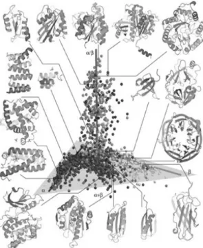
Topics 1 and 2 are discussed in sections 1.2 and 1.3, respectively. The large structural biology data currently available highlights the complexity of the protein fold problem [12]. Although the interactions determining the native state are the same for all proteins and this determines a restricted number of secondary structure elements, there is an enormous number of possible spatial arrangements (Figure 1.1). Moreover, protein size and oligomerization status offer further diversification potential. However, proteins occupy a discrete portion of the conformational space: the number of protein folds – the topological arrangements of secondary structure elements – is restricted (Figure 1.1).
Figure 1.1 – Three-dimensional representation of the protein conformational space. Each sphere
represents a protein fold family among compact globular proteins. The structures were placed in space according to their pair-wise structure alignments. The structures cluster according to SCOP protein
fold classes. Representative
structures are shown to highlight the structure variability and
different secondary structure
arrangements using the same restricted number of basic elements. From [12].
conformational space; second, the timescale of protein folding requires highly computer intensive calculations. Earlier studies based in minimalistic Gō-type models have been complemented by more realistic off-lattice models. Increasing computer power, including distributed computing projects such as Rosetta@home of the Foldit game [13], allow routine full
atomistic molecular dynamics and ab initio structure prediction studies.
1.2.Stabilization of the folded state
1.2.1. Overall view
The folding status (folded/unfolded) of proteins is dictated by the balance between two energetic parameters. On the one hand, the unfolded state has high conformational entropy because proteins are linear polymers of tens to hundreds of amino acid residues; on the other side, the multiplicity of interactions involving side chains, the backbone and the solvent constitute an enthalpic gradient towards the folded state. Folding is then driven by the enthalpy of interactions forming while interacting groups are brought into proximity. The thermodynamic forces responsible for the folding of proteins to their native conformations were first described by Kauzmann [15]: hydrophobic interactions, hydrogen bonds and electrostatic interactions [16-17]. These interactions are sometimes referred as “weak interactions” in protein folding. This is because of their small nominal magnitude. However, the very significant number of individual interactions in folded proteins accounts for a high overall energetic term which keeps the native protein structure. In some cases, the folded conformation is further stabilized by additional contributions arising from disulfide bridges, oligomerization, cofactor binding or post-translational modifications. Together, all these molecular interactions contribute to building up all levels of protein structure.
1.2.2. The hydrophobic effect
packed hydrophobic core surrounded by a polar shell facing the solvent [21]. The hydrophobic contribution to protein folding can be determined through mutagenesis [22] or by the free energy change of transferring an amino acid side chain from water to a hydrophobic solute [23]. Average stabilization values are 1.3 kcal/mol per buried methyl group or 1-2 kcal/mol per aromatic ring interaction [22]. The vast hydrophobic areas buried during protein folding account for the stabilizing importance of the hydrophobic effect.
1.2.3. Hydrogen bonds
Hydrogen bonds are also important stabilizing features in protein structures. The backbone and several amino acid side chains have hydrogen bond donor and acceptor groups. Further, the solvent can also be included in hydrogen bonding patterns. The contribution of hydrogen bonds to the overall protein stabilization is very relevant. About two thirds of all residues form peptide hydrogen bonds in the native state [24]. It is the involvement of the backbone in repetitive interactions that drives secondary structure formation [25]. The stabilization by hydrogen bond averages to 1.3 kcal/mol [26-27]. For geometric reasons, the same hydrogen bond donor or acceptor can participate in multiple hydrogen bonds, creating hydrogen bond networks.
1.2.4. Ion pairs
The most individually stabilizing interaction in protein folding is the electrostatic interaction between charged amino acid side chains, called ion pairs. Ion pairs are usually located at the protein surface, being favored by the aqueous polar environment. A single ion pair can contribute with up to 3-5 kcal/mol to stability [28]. This value is close to the typical stability of the
native versus the unfolded state (ΔGN-U), meaning that in some proteins
individual magnitude, the average number of ion pairs in proteins is small. Consequently, they are not major driving forces in protein stabilization [18].
1.2.5. Covalent modifications
Disulfide bridges are cross-links which effectively restrict the conformational space, decreasing protein conformational entropy and increasing protein stability [29-30]. The most significant effects occur with disulfides bridging distant polypeptide regions. For avoiding the formation of aberrant disulfides – which form off-pathway folding species – organisms have developed specialized enzymatic mechanisms [31]. Prokaryotes produce a family of disulfide bond proteins (Dsb) which assist oxidative protein folding – with the concomitant cysteine thiol oxidation – and correct aberrant disulfides [32]. In eukaryotes, the endoplasmic reticulum is the oxidative cellular compartment where disulfide formation is regulated by protein disulfide isomerase (PDI) and Ero1 [33].
Similarly to disulfide bridges, the binding of cofactors also cross-links different parts of the polypeptide and favors folding. Proteins can bind a broad range of organic or inorganic cofactors in a covalent or non covalent manner. For example, zinc finger domains can only fold to their native
structure in the presence of the metal ion [34]. Cytochrome c requires its
covalently bound heme for folding and function [35-36]. Calcium is frequently associated with conformational fine-tuning [37]. Also, the contribution from post-translational modifications such as glycosylation, phosphorylation and lysine methylation is relevant well [16].
1.2.6. Two state protein folding thermodynamics
The native state is then stabilized by the combination of multiple mutually supportive weak interactions. The outcome is that the folded conformations of proteins are only marginally stable even under the most favorable
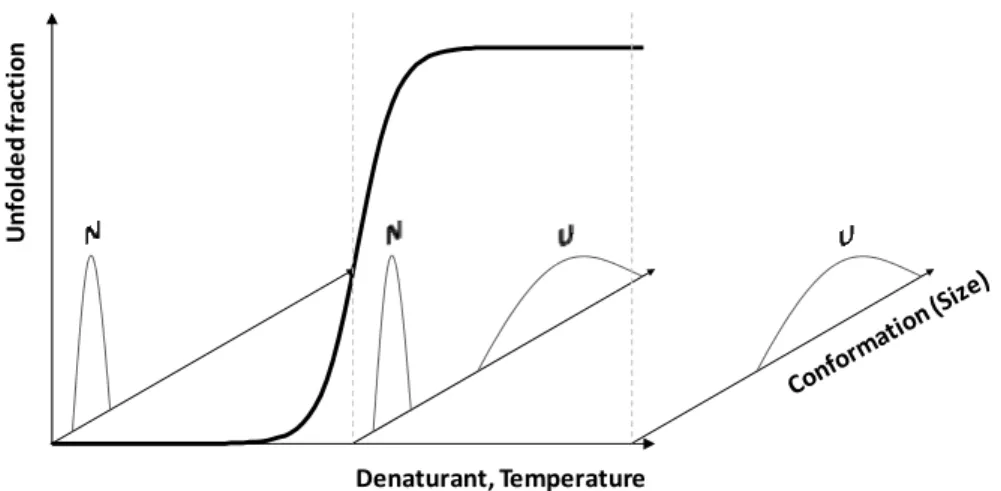
Figure 1.2 – Two state unfolding. Denaturing conditions (chemical denaturant or
temperature) unfold a two state folder in a cooperative manner. At any condition, only the native (N) and/or unfolded (U) state are detected and interconvert. The native state is a unique state while the unfolded state ensemble groups a broader set of conformations.
covalent interactions. No energetic component is preponderant in the overall stabilization. Even the simplest protein folding energetic potentials must include multiple energetic components to reproduce protein folding [17].
Small single domain proteins usually unfold reversibly [1] and according to a simple two state process. This means that the only conformations detectable are the native and the unfolded state. These two interconvert and no intermediately folded state is populated at detectable levels (Figure 1.2). Due to the cooperative nature of protein folding, such a protein is perturbed by an increasingly denaturing environment, the conformation initially changes very little [40]. Then, for a limited range of conditions, the protein unfolds completely. In this simple unfolding scenario, the same unfolding profile will be obtained independently of the technique used to probe it [40].
The thermodynamics of two state reversible unfolding is characterized as
a function of the native (N)/unfolded (U) equilibrium constant, :
=
from which the free energy variation is obtained
∆ − = − = − ( − )
Un
fo
ld
e
d
f
rac
ti
o
n
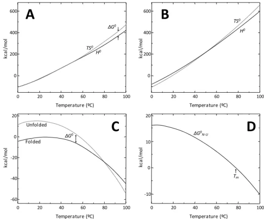
where R and T are the gas constant and absolute temperature, respectively. The thermodynamic characterization of lysozyme unfolding at pH 7 carried out by Privalov and co-workers [41] illustrates the complex relationship between the thermodynamic parameters that determine protein stability. The opposing effects of denatured state conformational entropy
(TS0) and native stabilizing interactions (H0) individually comprise several
hundred kcal/mol. However, both contributions have similar temperature trends and compensate each other, implying marginal protein stability. The
Gibbs free energy of folding exhibits a temperature dependence (∆ ( ))
Figure 1.3 – Temperature-dependent thermodynamic folding parameters for lysozyme at pH 7. The enthalpic (H) and entropic (TS) contributions are very large and of similar
magnitude for the folded (A) and unfolded (B) states. The most populated state is the lowest energy one (C). The energetic difference between the folded and unfolded states is small and
results in marginal protein stability (D). At the point where ΔGN-U is null, there is equal amount
of protein in both the folded and unfolded states. This temperature is referred to as the
midpoint denaturation or melting temperature (Tm). From [41].
0 20 40 60 80 100
0 200 400 600 Temperature (ºC) kc a l/ m ol
0 20 40 60 80 100
0 200 400 600 Temperature (ºC) kc a l/ m ol
0 20 40 60 80 100
-60 -40 -20 0 20 Temperature (ºC) kc a l/ m o l
0 20 40 60 80 100
TS0
H0
TS0
H0
Unfolded
ΔG0
ΔG0
Folded
expressed by the Gibbs-Helmholtz equation (Figure 1.3). If the heat capacity
(∆Cp ) and apparent enthalpy (∆H ) difference between the native and
the unfolded states is assumed temperature-independent, the equation has the following formula:
∆ ( ) = ∆ 1 − + ∆ ( − ) − ln ( )
The Gibbs-Helmholtz equation expresses the fact that ∆ is maximal
at some temperature and that there are two temperatures where it equals
zero (i. e. proteins can be denatured by heat or by cold). Heat denaturation
occurs because of the enthalpic compensation of stabilizing interactions by temperature. Cold denaturation is due to the decreased hydrophobic contribution at lower temperatures [42]. Experimentally, usually only heat denaturation is observed because the cold denaturation temperature is frequently below the water melting temperature. Nevertheless, this has been observed for a few proteins [43].
1.2.7. Engineering protein stability
Several studies have shown that the stability of naturally occurring proteins is not optimized and can be improved by mutagenesis [39, 44], sometimes paralleled with function enhancement [44-47]. However, in the extreme case, increased stability may generate a highly compact and stiff, non functional native state [39]. This suggests that marginal protein stability
is the evolutionary response to the need to achieve the native state (i. e. to
have a minimum energy state [1]) while preserving (i.)the protein dynamics
required for catalysis, conformational plasticity, protein recognition and proteolytic susceptibility [39], a key element in cellular protein turnover
[48]; and (ii.) an energetic buffer which allows protein evolution [49] (Figure
Figure 1.4 – Protein evolution. (A) Along evolutionary time, proteins accumulate mutations
which impact folding and stability. The marginal conformational stability of proteins implies that only a subset of all allowed mutations are tolerated. Mutations outside this neutral zone may are deleterious because they affect the protein’s ability to fold (destabilizing mutations) or to be functionally dynamic (over-stabilizing mutations). (B) Molecular chaperones rescue the folding of misfolded species and create an enhanced “neutral area” from where new functions may arise. From [49].
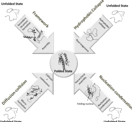
1.3.Protein folding models
Since the discovery that the protein native structure is determined by its amino acid sequence, much effort has been made in elucidating the mechanistics of protein folding. The first milestone in this field was set by Cyrus Levinthal in the late 1960s [50]. He elegantly pointed out that if a small 99 peptide bond protein would randomly sample just three rotamers of each of its 198 phi and psi angles in the typical timescale of molecular rotations –
1 picosecond - it would take 1075 years to fold – much more than the age of
the universe. However, proteins fold in a biologically relevant timescale (typically in the microsecond to second timescales [51]). This became known as Levinthal’s Paradox. It encloses a fundamental aspect of protein folding: the range of accessible conformations during folding is restricted. For solving this paradox, Levinthal postulated the existence of a pathway consisting of a well-defined and restricted sequence of protein conformational changes bridging the unfolded to the folded state [52] – the sequential model of protein folding. However, no mechanistic description to describe what the
A
Deleterious(Dynamics and regulations)B
Deleterious (Unfolding Aggregation Degradation)
New function
Chaperone buffering
S
ta
b
ility Ne
u
tr
a
l
Evolutionary change (time/mutations)
Ne
u
tr
a
l
folding pathway could be was put forward. Several models about protein folding have however been conceived since then (Figure 1.5).
In the late 1970s Levitt had already identified the caveat in Levinthal’s
paradox: the search for the native state is not unbiased. He noted that protein
folding could be reproduced in silico as a random search if native-like
interactions are considered to be on average more stabilizing than non-native ones. Thus, according to this random search model [53] the folding pathway is analogous to solving a jigsaw puzzle, where the order of the steps is not relevant but the end result is always the same native state [54]. More recently, the kinetics and thermodynamics of the folding of the villin headpiece domain has been successfully modeled using this formalism [55].
Kim and Baldwin proposed the framework model [53]. This model considers that secondary and tertiary structural elements form independently of one another. Secondary structure elements build up in the beginning of the folding reaction and progressively assemble into the native tertiary structure.
The hydrophobic collapse model [56] hypothesizes that folding initiates by a rapid polypeptide collapse driven by hydrophobic side chain self-association, resulting in the formation of an intermediate state devoid of
secondary structure and with non-native tertiary structure, i. e. a
Karplus and Weaver proposed the diffusion-collision model [61-62]. In this case, the folding protein is considered to be composed of several independent marginally stable secondary structure microdomains each one exhibiting fast conformational dynamics. During folding individual microdomains collide and eventually adhere and coalesce to give rise to the native tertiary structure.
The classical nucleation model proposes that the folding reaction is guided by the formation of a marginally stable nucleus containing correct secondary and tertiary structural elements. The place of nucleation in the folding reaction was matter of dispute: Wetlaufer proposed that nucleation would comprise the folding onset [63] and Baldwin proposed that nucleation would be the limiting factor [64]. In any case, the nucleus templates formation of further structure around it, restricts the available conformational space and speeds folding without implying the existence of folding intermediate states [65]. In the 1990s Monte Carlo simulations of a lattice model by Shakhnovich and co-workers [66] supported that nucleation limits folding. Once the nucleus is assembled, the native conformation is promptly formed. This discovery permanently associated the study of protein folding with that of the transition state. The nucleation model describes the folding of two state folders but fails to describe folding processes where intermediates accumulate [67].
In the 1990s, Fersht and co-workers established that the folding of chymotrypsin inhibitor 2 (CI2) – a 64 residue two state folder – could only be described by a new folding model: the nucleation-condensation model [68-70]. This model postulates the existence of a marginally stable nucleus composed mainly by adjacent residues early in folding. The rate limiting step is the eventual stabilization of this small nucleus by long range interactions. This extended nucleus is not formed in the transition state but represents the
Figure 1.5 - Protein folding models.
condensation of tertiary structure around it. Since the proposal of this model, other proteins were shown to adopt compatible folding kinetics [72].
1.4.Protein folding kinetics
Protein folding is a structural event. Nevertheless, it can be appropriately described by standard chemical reaction kinetics theory. The starting state – the unfolded state ensemble – is regarded as a “reagent” and the end state – the native state – as a “product”. The reaction mechanism, energetics and kinetics can be described by monitoring the interconversion of the two entities, possibly through some intermediate states. Like a reaction, protein
folding is a diffusive process, i. e. an unfolded polypeptide will spontaneously
acquire the most stable accessible conformational state given the solvent conditions through a stochastic search.
Folded State
Unfolded State
Unfolded State
Unfolded State Unfolded State
The classical way of studying protein folding in vitro is through
temperature-, pressure-, acid- or denaturant-induced renaturation [71]. The folded and unfolded states frequently exhibit distinct spectroscopic properties. Coupling renaturation (by manual or stopped flow mixing, temperature or pressure jumps) to spectroscopic detection allows monitoring the transition between the two states and describing folding [73]. Information about folding can also be obtained by studying the analogous unfolding transition. A summary of the main techniques used in studying protein folding – not just the kinetics – is given in Table 1.1.
The conformational search inherent to protein folding implies that only the lowest energy states (typically the native state) are unique and well
defined,allothers (e.g.theunfolded and transition states)beinginfact an
Table 1.1 – Experimental techniques useful for the study of protein folding. From [73].
Technique Timescale Information content
Intrinsic tryptophan fluorescence ≥ ns Tryptophan environment
Far UV CD ≥ µs Secondary structure content
Near UV CD ≥ µs Aromatic residue packing
Raman spectroscopy ≥ µs Solvent accessibility, aromatic residues’ conformation
Infrared spectroscopy ≥ ns Secondary structure content
ANS binding ≥ µs Hydrophobic surface exposure
FRET ≥ ps Molecular ruler
Fluorescence correlation
spectroscopy ≥ ps Diffusion, size and shape
Fluorescence anisotropy ≥ µs Shape and size
Small-angle X-ray scattering ≥ µs Radius of gyration
Absorbance ≥ ns Chromophore environment
Real-time NMR ≥ min Structure
Native-state hydrogen exchange h Global stability
Pulsed H/D exchange by NMR ≥ ms Solvent accessibility
Pulsed H/D exchange by ESI-MS ≥ ms Solvent accessibility
NMR relaxation ~ms Denatured state structure, conformational changes
Protein engineering Probe
ensemble of distinct states. Then, the aforementioned states are accurately named the unfolded state ensemble (USE) and the transition state ensemble (TSE). The reason for the occasional language simplification is due to the fact that these states are most frequently experimentally accessible though their statistically averaged properties. The TSE is defined as the set of conformations such that folding trajectories starting from each one of them have a 50% probability of either reaching to the folded state before unfolding and reaching the unfolded state before folding [74].
Protein folding is a cooperative process [75]: the establishment of native or native-like contacts facilitates further interactions. This speeds folding [76] and stabilizes the native state once it is formed, as full unfolding requires the cumulative loss of the interaction in the cooperative network. In the extreme case, as it is frequently found in small globular proteins, cooperativity leads to two state folding [77] where the only detected populated states are the native and the unfolded ones, which interchange between themselves during folding. In this situation, protein folding kinetics is monoexponential. The surprisingly fast protein folding rates are achieved through a combination of factors:
1. Presence of residual structure in the fully unfolded state which
restricts the accessible conformational space during folding;
2. The folding pathway comprises metastable intermediates which work
like hubs directing folding to the native state [78];
3. The funneled energy landscapes biases folding to the native state.
(pseudo-first order), an essential element of the stability and kinetics of proteins [75, 79].
In the framework of the success of the nucleation formalism in describing the folding of small globular proteins, the folding transition state has been routinely probed by a protein engineering method named Φ-value analysis [71]. It consists in analyzing the effect of single point mutations in the protein’s folding behavior and determining a thermodynamic parameter – the Φ-value – defined as
Φ =− ∆∆ ≈∆∆∆∆ ‡
where and are the folding rates of the mutant and wild type
proteins, respectively, ∆∆ is the change in folding free energy upon
mutation and ∆∆ ‡ is the change in activation free energy upon mutation
(Figure 1.6). The approximation is valid for non-disruptive (i. e. having a
Figure 1.6 – Φ-value analysis of protein folding. The putative folding energy diagrams for
two point mutants are sketched. (A) The mutation does not perturb the interactions and, consequently, the energetics of the selected amino acid residue in the unfolded nor in the transition states. Nevertheless, the mutation is destabilizing. The Φ-value is then zero. (B) The mutation affects a residue which is part of the folding nucleus. The transition state is then destabilized in the same amount as the native state and the Φ-value equals 1. From [71].
Proteins which were selected by evolution to fold into a biologically relevant structure are able to satisfy the local structure propensity of amino acid residues (dictated by their respective rotamers) and intramolecular interactions (responsible for stabilizing the native state) without contradicting each other. Of course this can only occur in native or native-like structures, guiding the folding process. This situation is called the “minimal frustration” of protein folding [81-82].
So, protein folding is a highly complex mechanism. Part of the complexity comes from the definition of the starting point, the unfolded state. Unlike the native state, the unfolded state is a broad collection of conformationally distinct states with high entropy – the denatured state ensemble. It contributes with structural heterogeneity to the starting state, and determines a lot of the random search for the native state. The inherent stochasticity of protein folding results in different parts of the protein being folded at different times in the folding reaction, almost independently of each
Umut U
‡mut ‡
Nmut
N
Umut U
‡mut
‡
Nmut
N ΔΔG‡-U
ΔΔGN-U
ΔΔG‡-U
ΔΔGN-U
A
B
other. These folding units forming in one single cooperative step are called
“foldons” [81-82]. Since every intermediate en route to the native state has a
different conformation, it also has a specific energy. The stochastic fluctuation between sequential folding intermediates gives rise to a distorted, rugged funnel, populated by a myriad of energetic basins (or kinetic traps, following a kinetic analysis) which stabilize misfolded intermediates and slow down folding. To overcome these traps, local or
global unfolding must occur. Some basins can be transposed within kBT.
Deeper ones can become conformational dead-ends from which the protein
cannot be rescued without external assistance (e. g. by molecular
chaperones), despite the favorable thermodynamics. Folding intermediates have been detected experimentally for many systems including hen lysozyme
[83], cytochrome c [84] among others.
The extreme case of kinetically-controlled folding is achieved for proteins whose native state – determined by its biological activity – is not the lowest energy one but rather a metastable higher energy state trapped by a large activation energy barrier. This is possible for proteins which include prosegments in the newly-synthesized polypeptide which are subsequently proteolytically excised. Such post-translational processing occurs in insulin [85], α-lytic protease [86], pepsin [87] and the serpins [88].
1.5.Protein folding energetics – the landscape view
conformations. The exact sequence of conformational transitions is stochastically determined – like the jigsaw puzzle concept [54]. However, since the Gibbs free energy (ΔG) of each conformation is different and the native state has the lowest energy – Anfinsen’s thermodynamic hypothesis – the energy landscape is biased towards the native state. The formation of stabilizing interactions precludes the sampling of competing conformations greatly restricting the accessible conformational space. On the other hand, incorrect local folds tend to be eliminated by more stable conformations arising in the fluctuations inherent to this diffusive process [82, 89]. The energetic gradient also implies that not all conformations are equally likely in the folding pathway, solving Levinthal’s paradox, and that native-like intermediates may act as hubs guiding folding [90]. This situation has been pictured by Onuchic and co-workers as a funneled energy landscape [91].
The folding funnel determines protein folding kinetics and thermodynamic properties. Its shape is a function of the medium
composition. Events such as solvent change, external perturbations (e. g.
temperature, pressure), protein association or ligand binding change the shape of the folding funnel and, ultimately, protein conformation. This is the thermodynamic basis for protein allostery, protein association, protein conformational changes and protein unfolding.
The bottleneck inherent to the folding funnel acts to speed folding. In small globular proteins this produces a single exponential folding kinetics (Figure 1.7) [77]. However, the many degrees of freedom in protein folding produce a folding funnel which is not smooth but rather rugged, with many
energetic – i. e. kinetic – traps. These local minima illustrate the situation
Figure 1.7 - A schematic folding funnel of acylphosphatase. The
great conformational space
accessible to the unfolded state is progressively restricted as the protein lowers its free energy through folding. The energetic gradient progressively biases the conformational search to the native state, although multiple folding pathways are still accessible. Once the folding transition state barrier, depicted as a saddle point, is overcome by the formation of the folding nucleus (depicted as yellow spheres in the structure) folding is fast and productive. From [96]. proteins shows that energetic traps have been smoothed by evolution rendering naturally occurring proteins “minimally frustrated” [89, 95].
Growing evidence supports the hypothesis that the folding energy landscape may in fact include additional folding funnels accounting for misfolded species [97]. Due to their simplicity, the folding of naturally occurring small single domain proteins is frequently associated with smooth energy landscapes, giving rise to single exponential folding kinetics. In the framework of landscape theory it is possible to envisage an extreme situation where a determined polypeptide sequence folds to the native state without encountering significant energetic barriers throughout the folding funnel (Figure 1.8B), a situation termed “downhill folding” [98]. In such a scenario,
folding kinetics is non-exponential and all conformers are en route to the
Figure 1.8 - Protein folding funnels. (A) Levinthal’s funnel. All folding pathways are equally
likely and there is no folding cooperativity in the search for the native state. (B) Downhill folding funnel. Smooth energy landscape without kinetic traps. (C) Folding funnel for a protein with an obligatory on-pathway folding intermediate. (D) Highly frustrated folding funnel, with many kinetic traps and alternative folding routes. From [100].
1.6.Conformational states – a unified view of protein folding
The highly complex interaction network in proteins coupled with the large conformational space accessible results in a series of conformational states being accessible to proteins. Unlike the classical view of protein folding whereby a polypeptide is synthesized in the ribosome in the unfolded state and eventually attains the monomeric or oligomeric native state, several intermediate or off-pathway states do exist and have been shown to be relevant in the biologic context of protein folding. Partially folded intermediates may expose hydrophobic patches which drive self-aggregation. Misfolded or aggregated protein may be recognized by cellular quality control systems and degraded. It is believed that, given appropriate conditions, every protein has the potential of acquiring the β-sheet rich aggregation-prone amyloid conformation, which may polymerize into fibrils (Figure 1.9) [96].
The exception to this panorama are proteins which, notwithstanding the possibility of having local persistent structure, the majority of the polypeptide chain is in a random coil-like conformation: the so-called intrinsically unstructured proteins or natively unfolded proteins. These proteins are not able to acquire a compact native state because of having a
highcharge density. Theseproteins arenotwithoutfunctional significance.
Figure 1.9 – Protein conformational states. From the moment a polypeptide is synthesized
in the ribosome, it can adopt a multitude of conformations apart from the unfolded and native states. Intermediately folded states may be aggregation- or degradation-prone. The β-sheet rich amyloid state is an aggregated state which is thought to be accessible to all proteins. Artificially, proteins can be superconcentrated and forced to adopt a crystalline state. From [96].
Their high conformational dynamics renders them promiscuous protein binders. So, these proteins fulfill mainly protein network integration roles, working like hubs binding proteins from different signaling pathways and contributing to signal transduction [101-104].
1.7.Protein folding in the cell
C-terminus – and the synthesis rate is slow (~2-4 amino acid residues per second in eukaryotic systems [105]), likely constituting the rate-limiting
factor in protein folding in vivo. Since protein folding is dictated by long
range interactions which may involve interactions between the N- and
C-terminus, especially in multidomain proteins (e. g. [106]), and shape the
folding energy landscape [107] non-native interactions may occur during protein synthesis. Secondly, protein biosynthesis occurs in the context of densely packed polysomes (Figure 1.10), further favoring interaction of aggregation-prone solvent-exposed hydrophobic residues. To minimize improper interactions, ribosomes are oriented around the mRNA molecule in a pseudohelical arrangement which minimizes the interactions between vicinal nascent chains [108]. The ribosome exit channel is hydrophilic [109] and favors co-translational folding [110] of at least α-helical elements [111-113]. Cell environment is highly crowded, with macromolecular concentrations reaching as much as 400 mg/ml [114-116]. The excluded volume effect favors protein misfolding and aggregation [116-118]. However, it also biases the folding landscape towards compact conformations [119], restricting the accessible conformational space and speeding folding in some situations [116-117, 120-121]. As a response, cells accumulate compatible solutes, also known as osmolytes [122], sometimes up to molar concentrations, which favor protein hydration and, consequently, hydrophobic burial and folding. To cope with the challenges of protein folding, biological systems have evolved a specialized protein quality control machinery (Figure 1.11) aimed at aiding or correcting folding – the molecular chaperones – or, ultimately, degrading terminally misfolded or aggregated proteins [123].
Molecular chaperones recognize and reversibly bind nascent polypeptides or non-native protein conformations (hydrophobic amino acid stretches
Figure 1.10 – Polysomes.
Several ribosomes transcribing a single mRNA molecule (in the center). The newly synthesized polypeptides are depicted as red or green threads on the exterior of the polysome. The high protein density favors aggregation of the
misfolded polypeptides. To
overcome this problem,
polypeptides exit the ribosomes at opposing faces. From [108].
Figure 1.11 - Protein quality control systems. As
the newly synthesized
protein exits the ribosome, surveillance mechanisms act to detect, correct and ultimately degrade misfolded species. Holding chaperones prevent protein aggregation; folding chaperones assist the
folding and unfolding
chaperones act on
Downstream-acting chaperones do not bind the ribosome and assist co- or post-translational folding. These include members of the Hsp70 family (DnaK in bacteria, Hsc70 in higher eukaryotes [125]) and further downstream systems like the chaperonins (Hsp60 family: GroEL in bacteria, thermosome in archaea and TRiC/CCT in eukarya [130-132]) and Hsp90 [133]. Broad-specificity chaperones such as members of the Hsp70 or chaperonin families primarily recognize hydrophobic amino acid side chains and promote their folding through ATP-dependent binding-release-rebinding cycles. If a protein is terminally misfolded and cannot be rescued by chaperone action, the polypeptide is degraded. In archaea, eukaryotes and some bacteria, the most important proteolytic system is the ubiquitin-proteasome pathway [134]. It involves tagging the misfolded protein with multiple ubiquitin chains through the sequential action of three ligases and subsequent proteolytic degradation by the proteasome, a multiprotein complex.
1.8.References
1. Anfinsen CB (1973) Principles that govern the folding of protein chains. Science181, 223-230
2. Yewdell JW (2005) Serendipity strikes twice: the discovery and rediscovery of defective ribosomal
products (DRiPS). Cell Mol Biol (Noisy-le-grand)51, 635-641
3. Chiti F & Dobson CM (2006) Protein misfolding, functional amyloid, and human disease. Annu Rev
Biochem75, 333-366
4. Qu BH, Strickland E & Thomas PJ (1997) Cystic fibrosis: a disease of altered protein folding. J Bioenerg Biomembr29, 483-490
5. Cheung JC & Deber CM (2008) Misfolding of the cystic fibrosis transmembrane conductance regulator
and disease. Biochemistry47, 1465-1473
6. Gersting SW, Kemter KF, Staudigl M, Messing DD, Danecka MK, Lagler FB, Sommerhoff CP, Roscher AA & Muntau AC (2008) Loss of function in phenylketonuria is caused by impaired molecular
motions and conformational instability. Am J Hum Genet83, 5-17
7. Gregersen N, Bross P & Andresen BS (2004) Genetic defects in fatty acid beta-oxidation and acyl-CoA
dehydrogenases. Molecular pathogenesis and genotype-phenotype relationships. Eur J Biochem271,
470-482
8. Blennow K, de Leon MJ & Zetterberg H (2006) Alzheimer's disease. Lancet368, 387-403
9. Uversky VN & Eliezer D (2009) Biophysics of Parkinson's disease: structure and aggregation of
alpha-synuclein. Curr Protein Pept Sci10, 483-499
10. Shastry BS (2003) Neurodegenerative disorders of protein aggregation. Neurochem Int43, 1-7
11. Dill KA, Ozkan SB, Shell MS & Weikl TR (2008) The protein folding problem. Annu Rev Biophys37,
289-316
12. Hou J, Sims GE, Zhang C & Kim SH (2003) A global representation of the protein fold space. Proc Natl
Acad Sci U S A100, 2386-2390
13. Cooper S, Khatib F, Treuille A, Barbero J, Lee J, Beenen M, Leaver-Fay A, Baker D, Popovic Z & players
![Table 1.1 – Experimental techniques useful for the study of protein folding. From [73]](https://thumb-eu.123doks.com/thumbv2/123dok_br/15769788.641156/40.892.229.775.494.923/table-experimental-techniques-useful-study-protein-folding.webp)