ÓRGÃO OFICIAL DA SOCIEDADE PORTUGUESA DE REUMATOLOGIA
302 ARTIGO DE REvISÃO
1. Serviço de Reumatologia do Centro Hospitalar de Lisboa Ocidental, Hospital de Egas Moniz, EPE
2. CEDOC - Faculdade de Ciências Médicas, Universidade Nova de Lisboa
Denosumab: recent update in
postmenopausal osteoporosis
ACTA REUMATOL PORT. 2012;37:302-313 Inês Silva1, Jaime C Branco1,2
AbstrAct
Postmenopausal osteoporosis is a major concern to public health. Fractures are the major clinical consequen -ce of osteoporosis and are associated with substantial mor bidity, mortality and health care costs. Bone strength determinants such as bone mineral density and bone quality parameters are determined by life-long remodeling of skeletal tissue. Receptor activator of nuclear factor-kB ligand (RANKL) is a cytokine essen-tial for osteoclast differentiation, activation and survi-val. Denosumab (Prolia®) is a fully human monoclo-nal antibody for RANKL, which selectively inhibits os-teoclastogenesis, being recently approved for the treat-ment of postmenopausal osteoporosis in women at a high or increased risk of fracture by the FDA in the Uni-ted Sates and by the European Medicines Agency in Eu-rope since June 2010. FREEDOM, DECIDE and STAND are the phase 3 trials comparing denosumab with placebo and alendronate in postmenopausal os-teoporosis. The authors aim to update denosumab role in postmenopausal osteoporosis with a physiopatholo-gical review.
Keywords:RANK; RANKL; Osteoprotegerin; Denosu-mab; Postmenopausal osteoporosis.
IntroductIon
Osteoporosis (OP) is a skeletal disease associated with an imbalance in bone resorption and formation, which turns to a loss of bone mass and deterioration of bone
microarchitecture1. This results in low bone mineral
density (BMD) and poor bone quality, reduced bone
strength and increased risk of fractures2. Is a
worldwi-de public health problem with serious consequences in personal suffering and economical costs3,4. Clinical
tools to diagnose OP and predict fracture risk are avai-lable but patients who are at risk for fracture or with a previous fracture are very often not identified or trea-ted3,5-8. The bone remodeling unit (BRU) includes a
se-quence of events, during which osteoclasts (OC) resorb bone over a period of 3 weeks, creating cavities that are termed as remodeling space. The resorption is follo-wed by osteoblast (OB) activation and osteoid forma-tion, filling the cavities over a period of 3 months. When the matrix synthesis is finished OB become em-bedded in the matrix as osteocytes (that will function
as mechanoreceptors)3,9. Bone remodeling permits the
repair of microdamage, maintains normal skeletal mass and participates in regulation of systemic calcium ho-meostasis9,10-13. As bone resorption/formation is tightly
coupled, inhibition of resorption eventually results in inhibition of formation9(Figure 1). OP therapy may be
classified as antiresorptive (estrogens, bisphosphonates, calcitonin and raloxifene) or anabolic [teriparatide (re-combinant human parathyroid hormone PTH1-34) or PTH1-84)]. Strontium ranelate appears to have both functions3,9. Research is focusing on drugs that target
the remodeling cycle acting in OC, OB and osteocytes or molecules that control the signaling pathway for cell functioning and gene transcription9. Examples of
stu-dies on way include Glucagon-like peptide 214, cathepsin K inhibitors15, PTH1-2816, calcium-sensing
receptors17, Wingless (Wnt)/ -catenin pathway18,
scle-rostin and Dickkopf-119, activin (fusion protein
ACE--011)20. However, the discovery of the receptor
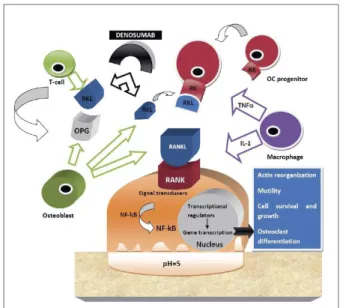
activa-tor of nuclear facactiva-tor KB ligand (RANK ligand, RANKL) osteoprotegerin (OPG) and RANK as a RANKL/OPG/ /RANK signaling pathway for the bone balance brought advances to the understanding of healthy bone turno-ver, and to osteolytic and destructive bone diseases like OP, rheumatoid arthritis (RA), Paget’s disease of bone
identi-ÓRGÃO OFICIAL DA SOCIEDADE PORTUGUESA DE REUMATOLOGIA
303 inÊs silva e col.
fied as a potential target for therapeutic intervention in the treatment of these diseases. Possible strategies to down-regulate RANKL include inhibition of RANKL production, stimulation of endogenous OPG, and ad-ministration of exogenous OPG, soluble RANK or
an-tibody to RANKL2 (Figure 2). Denosumab (Prolia®,
AMG 162; Amgen Inc., Thousand Oaks, California, USA) is a fully human monoclonal antibody (IgG2 im-munoglobulin isotype) with a high affinity and speci-ficity for RANKL2,3,11, currently approved by Food and
Drug Administration (FDA) and European Medicines Agency (EMA) for clinical use in postmenopausal wo-men OP with high risk of fracture and for the treat-ment of bone loss associated with hormone ablation in men with prostate cancer11,21.
This manuscript reviews Denosumab pharmacolo-gical and clinical data in postmenopausal osteoporo-sis, with a previous physiopathology overview.
AduLt sKELEton
PhysIoLoGy
The adult human skeleton is a metabolically organ with a coupled bone turnover that maintains the equili-brium in the trabecular and cortical bone. The total
amount of bone loss is proportional to the number of BRUs activated at the bone surface at a given time10. In
the healthy adult, more than 1 million BRUs are active and up to 5-10% of existing bone is replenished an-nually. Full skeletal regeneration is accomplished once
every decade21. High bone turnover diminishes bone
strength independently of BMD, because excessive num ber of resorption cavities act as areas of stress that may be a source of microcracks2,3,11. This high turnover
can eventually result in osteopenia or osteoporosis3.
rAnKL/oPG/rAnK sIGnALInG PAthwAy
OB are mononuclear cells responsible for the deposi-tion of bone matrix and for OC reguladeposi-tion. They ori-ginate from mesenchymal stem cells (MSC) by the
ac-FIGurE 1. Bone remodeling sequences in healthy individuals. The bone remodeling unit (BRU) includes a sequence of events, during which osteoclasts (OC) resorb bone over a period of 3 weeks, creating cavities that are known as remodeling space. The resorption is followed by osteoblast (OB) activation and osteoid formation, filling the cavities over a period of 3 months. When the matrix synthesis is finished OB become embedded in the matrix as osteocytes (that will function as mechanoreceptors)3,9. Legend: BRU – bone
remodeling unit; CL – cement line; LC – lining cells; OB – osteoblast; OC – osteoclast; OS – osteoid
ÓRGÃO OFICIAL DA SOCIEDADE PORTUGUESA DE REUMATOLOGIA
304 Denosumab: upDate
tion of transcription factors like core binding factor α1 (Cbfa-1) also known as Runx2, osterix (Osx), activa-ting transcription factor 4 (ATF4), and bone
morpho-genic proteins (BMP) as BMP422. OC are derived from
mononuclear precursors in the myeloid lineage of he-matopoietic cells that also originate macrophages. Ma-crophage-colony stimulating factor (M-CSF) expres-sion by osteoblastic stromal cells is required for pro-genitor cells to differentiate into OC, but is unable to complete this process by its own23. The principal final
mediator of osteoclastic bone resorption is RANKL, highly expressed by OB and T cells (mainly T helper
cells 17)3. RANK is located on the cell membrane of
OC and pre-OC and is a receptor for RANKL. RANKL/RANK binding stimulates OC differentiation and survival, resulting in increased bone resorp-tion3,11,21. OPG is a “decoy receptor” for RANKL,
pre-venting its interaction with RANK, OC differentiation,
activation of mature OC and permits OC apoptosis3,21
(Figure 2). RANKL/OPG ratio represents an important determinant of bone resorption. This ratio is decreased by estrogens, interleukin-4 (IL-4), IL-13, interferon-γ
(IFN-γ), transforming growth factor- (TGF- ), and in-creased by glucocorticoids, PTH, PTH-related protein, prostaglandins, IL-1, IL-17, tumor necrosing factor-α
(TNF-α), 1,25-dihydroxyvitamin D (1,25vitD) and
BMP23,11,21. The precise role of the RANKL/OPG/RANK
signaling pathway in regulating bone remodeling and mass was validated by gene studies in mice, with ge-netic RANKL or its receptor deficiency and OPG over-expression resulting in osteopetrosis, and genetic OPG
ablation resulting in osteoporosis24-27. The Wnt
signa-ling in OB is also a source of OPG regulation. This pathway integrated with RANKL/OPG/RANK enabled
to understand the normal and diseased bone11. Many
familiar bone diseases are mediated by RANKL/OPG/
/RANK pathway21(Table I). Evidence is accumulating
that bone remodeling is modulated through the inte-raction of OC and OB with immune cells, cytokines and circulating hormones28. It was demonstrated that
activated T cells directly stimulate osteoclastogenesis through RANKL, and RANKL prolongs the survival of dendritic cells and thereby increases T cells activity29.
Infection and malignancy concerns have been raised by inhibiting RANKL, however it seems that RANKL/ /OPG/RANK system does not have an essential role in immune function of adults, who already have a fully
developed immune system30.
PostmEnoPAusAL ostEoPorosIs
Estrogen is a positive regulator of OPG expression and its decrease in postmenopausal women are associated with increased RANKL expression, which shifts the bone remodeling balance toward the bone resorp-tion31. The incidence of postmenopausal OP is growing
due to changing demographics and increasing life ex-pectancy, which will also increase its economic and so-cial burden fracture-related31,32. OP fractures occur mo
-re f-requently in women, its f-requency inc-reases with age, and spine, hip and wrist are the skeletal sites ty-pically associated to OP31. Vertebral fractures may
re-sult from relatively mild trauma in osteroporosis and
tAbLE I21. rEvIEw oF thE dIsEAsEs mEdIAtEd by dIsruPtIons In rAnKL/oPG/rAnK sIGnALInG PAthwAy
RANKL/OPG/RANK-mediated diseases Mechanism(s)
Postmenopausal osteoporosis ↑ bone marrow stromal cell expression of RANKL
Primary hyperparathyroidism ↑ RANKL, ¯ OPG expression by osteoblasts
Bone Paget’s disease ↑ stromal cell expression of RANKL
Rheumatoid Arthritis ↑ RANKL expression by synoviocytes and T cells
Periodontal bone loss ↑ RANKL expression by activated T cells
Myeloma bone disease ↑ RANKL expression by myeloma cells
Osteolytic bone metastases ↑ RANKL expression by tumour cells
Humoral hypercalcaemia of malignancy Parathyroid hormone related protein (PTHrP) mediated:
↑ RANKL, ØOPG by osteoblasts
Familiar expansile osteolysis Activating mutations of the RANK gene
Idiopathic hyperphosphatasia (Juvenil Paget’s disease) Inactivating mutations of the OPG gene
ÓRGÃO OFICIAL DA SOCIEDADE PORTUGUESA DE REUMATOLOGIA
305 inÊs silva e col.
are associated to an increased risk for subsequent frac-ture. Hip fractures are associated with worst outcomes and secondary complications, and their risk increases in women from the age of 70 years31-33. Healthcare costs
due to OP are difficult to calculate as they include the costs of acute hospital care, loss of working days for fa-mily carers, longterm care and medication31-33. The
In-ternational Osteoporosis Foundation (IOF) estimated that the number of osteoporotic fractures in 2000 was 3.79 million and the total direct costs resulting from these fractures was estimated at €31.7 billion, which is expected to increase to €76.7 billion by 205034.
Pre-vention of osteoporosis-related fractures appears to be essential to the quality of life and independence of pos-tmenopausal women and prevent the economic bur-den32-34.
ostEoPorosIs trEAtmEnt: dEnosumAb
As A nEw oPtIon
The ideal anti-osteoporotic agent would have to meet the following favorable biological properties: a rapid onset action, long duration effect, patient’s complian-ce and persistencomplian-ce, documented cost-effectiveness and excellent safe profile10,11. Denosumab is a fully human
monoclonal immunoglobulin G2 antibody that binds with high affinity and targets the activity of human RANK ligand , preventing it to interact with RANK on the OC surface, which turns into the disruption of cel-lular signaling of bone resorption OC-mediated3,10,11,31.
Denosumab inhibits numerous aspects of OC diffe-rentiation and function (fusion, diffediffe-rentiation, at-tachment to bone, activation and survival), by inhibi-ting the intracellular signal pathways that are activated
by the RANKL /RANK binding10. Denosumab does not
bind to murine RANKL1, and does not cross-react with other human proteins of similar structure to RANKL,
such as TNF-α, TNF- , TNF-related
apoptosis-indu-cing ligand (TRAIL) or CD40 ligand11,31.
PhArmAcoKInEtIcs And mEtAboLIsm
The pharmacokinetics of denosumab is nonlinear with dose. Although the absorption, bioavailability, distri-bution, and elimination are not well defined, studies with similar IgG antibodies showed that subcutaneous denosumab is absorbed by the lymphatic system with subsequent drainage into the vascular system2,3. The
bioavailability is probably 50-100% with a distribu-tion that is about the same as the plasma volume, the
clearance is probably by the reticuloendothelial sys-tem and no significant amount of denosumab seems to
be filtered and excreted by the kidneys35.
Subcuta-neous administration is characterized by 3 stages: a prolonged absorption phase with the maximum serum concentration (at 5-21 days post-dose) increasing dis-proportionately than the increase in dose; a long du-ration phase with half-life of a maximum of 32 days; a rapid terminal phase when serum concentration is
lo-wer than 1000ng/ml36. The long duration of
denosu-mab activity is probably due to a combination of a long half-life and a very potent antiresorptive effect at ear-ly stages of OC differentiation2,10. A feature that
dis-tinguishes denosumab from biphosphonates is the ra-pid reversibility of its antiresorptive effect once it has been eliminated and OC regeneration has occurred, since it is not incorporated in the bone matrix as the biphosphonates3. Its capacity to reduce bone
resorp-tion was measured by serum C-telopeptide (CTX-1), by 82% within 72 hours post-dose, with a sustained ef-fect during the 6-month dosing interval37.
PrE-cLInIcAL studIEs
The preclinical studies evaluating the RANKL/OPG/ /RANK role in bone showed: RANKL induces OC-like cells formation in cell cultures38, recombinant OPG
in-hibits OC differentiation in a dose-dependent
man-ner39, osteotropic hormones and cytokines regulate
RANKL and OPG expression in human-derived OB
cell lines40, OPG knockout mice develop OP and
fra-gility fractures41, RANKL and RANK knockout mices
have osteopetrosis with the total absence of OC24,25,42,43.
Human monoclonal antibodies (mAbs) are the fastest--growing category of mAb therapeutics entering clini-cal studies44. Murine antibodies are easier to produce,
but are limited by safety issues and diminished effica-cy owing to the immunogenicity of the mouse-derived
protein sequences44,45. One path started to develop
mAbs containing a combination of rodent-derived and human-derived sequences, resulting in chimeric and humanized mAbs. During the 2000s, human mAbs stand for 45% of the mAb candidates in the clinic and 88 are now in clinical development. Denosumab is one of the 7 mAbs currently approved for marketing in the
United States44-46. Denosumab was generated by
ÓRGÃO OFICIAL DA SOCIEDADE PORTUGUESA DE REUMATOLOGIA
306 Denosumab: upDate
concern that AMG 161 could have a toxic profile cau-sed by death of RANKL-producing cells. It was con-verted to noncytotoxic IgG2 mAb, AMG 162, with high affinity for human RANKL45,48. The difficulty of
AMG 162 to recognize rodent RANKL has complica-ted pre-clinical studies and the relevant data were
ob-tained from cynomolgus monkeys45. The majority of
the preclinical studies in RANKL inhibition in mice and rats used other agents than denosumab. The most commonly used were fusion molecules of recombinant OPG or RANK and the Fc fragment of IgG, called
OPG--Fc or RANKOPG--Fc, respectively3. These studies
de-monstrated that recombinant OPG prevents bone loss
in ovariectomized (OVX) rats39, recombinant OPG
de-creases OC differentiation in normal mice, resulting in non-lethal osteopetrosis39, recombinant OPG prevents
bone loss in mice with low BMD that over-express
TNF-α49, RANKL inhibition increases bone
minerali-zation and improves mechanical strength in the femur of young male rats50, in aged OVX rats a combination
of rat OPG and PTH increased BMD more than either agent alone51.
In the cynomolgus monkeys the preclinical studies showed: 5-year-old male monkeys treated with deno-sumab had an increased bone mass and improved bone strength in femur and lumbar vertebral bodies52,
skeletal benefits were demonstrated with the RANKL inhibition in RA models53, ovariectomy models54,
mul-tiple myeloma models55and inflammatory bowel
di-sease models56. Knocking technology has been
recen-tly used to create a genetically engineered mouse ex-pressing a chimeric form of RANKL (human/murine) that is bound and neutralized by denosumab, which has been studied for evaluating the skeletal effects of denosumab in different circunstances57,58.
cLInIcAL studIEs on
PostmEnoPAusAL ostEoPorosIs
The first study of RANKL inhibition in humans was conducted with OPG-Fc, a phase 1 randomized, dou-ble-blind, placebo-controlled, sequential dose escala-tion study in healthy postmenopausal women. Each of the 52 subjects received a single subcutaneous dose of OPG-Fc 0.1, 0.3, 1.0, 3.0 mg/kg or placebo. It was shown that a single dose of OPG-Fc caused a rapid, reversible and dose-dependent suppression of bone resorption, with no adverse events (AEs) or neutrali-zing antibodies59. This provided support for further
clinical studies.
PHASE1: a phase 1 study was conducted in 49 heal thy
postmenopausal women who received a single subcu-taneous dose of denosumab 0.01, 0.03, 0.1, 0.3 and 3.0 mg/kg or placebo. All the cohorts had a follow-up of 6 months, except the highest doses (0.1, 0.3 and 3.0 mg/kg) that were followed 9 months. Pharmaco-logical effect was assessed by urinary N-telopeptide (NTX) and seric serum bone-specific alkaline phos-phatase (BSAP) levels. Rapid and reversible NTX duction was sustained for 6 months. Later BSAP re-duction occurred with a lesser magnitude. Denosu-mab was well tolerated with no drug-related serious adverse events (SAEs) reported or study discontinua-tion due to AEs. Infectious events were similar in both groups (33% in placebo versus 38% in denosumab). Two hospitalizations: severe undetermined abdominal pain (in the 0.01mg/kg group) and a cholecystitis (in the 0.1 mg/kg group). Mild transient dose-dependent decrease in albumin-adjusted serum calcium and dose-dependent early increase in PTH levels36(Table II).
PHASE2: a randomized phase 2 study was performed
ÓR
GÃO
OFI
CI
AL
D
A
S
OCI
ED
AD
E
P
ORT
U
GU
ES
A
D
E
REU
MA
T
OL
OGI
A
3
0
7
in
Ês
sil
v
a
e
col.
tAbLE II. rEsumE oF thE PhAsE I And II trIALs oF dEnosumAb In PostmEnoPAusAL womEn
Number and
Study mean age of Study Study
Authors phase patients/subjects characteristics duration Primary endpoints Key efficacy results Key safety profile Bekker et al36 I 49 healthy PMW; RT, DB, SD, DE 6 or 9 Bone antiresorptive Dose-dependent, activity and No related SAEs, no
59.6 years months depending on dosing safety rapid (within 12h), clinical significantly profound (up to 84%) laboratory changes and and sustained (up to 6
months) decrease in bone turnover
McClung II 412 PMW with RT, PC, dose-ranging, 12 months Percent change from Increased BMD (LS: 3.0 to No significant et al37 low BMD; also included an baseline in BMD at LS, 6.7% compared with a differences between
63 years open-label ALN group changes in BTMs decrease of 0.8% in placebo) the ALN, denosumab
(70mg orally, once and decreased BTMs (within and placebo profiles
weekly) 72h) compared to placebo
Lewiecki II 412 PMW with Extension study of 24 months Percent change from Continuing increases in BMD 6 cases of infections et al61 low BMD; McClung et al; baseline in BMD at LS, (4.13% to 8.89% compared associated to
63 years same design changes in BTMs with – 1.18% with placebo) hospitalization in the and maintained suppression denosumab group;
of BTMs similar AEs
Miller and II 412 PMW with Extension study of 48 months Percent change from Significant increases in BMD Similar AEs, more et al62 low BMD; McClung et al; study baseline in BMD at LS, (LS: 9.4% to 11.8% and infections as SAEs in
63 years design reviewed, changes in BTMs TH: 4.0% to 6.1%) and the denosumab group modification of the New endpoints: effects sustained suppression of
denosumab regimens; of discontinuing and BTMs; discontinuing therapy in the ALN group no reinitiating denosumab for 24 months decreased additional ALN for the on bone density BMD to baseline levels; 24 months period and remodeling retreatment for 12 months
extension increased LS BMD by 9% from
original baseline values.
Seeman II 247 osteopenic, DB; patients 12 months Evaluation of the Better effects with Incidence of AEs
et al68 osteoporotic randomized to denosumab and ALN denosumab than ALN similar in both
PMW; 60.6 years denosumab, ALN effects on cortical and groups
or placebo trabecular
microarchi-tecture with pQCT
ÓR GÃO OFI CI AL D A S OCI ED AD E P ORT U GU ES A D E REU MA T OL OGI A 3 0 8 De n osu ma b: u pDa te B M D to a n e x te n d s im ila r to th at o b se rv ed w ith d en o su m ab in iti al tr ea tm en t. B y m on th 4 8 , B M D in -cr ea se d 9 % a t t h e l u m b ar sp in e a n d 3 ,9 % a t t h e t ot al h ip fr om b as eli n e an d th e B TM va lu es w er e s im ila r t o th os e of th e con tin u ou s t re at m en t gr ou p . In 3 .2 % of d en os u m ab p a-tie n ts oc cu rr ed in fe ct ion s, all w er e co m m o n c o m m u n ity -a cq u ire d (n o op p or tu n is tic in fe ct ion s) , w ith r e-q u ire d h os p ita liz at ion s ol ve d w ith st an d ar d a n tib io tic s. T h is o rig in al st u d y w as e x te n d ed 4 a d d iti o n al ye ar s w ith a ll p at ie n ts s w itc h ed to d en os u m ab 6 0 m g/ 6 m on th s s u b -cu ta n eou s a n d a fu rth er a n aly sis 2 ye ar s af te r (6 y ea rs o f d en o su m ab tr ea tm en t) r ep o rt ed a n B M D in -cr ea se of 1 3 .3 % a t t h e l u m b ar s p i-n e w ith s u st ai n ed s u p p re ss io n o f B TMs 3 7 ,6 0 -6 2 (T ab le II) . P H AS E 3 : T R E A T M E NT OF P OS T M E NOP AU S AL O S T E O P O R O S IS “F ra ct u re R ed u ct io n E va lu at io n o f D en o su m ab in O ste o p o ro sis E ve ry 6 M o n th s” ( F R E E D O M )6 3 , r an d o -m iz ed 7 6 6 8 p o st m en o p au sa l w o -m en w it h o st eo p o ro si s in to 2 gr o u p s: p la ce b o o r su b cu ta n eo u s d en o su m ab 6 0 m g/ 6 m o n th s. P ri-m ar y en d p o in t w as a r ed u ct io n in th e in cid en ce o f n ew r ad io gr ap h ic ve rte b ra l f ra ct u re s i n a 3 -y ea rs p e-rio d . S ec o n d ar y e n d p o in ts w er e r e-d u ct io n in h ip a n d o th er n o n ve rte -b ra l f ra ct u re s a n d c h an ge s i n B M D an d B T M s. D en o su m ab g ro u p p re -se n te d a 6 8 % re d u ct io n in n ew v er -te b ra l fr ac tu re ri sk c o m p ar ed to p la -ce b o ( 2 .3 % v s. 7 .2 % ; p < 0 .0 0 0 1 ), 4 0 % r ed u ct io n in th e h ip fr ac tu re (0 .7 % d en o su m ab v s. 1 .2 % p la ce -bo ; p =0.036), an d 20% r ed u ct io n in th e n o n ve rt eb ra l f ra ct u re s ris k (6 .5 % d en o su m ab v s. 8 .0 % p la ce -b o ; p = 0 .0 1 1 ). D en o su m ab g ro u p sh o w ed a s ig n ifi ca n tly in cr ea se in B M D a t a ll sk ele to n , m ain ly in th e
tAbLE III. rEsumE oF thE PhAsE III trIALs oF dEnosumAb In PostmEnoPAusAL ostEoPorosIs trEAtmEnt
Number and mean Baseline Study
Study age of patients T-score Study characteristics duration Primary endpoints Key efficacy results Key safety profile FREEDOM63 7868 osteoporotic < -2.5 at LS RT, pivotal, international; 36 months Reduction in incident Denosumab reduced Similar AEs and
PMW; 72.3 years or TH and largest phase III trial; morphometric new the risk of vertebral SAEs compared to ≥-4.0 at both denosumab vs placebo; vertebral fracture (68%), hip (40%) placebo
sites treatment of PMO and nonvertebral
(20%) fractures
DECIDE65 1189 PMW naïve ≤-2.0 at LS Multi-center, DB, non- 12 months Changes in BMD at Greater increases in Similar overall rates with low BMD; or TH -inferiority; head-to-head the TH, FN, LS and BMD at all measured of AEs and SAEs in
64.4 years comparison denosumab DR from baseline skeletal sites in both groups
vs ALN denosumab group
STAND66 504 osteopenic- <-2.0 to ≥ -4.0 Multi-center, DB, non- 12 months TH BMD, bone Transition to Similar overall rates -osteoporotic PMW at LS or TH -inferiority; head-to-head remodeling and safety denosumab increased of AEs and SAEs in
pretreated with comparison denosumab at 12 months BMD (1.9% both groups
ALN; 67.6 years vs ALN denosumab vs 1.05%
ALN) and reduced BTMs to a greater extent than continued ALN
ÓRGÃO OFICIAL DA SOCIEDADE PORTUGUESA DE REUMATOLOGIA
309 inÊs silva e col.
forearm. No differences of total incidence of AEs or SAEs were reported in both groups. The incidence of se-rious infections was similar in both groups. Infections resulting in death were 0.2% in both groups. Few de-nosumab patients were diagnosed endocarditis (n=3), pancreatitis (n=8), eczema was reported in 3% of de-nosumab group versus 1.7% in placebo group (p<0.001). Cellulitis as an SAE occurred in 0.3% (12/3886) in the denosumab group versus less than 0.1% (1/3876) in the placebo group (p=0.002). This study was extended for 2 years with all patients swit-ched to open-label denosumab 60 mg/6 months (with a significantly CTX-1 reduction and BMD increase64),
and then it was extended 5 more years to complete a to-tal of 10 years of denosumab exposition. Authors con-cluded that denosumab given subcutaneously twice yearly for 36 months reduced the risk of new vertebral, nonvertebral and hip fractures in women with OP.
HEAD-TO-HEAD COMPARISON WITHALENDRONATE
“Determining Efficacy: Comparison of Initiating
De-nosumab Versus Alendronate” (DECIDE)65, a
double--blind, double-dummy noninferiority 1-year study to compare the effects of denosumab and alendronate on BMD and BTM in naïve postmenopausal women (n=1189) with T-score of lumbar spine or total hip <-2. Patients were randomized to receive subcutaneous denosumab 60mg/6months plus oral placebo/week or oral alendronate 70mg/week plus subcutaneous place-bo/6months. The primary endpoint was percent chan-ge from baseline in total hip BMD at month 12 and the secondary endpoints were percent change in femoral neck, trochanter, lumbar spine and one-third distal ra-dius BMD, and changes in BTM. Denosumab group showed a greater BMD increase at the total hip versus placebo (3.5% vs. 2.6%; p<0.0001) and a greater re-duction in BTMs compared to alendronate. The overall incidence of AEs, SAEs, infections or neoplasms was similar between both groups. Authors concluded that denosumab was associated with both significantly more reduction in bone resorption and greater gains in BMD at all measured skeletal sites compared to alen-dronate.
TRANSITIONING FROMALENDRONATE TODENOSUMAB
“Study of Transitioning from Alendronate to
Denosu-mab” (STAND)66, a randomized, blind,
doubledummy, parallelgroup, 1year study in postmenopau -sal women (n=504) previously treated with alendro-nate at least 6 months, and with a lumbar spine or
to-tal hip T-score of -2.0 to -4.0. The primary endpoint was percent change in BMD at the total hip at 12 months for denosumab compared to alendronate. The study design allowed testing the primary endpoint for superiority if noninferiority was demonstrated. At 12 months denosumab group presented a significantly greater increase in BMD at the total hip (denosumab 1.9% vs. alendronate 1.05%; p<0.0001), lumbar spi-ne, and distal one-third radius, compared with conti-nuing of alendronate. AEs and SAEs were similar in both groups. Authors concluded that postmenopausal women with low BMD can be safely transitioned from weekly oral alendronate to 6-monthly subcutaneous denosumab to achieve an incremental increase in bone mass.
FurthEr cLInIcAL studIEs
Histology and Histomorphometry with Denosumab Reid et al67, collected iliac crest biopsies from
FREE-DOM and STAND populations, after 12, 24 and 36 months of denosumab exposition. In the FREEDOM study median eroded surface was reduced by greater than 80% and OC were absent from greater than 50% of biopsies in the denosumab group. It was shown a reduction in fractures in the same cohort of patients in whom there was such a clear histological evidence of reduced turnover. In STAND study, the histomorpho-metry indicates that denosumab 60mg/6months pro-duces greater inhibition of turnover than occurs with alendronate 70mg/week, but without evidence of any untoward interaction after the transition from alen-dronate to denosumab.
EFFEcts oF dEnosumAb on bonE
mIcroArchItEcturE
Seeman et al68, conducted a double-blind pilot study to
ÓRGÃO OFICIAL DA SOCIEDADE PORTUGUESA DE REUMATOLOGIA
310 Denosumab: upDate
dEnosumAb In PAtIEnts wIth rEnAL
ImPAIrmEnt
Block et al69, conducted a phase 1 study to evaluate the
pharmacokinetics, pharmacodynamics and safety of a single dose of 60mg subcutaneous denosumab in 55 patients with different degrees of renal function. Re-sults showed that the renal impairment did not affect the denosumab pharmacokinetics and pharmacody-namics, and no dose adjustment is necessary in im-pairment renal function. About 30% of patients with severe renal function and hemodialysis showed symp-tomatic hypocalcaemia.
bEsIdE PostmEnoPAusAL ostEoPorosIs
trEAtmEnt
PREVENTION OF POSTMENOPAUSAL OSTEOPOROSIS
“Denosumab Fortifies Bone Density” (DEFEND)70, was
a randomized, placebo-controlled, trial to evaluate ef-ficacy and safety of denosumab in postmenopausal wo-men with osteopenia.
Denosumab is currently being studied for other in-dications as RA bone erosions, bone loss associated with androgen deprivation therapy, bone loss associa-ted with aromatase inhibitor therapy and for the treat-ment of bone metastases. However these studies are beyond the scope of this paper.
sAFEty And toLErAncE
In the DEFEND, DECIDE, and STAND studies, AEs and SAEs, including infections and malignancies, were
similar in both groups33. In DECIDE the most common
types of infections were nasopharyngitis, influenza and upper respiratory tract infections. In STAND were
na-sopharyngitis and bronchitis1. In the FREEDOM study
the only significant AE were cellulitis and eczema71,72.
The longest exposure to denosumab reported to date is 6 years. No significant differences in infections,
neo-plasms were reported as SAEs for denosumab11. There
were no denosumab-related cases of osteonecrosis of jaw, fracture repair problems, changes in white blood cells counts, T, B, or natural killer cell counts, immu-noglobulins or antibodies to denosumab1,11.
cost-EFFEctIvEnEss ProFILE
Affordability of a drug therapy is a concern for healthca -re system and patients73. Hiligsmann et al74, assessed
the potential cost effectiveness of denosumab in the treatment of postmenopausal osteoporotic women in an updated version of a validated Markov
microsimu-lation model populated with cost and epidemiological data from Belgium and a base-case population defined from FREEDOM. It was concluded that denosumab is
cost effective compared with no treatment74and oral
bisphosphonates75for postmenopausal Belgian women
similar to FREEDOM population. Additional publis-hed studies with data from England, Wales and Sweden concluded that denosumab has a higher probability of being cost-effective in some patients subgroups76,77. A
cost-utility analysis of denosumab versus standard care (alendronate and colecalciferol) in the treatment of post-menopausal osteoporosis in Portugal concluded that denosumab is a cost-effective therapeutic strategy with an Incremental Cost-effectiveness Ratio (ICER)
OF 14.887 € per Quality Adjusted Life Year (QALY)
gained. The analysis was undertaken from a NHS pers-pective, efficacy data for denosumab was taken from FREEDOM and the comparator was taken from a meta--analysis conducted by National Institute for Health and Clinical Excellence (NICE). Epidemiological Por-tuguese data were complemented with Swedish data whenever the former were unavailable. The model
pre-tAbLE Iv. rEPortEd dIFFErEncEs bEtwEEn
dEnosumAb And thE bIPhosPhonAtEs
Denosumab compared to biphosphonates
• Denosumab blocks osteoclasts formation, function and survival while biphosphonates cause loss of resorptive function but “disabled” osteoclasts may persist78
• Exerts its effect from within the extracellular fluid67 Rapid offset of action at about 6 months11,57 • Induces more rapid and greater reduction in bone
remodeling63
• Superior pharmacokinetic properties (better distribution, no excretion of kidneys)10 • Greater increase in bone mineral density64
• Positive effect on both cancellous and cortical bone3,72 • Better strength indexes and geometrical bone variables67,68 • Completely reversible effect and no accumulation in
bone3
• Possible non-blunting effect on subsequent anabolic therapy10
• Better patient’s convenience and compliance1 • Denosumab was the first anti-bone resorbing agent
ÓRGÃO OFICIAL DA SOCIEDADE PORTUGUESA DE REUMATOLOGIA
311 inÊs silva e col.
dicts that relative to the comparator, denosumab would prevent 12 hip, 22 vertebral, 2 wrist and 1 other os-teoporotic fractures, per 1000 patients, over a 10 years period, and the probability of cost-effectiveness was between 91% and 64%78,79.
dIFFErEncEs bEtwEEn dEnosumAb And
thE bIPhosPhonAtEs
The effect of RANKL inhibition is quite unique among antiresorptive agents. Table IV shows how denosumab differs from the effects of byphosphonates on bone in several aspects11,31,67,81.
concLusIon
In postmenopausal women with osteoporosis denosu-mab reduced the risk of vertebral and nonvertebral fractures versus placebo. In those with low BMD or OP it increased BMD and decreased BTMs more than alen-dronate, both in treatment-naïve and who switched from alendronate. Denosumab was safety and well to-lerated, being cost-effective.
corrEsPondEncE to
Inês Maria Crispim Gomes da Silva
Alta do Lumiar, Rua Helena Vaz da Silva nº4 3ºD 1750-429 Lisboa
E-mail: [email protected]
rEFErEncEs
1. Moen AD, Kean SJ. Denosumab: a review of its use in the treat-ment of postmenopausal osteoporosis. Drugs Aging 2011;28:63-82.
2. Lewiecki EM. Denosumab update. Current Opinion in Rheu-matology 2009;21:369-373.
3. Lewiecki EM. Denosumab: an investigational drug for the ma-nagement of postmenopausal osteoporosis. Biologics: Targets and Therapy 2008;2:645-653.
4. Burge R, Dawson-Hughes B, Solomon DH. Incidence and eco-nomic burden of osteoporosis-related fractures in the United States, 2005-2025. J Bone Miner Res 2007;22:465-475. 5. Kanis JA, Borgstrom F, De Laet C, et al. Assessment of fracture
risk. Osteoporos Int 2005;16:581-589.
6. Solomon DH, Morris C, Cheng H, et al. Medication use pat-terns for osteoporosis: an assessment of guidelines, treatment ra-tes, and quality improvement interventions. Mayo Clin Proc 2005;80:194-202.
7. Johnell O, Kanis JA, Odén A, et al. Fracture Risk following an osteoporotic fracture. Osteoporos Int 2004;15:175-179. 8. Feldstein A, Elmer PJ, Orwoll E, Herson M, Hillier T. Bone
mine-ral density measurement and treatment for osteoporosis in older individuals with fractures – a gap in evidence-based practice gui-deline implementation. Arch Intern Med 2003;163:2165-2172.
9. Deal C. Future therapeutic targets in osteoporosis. Current Opi-nion in Rheumatology 2009;21:380-385.
10. Charopoulos I, Orme S, Giannoudis P. The role and efficacy of denosumab in the treatment of osteoporosis: an update. Expert Opin Drug Saf;doi:10.1517/ard2010.
11. Geusens P. Emerging treatments for postmenopausal osteopo-rosis – focus on denosumab. Clinical Interventions in Aging 2009;4:241-250.
12. Anastasilakis AD, Toulis KA, Goulis DG, et al. Efficacy and sa-fety of denosumab in postmenopausal women with osteopenia or osteoporosis: a systematic review and a meta-analysis. Horm Metab Res 2009;41:721-729.
13. Bolland MJ, Grey AB, Gamble GD, Reid IR. Effect of osteopo-rosis treatment on mortality: a meta-analysis. J Clin Endocrinol Metab 2010;95:1174-1181.
14. Henriksen DB, Alexandersen P, Hartmann B, et al. GLP-2 sig-nificantly increases hip BMD in postmenopausal women: a 120-day study. J Bone Miner Res 2007;22(suppl1):S37.
15. Gauthier JY, Chauret N, Cromlish W, et al. The discovery of oda-nacatib (MK-0822), a selective inhibitor of cathepsin K. Bioorg Med Chem Lett 2008;18:923-928.
16. Okazaki M, Potts JT, Gardella TJ. Identification and optimiza-tion of residues in PTH and PTHrP that determine altered mo-des of binding to the PTH/PTHrP receptor. J Bone Miner Res 2008;23:S103.
17. Brown EM. The calcium-sensing receptor: physiology, pathop-hysiology and CaR-based therapeutics. Subcell Biochem 2007;45:139-167.
18. Baron R, Rawadi G. Targeting the Wnt/beta-catenin pathway to regulate bone formation in the adult skeleton. Endocrinology 2007;148:2635-2643.
19. Padhi D, Stouch B, Fang L. Antisclerostin antibody increases markers of bone formation in healthy postmenopausal women. J Bone Miner Metab 2007;22 (Suppl 1):S37.
20. Ruckle J, Jacobs M, Kramer W. A single dose of ACE-011 is as-sociated with increases in bone formation and decreases in bone resorption markers in healthy postmenopausal women. J Bone Miner Res 2007;22 (Suppl 1):S38.
21. Romas E. Clinical applications of RANK-ligand inhibition. In-ternal Medicine Journal 2009;39:110-116.
22. Caetano-Lopes J, Canhão H, Fonseca JE. Osteoblasts and bone formation. Acta Reumatol Port 2007;32:103-110.
23. Boyce BF, Xing L. Functions of RANKL/RANK/OPG in bone mo-deling and remomo-deling. Archives of biochemistry and biophy-sics 2008;473:139-146.
24. Kong YY, Yoshida H, Sarosi I. OPG is a key regulator of osteo-clastogenesis, lymphocyte development and lymph-node orga-nogenesis. Nature 1999;397:315-323.
25. Li J, Sarosi I, Yan XQ. RANK is the intrinsic hematopoietic cell surface receptor that controls osteoclastogenesis and regulation of bone mass and calcium metabolism. Proc Natl Acad Sci U S A 2000; 97:1566-1571.
26. Bucay N, Sarosi, Dunstan CR. Osteoprotegerin-deficient mice develop early onset osteoporosis and arterial calcification. Ge-nes Dev 1998;12:1260-1268.
27. Mizuno A, Amizuka N, Irie K. Severe osteoporosis in mice lac-king osteoclastogenesis inhibitory factor/osteoprotegerin. Bio-chem Biophys Res Commun 1998;247:610-615.
ÓRGÃO OFICIAL DA SOCIEDADE PORTUGUESA DE REUMATOLOGIA
312 Denosumab: upDate
29. Xing L, Schawrz EM, Boyce BF. Osteoclast precursors, RANKL/RANK, and immunology. Immunol Rev 2005;208:19-29. 30. Stolina M, Dwyer D, Morony S. Rats and mice overexpressing soluble OPG have high bone mass but no alteration in immu-nological parameters or lymphocyte function. Arthritis Rheum 2005;52:S708.
31. Singer A, Grauer A. Denosumab for the management of pos-tmenopausal osteoporosis. Postgraduate Medicine 2010;122: 176-187.
32. Reginster JY. Antifracture efficacy of currently available thera-pies for postmenopausal osteoporosis. Drugs 2011;71:65-78. 33. Lewiecki EM. Denosumab for joints and bones. Current
Rheu-matology Reports 2009;11:196-201.
34. Reginster JY, Burlet N. Osteoporosis: a still increasing preva-lence. Bone 2006;38(2 Suppl 1):S4-9.
35. Lobo ED, Hansen RJ, Balthasar JP. Antibody pharmacokinetics and pharmacodynamics. J Pharm Sci 2004;93:2645-2668. 36. Beckker PJ, Holloway DL, Rasmussen AS, et al. A single-dose
placebo-controlled study of AMG 162, a fully human mono-clonal antibody to RANKL, in postmenopausal women. J Bone Miner Res 2004;19:1059-1066.
37. McClung MR, Lewiecki EM, Cohen SB, et al. Denosumab in postmenopausal women with low bone mineral density. N Eng J Med 2006;354:821-831.
38. Matsuzaki K, Udagawa N, Takahashi N, et al. Osteoclast diffe-rentiation factor (ODF) induces osteoclast-like cell formation in human peripheral blood mononuclear cell cultures. Biochem Biophys Res Commun 1998;246:199-204.
39. Simonet WS, Lacey DL, Dunstan CR, et al. Osteoprotegerin: a novel secreted protein involved in the regulation of bone den-sity. Cell 1997;89:309-319.
40. Hofbauer LC, Khosla S, Dunstan CR, et al. Estrogen stimulates gene expression and protein production of osteoprotegerin in human osteoblastic cells. Endocrinology 1999;140:4367-4370. 41. Bucay N, Sarosi I, Dunstan CR, et al. Osteoprotegerin-deficient mice develop early onset osteoporosis and arterial calcification. Genes Dev 1998;12:1260-1268.
42. Lacey DL, Tan HL, Lu J, et al. Osteoprotegerin ligand modula-tes murine osteoclast survival in vitro and in vivo. Am J Pathol 2000;157:435-448.
43. O’Brien EA, Williams JH, Marshall MJ. Osteoprotegerin ligand regulates osteoclast adherence to the bone surface in mouse cal-varia. Biochem Biophys Res Commun 2000;274:281-290. 44. Nelson AL, Dhimolea E, Reichert JM. Development trends for
human monoclonal antibody therapeutics. Drug Discovery 2010;9:767-774.
45. Schwarz EM, Ritchlin CT. Clinical development of anti-RANKL therapy. Arthritis Research and Therapy 2007:9(Suppl 1):S7(doi:10.1186/ar2171).
46. Scallon Bj, Moore Ma, Trinh H, Knight DM, Ghrayeb J. Chime-ric anti-TNF-alpha monoclonal antibody cA2 binds recombi-nant transmembrane TNF-alpha and activates immune effector functions. Cytokine 1995:7:251-259.
47. Kostenuik PJ. Osteoprotegerin and RANKL regulate bone re-sorption, density, geometry and strength. Curr Opin Pharma-col 2005;5:618-625.
48. Biot J, Fasano C, Santos CD. D’orthoclone au dénosumab – l’ex-pansion des anticorps monoclonaux à des fins thérapeutiques. Medicine Sciences 2009;25:1177-1182.
49. Schett G, Redlich K, Hayer S, et al. Osteoprotegerin protects against generalized bone loss in tumor necrosis
factor-transge-nic mice. Arthritis Rheum 2003;48:2042-2051
50. Ross AB, Bateman TA, Kostenuik PJ, et al. The effects of osteo-protegerin on the mechanical properties of rat bone. J Mater Sci Mater Med 2011;12:583-588.
51. Kostenuik PJ, Capparelli C, Morony S, et al. OPG and PTH-(1--34) have additive effects on bone density and mechanical strength in osteopenic ovariectomized mice. Bone 2004;142: 4295-4304.
52. Atkinson JE, Cranmer P, Saunders T. Denosumab (AMG 162), a fully human RANKL antibody, increases bone mass and bone strength in cynomolgus monkeys. J Bone Miner Res 2005;20(Suppl 1):S294.
53. Redlich K, Hayer S, Maier A, et al. Tumor necrosis factor alpha-mediated joint destruction is inhibited by targeting osteoclasts with osteoprotegerin. Arthritis Rheum 2002;46:785-792. 54. Kostenuik PJ, Bolon B, Morony S, et al. Gene therapy with
hu-man recombinant osteoprotegerin reverses established osteo-penia in ovariectomized mice. Bone 2004;34:656-664. 55. Vanderkerken K, De Leenheer E, Shipman C, et al. Recombinant
osteoprotegerin decreases tumor burden and increases survival in a murine model of multiple myeloma. Cancer Res 2003;63:287-289.
56. Byrne FR, Morony S, Warmington K, et al. CD4+CD45RBHi T cell transfer induced colitis in mice is accompanied by osteo-penia which is treatable with recombinant human osteoprote-gerin. Gut 2005;54:78-86.
57. Kostenuik PJ, Warmington K, Grisanti M, et al. RANKL inhibi-tion with (denosumab) AMG 162, a fully human MAb, causes sustained suppression of bone resorption and increased BMD in knockin mice expressing humanized RANKL. J Bone Miner Res 2005;20 (Suppl 1):S261.
58. Ferrari SL, Pierroz D, Bonnet N. A comparison of denosumab and alendronate in combination with PTH in ovariectomized knockin mice expressing humanized RANKL. J Bone Miner Res 2007;22(Suppl 1):S37.
59. Beckker PJ, Holloway D, Nakanishi A, et al. The effect of a sin-gle dose of osteoprotegerin in postmenopausal women. J Bone Miner Res 2001;16:348-360.
60. Bogado CE, Zanchetta MB, Boailchuk JA, Massari FE, Zanchet-ta JR. Denosumab: what’s new?. Curr Osteoporos Rep 2011;9:12-19.
61. Lewiecki EM, Miller PD, McClung MR, et al. Two-years treat-ment with denosumab (AMG 162) in a randomized phase 2 study of postmenopausal women with low BMD. J Bone Miner Res 2007;22:1832-1841.
62. Miller PD, Bolognese MA, Lewiecki EM, et al. Effect of denosu-mab on the bone density and turnover in postmenopausal wo-men with low bone mass after long-term continued, disconti-nued, and restarting of therapy. A randomized blinded phase 2 clinical trial. Bone 2008;43:222-229.
63. Cummings SR, Martin JS, McClung MR, et al. Denosumab for prevention of fractures in postmenopausal women with osteo-porosis. N Eng J Med 2009;361:756-765.
64. Papapoulos S, Chapurlat R, Brandi ML, et al. Five-year denosu-mab treatment of postmenopausal women with osteoporosis: re-sults from the first two years of the freedom trial extension. JBMR;2010; DOI-10.1002/jbmr.251.
ÓRGÃO OFICIAL DA SOCIEDADE PORTUGUESA DE REUMATOLOGIA
313 inÊs silva e col.
2009;24:153-161.
66. Kendler DL, Roux C, Benhamou CL, et al. Effects of denosumab on bone mineral density and bone turnover in postmenopau-sal women transitioning from alendronate therapy. J Bone Mi-ner Res 2010;25:72-81.
67. Reid IR, Miller PD, Brown JP, et al. Effects of denosumab on bone histomorphometry: the FREEDOM and STAND studies. J Bone Miner Res 2010;25:2256-2265.
68. Seeman E, Delmas PD, Hanley DA, et al. Microarchitectural terioration of cortical and trabecular bone: differing effects of de-nosumab and alendronate. J Bone Miner Res 2010;25:1886--1894.
69. Block G, Bone HG, Fang L. A single dose study of deonsumab in patients with various degrees of renal impairment. Am J Kid Dis 2010;55:A46.
70. Bone HG, Bolognese MA, Yuen CK, et al. Effects of denosumab on bone mineral density and bone turnover in postmenopau-sal women. J Clin Endocrinol Metab 2008;93:2149-2157. 71. Iqbal J, Sun L, Zaidi M. Denosumab for the treatment of
osteo-porosis. Curr Osteoporos Rep 2010;8:163-167.
72. Pageou SC. Denosumab. Landes Bioscience 2009;1:210-215. 73. Lewiecki EM. Clinical use of denosumab for the treatment for
postmenopausal osteoporosis. Current Medical Research and Opinion 2010;26:2807-2812.
74. Hiligsmann M, Reginster JY. Potential cost-effectiveness of de-nosumab for the treatment of postmenopausal osteoporotic wo-men. Bone 2010;47:34-40.
75. Hiligsmann M, Reginster JY. Cost Effectiveness of Denosumab Compared with Oral Bisphosphonates in the Treatment of Post-Menopausal Osteoporotic Women in Belgium. Pharmacoeco-nomics 2011;29:895-911.
76. Scotland G, Waugh N, Royle P, et al. Denosumab for the pre-vention of osteoporotic fractures in post-menopausal women: a NICE Single Technology Appraisal. Pharmacoeconomics 2011; Epub ahead of print Aug 20.
77. Jönsson B, Ström O, Eisman JA, et al. Cost-effectiveness of De-nosumab for the treatment of postmenopausal osteoporosis. Os-teoporos Int. 2011 Mar;22:967-982. Epub 2010 Oct 9. 78. Pereira JA, Cristino J, Perelmann J, et al. O uso de recursos e
cus-tos dos diferentes tipos de fracturas osteoporóticas em Portugal – estimativas na perspectiva do Serviço Nacional de Saúde e do Estado. Acta Reumatol Port 2011;36:31(sup).
79. Cristino J, Canhão H, Perelman J, Santos C, Pereira J. Cost-uti-lity analysis of denosumab versus standard care in the treatment of post-menopausal osteoporosis in Portugal. The Journal of The International Society for Pharmacoeconomics and Outco-mes Research 2011;14:A128.
80. Baron R, Ferrari S, Graham R, Russel G. Denosumab and bi-phosphonates: differents mechanisms of action and effects. Bone 2011;48:677-692.