Cop
yright
© ABE&M t
odos os dir
eit
os r
eser
vados
.
Cop
yright
© ABE&M t
odos os dir
eit
os r
eser
vados
.
Bone markers and
osteoporosis therapy
Marcadores de turnover ósseo e tratamento da osteoporose
Francisco Bandeira1, Aline G. Costa2, Manoel Aderson Soares Filho1, Larissa Pimentel1, Lourena Lima1, John P. Bilezikian2
ABSTRACT
Several factors are involved in determining bone quality including bone density, bone turnover, the extent of trabecular bone connectivity, cortical porosity and geometry. Metabolically acti-ve and in a continuous process of remodeling, approximately 20% of bone tissue is renewed annually. Bone turn over markers (BTM) are frequently used in clinical trials and to provide valid information about the effectiveness of osteoporosis treatment, relecting the state of bone metabolism and its response to treatment, although they are not useful alone to estimate bone loss. In this review the behavior of BTM from different clinical trials or different osteoporotic drugs will be addressed. Arq Bras Endocrinol Metab. 2014;58(5):504-13
Keywords
Bone markers; osteoporosis; bone density
RESUMO
Diversos fatores estão envolvidos na determinação da qualidade óssea, incluindo a densidade óssea, a remodelação óssea, a extensão da conectividade do osso trabecular, porosidade cortical e geometria. Metabolicamente ativo e, em um processo contínuo de remodelação, cerca de 20% do tecido ósseo é renovado anualmente. Por sua vez, marcadores de turnover ósseo (BTM) são frequentemente utilizados em estudos clínicos e fornecem informações válidas sobre a eicácia do tratamento da osteoporose, o que relete o metabolismo ósseo e sua resposta ao tratamento, embora eles não sejam úteis somente para estimar a perda óssea. Nesta revisão, o comportamento dos BTM em ensaios clínicos diferentes e com diferentes drogas osteoporóticas será abordado.
Arq Bras Endocrinol Metab. 2014;58(5):504-13
Descritores
Marcadores ósseos; osteoporose; densidade óssea
1 Division of Endocrinology,
Diabetes and Bone Diseases, Agamenon Magalhães Hospital, Brazilian Ministry of Health, University of Pernambuco Medical School, Recife, PE, Brazil
2 Metabolic Bone Diseases
Unit, Division of Endocrinology, Department of Medicine College of Physicians and Surgeons, Columbia University, New York, NY, United States
Correspondence to:
Francisco Bandeira Disciplina de Endocrinologia, Faculdade de Ciências Médicas, Universidade de Pernambuco Av. Rui Barbosa, 1435 52050-450 – Recife, PE, Brasil [email protected] Received on Mar/27/2014 Accepted on May/15/2014
DOI: 10.1590/0004-2730000003384
INTRODUCTION
B
one is a speciic type of tissue composed primarily of type I collagen impregnated with minerals in the form of hydroxyapatite crystals. Several factors are involved in determining bone quality including bone density, bone turnover, the extent of trabecular bone connectivity, cortical porosity and geometry (Figure 1) (1). Metabolically active and in a continuous process of remodeling, approximately 20% of bone tissue is renewed annually (2). This complex process begins at birth and is maintained throughout life. The active cel-lular participants in this process, often conigured asmultiple multicellular units, are osteoclasts, osteoblasts and osteocytes.
Cop
yright
© ABE&M t
odos os dir
eit
os r
eser
vados
.
RANKL expression increased by: IL-1, IL-7, IL-17, TNF-alpha Prostaglandin E2, decreased by 17beta-estradiol
RANKL expression increased by: IL-1, TNF-alpha
OPG production increased by: Glucocorticoid
Troglitazone
Smooth muscle cell
Endothelial cell Endothelial cell
RANK
OPG
OPG
RANKL
RANKL expression increased by: IL-1, IL-11, IL-17, TNF-alpha Prostaglandin E2, decreased by 17beta-estradiol
OPG
Immune system
Mature osteoclast
RANK RANK
Stromal cell or osteoblast
Osteoclast precursor
RANK RANKL
RANKL
Dendritic cell
RANKL T cell
T cell
Vascular system
Skeletal system
OPG
Bone strength = “Bone quality”
Moment of inertia (cortical geometry) Trabecular bone volume Trabecular geometry Trabecular conectivity
Architecture Turnover Material properties Microdamage
Figure 1. Factors related to bone quality.
Figure 2. The process of bone resorption: the RANK/RANKL/OPG system.
Osteoporosis can be primary or secondary to va-rious conditions such as hypogonadism, hyperthyroi-dism, skeletal metastases, multiple myeloma, anticon-vulsants, corticosteroids, and alcohol abuse (Table 1). The prevalence of osteoporosis increases with age and is accelerated in women by the menopause (5). The aging process is associated with bone loss even before the menopause sets in, suggesting that factors related to cellular aging, apart from estrogen deiciency, are im-portant (6). In assessing the dynamics of bone loss and, with therapy, bone gain, bone resorption and bone for-mation markers can be used, alone or together (7). In clinical practice, for reasons of cost, sometimes a single marker will be used. Since in most cases bone formation mirrors bone resorption, and vice versa, a single bone
turnover marker could be used. With regard to bone formation, the production phase of the collagen matrix coincides with increased alkaline phosphatase activity, while mineralization process coincides closely with os-teocalcin (Tables 2 and 3). In contrast, bone turn over markers (BTM) are frequently used in clinical trials and to provide valid information about the effectiveness of osteoporosis treatment, relecting the state of bone me-tabolism and its response to treatment, although they are not useful alone to estimate bone loss (8).
The aim of this work is to provide an available lit-erature from PubMed that reports bone markers data in response to osteoporosis therapy.
CHANGES IN BTM WITH BISPHOSPHONATE
TREATMENT
In regard to alendronate, the Fracture Intervention Trial (FIT) showed that the group treated with alen-dronate had a mean decrease in urinary NTX by 41.9% and 52.4 % in CTX after 3 years of treatment (9,10).
Cop
yright
© ABE&M t
odos os dir
eit
os r
eser
vados
.
Table 1. Causes of osteoporosis
Primary causes Secondary causes
Postmenopausal Hyperthyroidism, hyperparathyroidism, Cushing’s syndrome, diabetes mellitus, acromegaly, adrenal insuficiency Senile osteoporosis Anorexia nervosa, bulimia nervosa, athelete’s amenorrhea, hyperprolactinemia, panhypopituitarism
Idiopathic osteoporosis Osteogenesis imperfecta, Gaucher’s disease, hemochromatosis, homocystinuria, Marfan syndrome, Ehlers-Danlos syndrome, porphyria, Turner syndrome, Klinefelter syndrome, Menkes syndrome
Idiopathic juvenile osteoporosis Inlammatory bowel disease, malabsorption syndromes, celiac disease, primary biliary cirrhosis, gastrectomy, total parenteral nutrition, bariatric surgery
Multiple myeloma, leukemia, lymphoma, sickle cell anemia, thalassemia, hemophilia, mastocytosis, polycythemia Calcium, magnesium, phosphorus and vitamin D deiciency
Use of corticosteroids, excess thyroid hormones, heparin, warfarin, chemotherapy, cyclosporine, methotrexate, anticonvulsants, lithium, aluminum hydroxide, GnRH analogues, anti-retrovirals
Prolonged immobilization, relex sympathetic dystrophy, rheumatoid arthritis, systemic lupus, ankylosing spondylitis, pregnancy, chronic renal failure
Renal tubular acidosis, COPD, chronic liver disease, sarcoidosis, amyloidosis, transplantation, excessive alcohol intake
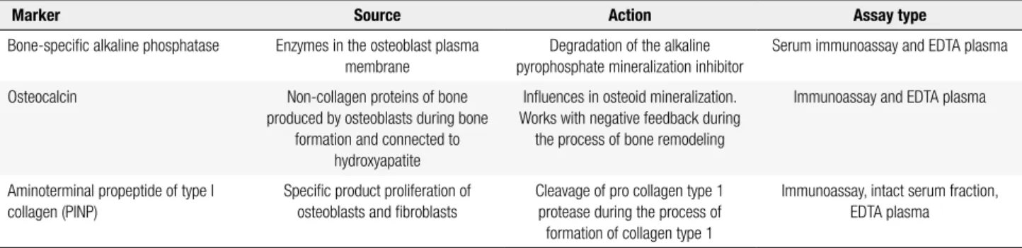
Table 2. Main bone formation markers
Marker Source Action Assay type
Bone-speciic alkaline phosphatase Enzymes in the osteoblast plasma membrane
Degradation of the alkaline pyrophosphate mineralization inhibitor
Serum immunoassay and EDTA plasma
Osteocalcin Non-collagen proteins of bone produced by osteoblasts during bone
formation and connected to hydroxyapatite
Inluences in osteoid mineralization. Works with negative feedback during the process of bone remodeling
Immunoassay and EDTA plasma
Aminoterminal propeptide of type I collagen (PINP)
Speciic product proliferation of osteoblasts and ibroblasts
Cleavage of pro collagen type 1 protease during the process of
formation of collagen type 1
Immunoassay, intact serum fraction, EDTA plasma
Table 3. Main bone resorption markers
Marker Source Action Assay type
C-telopeptide (CTX)* Isomerization of aspartyl beta that occurs in mature collagen
Cleavage of type 1 collagen by Cathepsin K in bone resorption
Immunoassay, measurable serum/urine, EDTA plasma
N-telopeptide amino-terminal portion of type 1 collagen
Bone collagen type 1 Cleavage of type 1 collagen by Cathepsin K in bone resorption
Immunoassay, urine, serum, EDTA plasma
Receptor activator Kappa-B ligand (RANKL)
Produced by osteoblasts, activated by B and T cells
Binds to RANK, which is expressed on osteoclasts and their precursors, stimulating their differentiation and
activation
Immunoassay or soluble forms in serum
Osteoprotegerin Secreted by osteoblasts RANKL receptor, reduces bone resorption by binding to RANK and
prevents osteoclastogenesis
Immunoassay in serum
Sclerostin Secreted mainly by osteocytes Antagonist of Wnt signaling, inhibits bone formation
Immunoassay in serum or plasma
* Beta CTX more useful in osteoporosis.
levels of P1NP (9). However, this association was not found for vertebral fractures. Similar results were found using collagen type I c-telopeptide (CTX) and bone-speciic alkaline phosphatase (BSAP). These results suggest that baseline BTM levels can inluence the ef-fectiveness of treatment with alendronate, especially at non-vertebral sites. Among patients without
osteopo-rosis, higher baseline levels of the 3 BTMs were associ-ated with a greater increase in hip bone mineral density (BMD) after treatment; in those with osteoporosis, higher baseline levels of P1NP were associated with a greater increase in vertebral BMD (10).
Cop
yright
© ABE&M t
odos os dir
eit
os r
eser
vados
.
in one or more BTMs were associated with large re-ductions in vertebral, non-vertebral and hip fractures. The greater the reduction in BTM, the lower the risk of fracture. This study showed that women in the alen-dronate group with a reduction of at least 30% of BSAP, had a lower risk of non-vertebral and hip fractures. This effect was as strong as the anti-fracture effect observed with changes in BMD in 1 year (11).
In the Vertebral Eficacy with Risedronate Therapy (VERT) study, it was observed that BSAP levels decline and reach a nadir of -33% from baseline values by year 3 (12). Postmenopausal women with at least one ver-tebral fracture who showed reductions in urinary CTX (mean -60% ) and NTX (mean -51%) at 3 and 6 months of treatment with risedronate, were signiicantly asso-ciated with reduction of vertebral and non-vertebral fractures risk after 3 years (12). In a subanalysis of the MOVER (MOnthly intraVenous ibandronatE ver-sus daily oral Risedronate) study, it was observed that the relative change in mean CTX and collagen type I cross-linked N-telopeptide (NTX) levels, in relation to the baseline, was similar with ibandronate 1 mg and risedronate, with a decrease in these levels at 3 months which remained below baseline levels by the end of the study. After 6 months, the mean reduction using 1 mg ibandronate was -67 and -53% for CTX and NTX, res-pectively (13).
Regarding zoledronic acid, data from the Health Outcomes and Reduced Incidence with Zoledronic Acid Once Yearly (HORIZON) study demonstrated that serum CTX, BSAP and urinary NTX levels were 59%, 30% and 58% lower, respectively, in the group treated with zoledronic acid compared to placebo. The effects of zolendronic acid on BTMs were similar to the other bisphosphonates (14).
In a study of postmenopausal women with low bone mass, using a different doses of zoledronic acid, there was a nadir in bone resorption markers in 1 month, with an average decrease of 65-83% in serum CTX and 50-69% in urinary NTX (15). These decreases tend to be dose-dependent, in agreement with previous reports that higher doses of bisphosphonates increase the dura-tion of drug acdura-tion. The reducdura-tion in BTMs remained at the end of the study (month 12), with an average de-crease of 49-52% in CTX and 54-65% in NTX. Serum osteocalcin and BSAP showed similar responses wi-thout strong apparent decline in one month, but with persistent suppression at the end of the study.
CHANGES IN BTM WITH STRONTIUM RANELATE
There is much controversy about BTMs during treat-ment with strontium ranelate (SR) (16, 17). In fact, the mechanism of action of SR is unknown. Although the presence of SR itself on bone matrix, leading to preser-vation of some determinants of bone material quality inluencing the intrinsic properties on bone tissues and an effect on the calcium-sensing receptor have all been proposed (18). In a recent post-hoc analysis of the core studies with SR, involving 2,373 women with postme-nopausal osteoporosis treated with SR or placebo, after 3 months of treatment, BSAP increased by 9.6% , se-rum C-propeptide of type I collagen (PICP) in 9.9%, s-CTX was reduced by 5.9%. Considering the technical and biological variability of BTM measurements, the-se changes could not be ascertained as signiicant in a given patient. On the other hand, the increments in BMD may be remarkable. After 3 years, mean BMD increased by 14.4% in the lumbar spine, 5.5% at the femoral neck and 7.1% at total hip. Multiple regres-sion analysis showed that changes in bone formation markers (PICP and BSAP), but neither in s-CTX nor u-NTX I, were signiicantly associated with increased BMD at the lumbar spine and femoral neck (19). The responses of BTM in women previously treated with long term bisphosphonates have been evaluated in re-cent studies and compared to those women who never took any osteoporosis medication before SR. Middle-ton and cols. (20) showed a positive, although delayed, responses after 2 years which were more pronounced in women previously treated with a bisphosphonate: se-rum CTX +61%, sese-rum P1NP +55% and sese-rum BSAP +46%. This data are in accordance with our own data on short-term changes and long-term changes in BMD in response to SR in postmenopausal women previously treated with long-term bisphosphonates. We observed a mean increase of 53.7% in serum CTX and 30.7% in osteocalcin levels and these changes were associated with a mean increase in lumbar spine BMD of 4.8% after 2.5 years of treatment with SR (21,22).
Cop
yright
© ABE&M t
odos os dir
eit
os r
eser
vados
.
more pronounced with tereparatide. Likewise, corti-cal porosity was also improved with both agent. Serum PINP increased at irst month with tereparatide, but it wasn’t measured at this time in SR group. As expected for bisphosphonate-naïve pacients bone turnover mark-ers decreased 3 and 6 months in the SR group (23).
CHANGES IN BTM WITH DENOSUMAB
Denosumab is a human monoclonal antibody with high afinity and speciicity for human RANK-L. By binding to RANK-L, denosumab prevents the interaction of RANK-L with RANK and, thus, inhibits bone resorp-tion. The reduction in resorption markers CTX, NTX and formation markers P1NP and BSAP is expected to occur as with any other potent anti-resorptive agent.
In the FREEDOM (Fracture REduction Evaluation of Denosumab in Osteoporosis every 6 Months) study, a clinical trial of postmenopausal women using Deno-sumab (60 mg every 6 months for 3 years) or placebo, there was a suppression of BTM within 12 hours after administration of the drug which persists until the next dose. Denosumab reduced bone resorption by an aver-age of 86% within 1 month, which is a greater reduc-tion seen than with other anti resorptive drugs. The de-crease in serum CTX was more pronounced and faster than the decreases in serum P1NP and BSAP. There was a signiicant correlation between serum CTX re-duction and increased BMD (24).
In FREEDOM, the median values for serum CTX were 0.049 g/mL (-90%) at day 10 and 0.131 ng/mL at mounth 6, after the irst extension dose (the seventh dose of denosumab). For serum P1NP, median values were 19.0 µg/L (-51%) at day 10 and 13.0 µg/L at mounth 6. In the 6 years of denosumab treatment without descon-tinue the drug, the median BTM values remained below the median values observed at FREEDOM baseline (25).
Taken together, these data suggest that biochemical markers of bone turnover are useful tools to assess the therapeutic effects of anti-catabolic or anti-resorptive osteoporosis medications and that serial measurements can help to decide whether a patient is responding or not to a speciic treatment.
CHANGES IN BTMS WITH OSTEOANABOLIC
THERAPY
Anabolic agents represent an important approach to the treatment of osteoporosis. They target bone
for-mation which distinguishes them from the mechanism of action of most of the drugs that we have available which have their primary action to inhibit bone resorp-tion. Parathyroid hormone (PTH) is currently the only approved osteoanabolic therapy for osteoporosis. It is available as the full-length molecule PTH (1-84) in Eu-rope and as the recombinant amino-terminal fragment PTH (1-34) (teriparatide) worldwide. Another promi-sing therapeutic osteoanabolic class is targeted against sclerostin. Monoclonal antibodies against sclerostin, romosozumab and blosozumab, are currently being developed and have advanced to clinical trials.
PTH
Cop
yright
© ABE&M t
odos os dir
eit
os r
eser
vados
.
notion that the adult human skeleton can model is a re-lative new one, because it was previously thought to be demonstrable only in the growing human skeleton. Es-timates have placed the percentage of bone accrual due to PTH-induced modeling at about 30%. When bone remodeling is stimulated, there is still greater bone for-mation than bone resorption, thus maintaining the ana-bolic window at a period of time beyond the modeling events (31).
The pattern of change in bone turnover markers with weekly teriparatide (56.5 ug/week) appears to be different from daily use of the drug. While changes in bone formation markers with weekly teriparatide are similar to the daily regimen (32,33), the u-NTX, re-lecting bone resorption falls, after an initial tendency to rise. Black and cols. have reported similar results us-ing weekly PTH (1-84) (34).
Many patients who are switched to PTH therapy have been taking an antiresorptive agent. A sub-group analysis of the EUROpean Study of FORSteo (EUROFORS) stratiied patients by previous antire-sorptive therapy (alendronate, risedronate, etidronate and non-bisphosphonate) before the switch to teripa-ratide. Regardless of previous antiresorptive use, bone formation markers P1NP and BSAP increased in all groups after 1 month of teriparatide treatment and con-tinued to rise through 6 months, although the incre-ment change varied according to previous antiresorp-tive treatment (35). In another study, Miller and cols. compared the effect of previous exposure to risedronate or alendronate in terms of subsequent effects of teripa-ratide (36). The hypothesis of this study was that the more potent effects of alendronate on bone turnover markers would lead to a slower onset of effect on bone formation markers, in comparison to risedronate. The results of this trial were consistent with this hypothesis.
In concept, the combination of an osteonabolic and an antiresorptive agent could be more beneicial than monotherapy with either drug alone, because the even-tual increase in bone resorption with teriparatide, which is believed to limit PTH’s effects, would be controlled by the antiresorptive agent. However, most of the stu-dies that have utilized combination therapy with PTH (1-84) or teriparatide and an antiresorptive have not been promising. Using alendronate as the antiresorp-tive agent, Black and cols. (37) studied postmenopausal women with 100 ug of PTH (1-84) while Finkelstein and cols. (38) studied men with 40 ug of teriparatide. In both cases, BTMs in the combination therapy group
mirrored the effects of monotherapy with alendronate, with reductions in bone formation and bone resorp-tion markers. It appeared that the potent antiresorp-tive effects of alendronate interfered with the potential actions of PTH (1-84) or teriparatide in this setting. This premise was tested by Deal and cols. (39) utili zing a less potent antiresorptive agent, raloxifene. In this study, the combination therapy arm showed an increase in bone formation markers similar to teriparatide alone. The increases in bone resorption markers, however, were reduced in comparison to teriparatide alone (39). The combination of teriparatide and risedronate in men also showed that BTMs mirrored the BTMs changes in the teriparatide-alone arm (40). The raloxifene (37) and risedronate (38) combination studies were small, and further data are needed, but they strengthened the idea that the anabolic window could be expanded by using combination therapy with teriparatide and an an-tiresorptive drug that does not have potent effects on bone resorption. Enforcing this notion is the combina-tion therapy study of teriparatide and zoledronic acid. The pattern of change in BTMs with this combination is different from the combinations previously described. With combination therapy, there was a rapid, but tran-sient, reduction in CTX during the irst 2 months, with a subsequent and gradual increase with levels remaining above baseline for last 6 months of the study. Levels of both markers were signiicantly lower with combina-tion therapy versus teriparatide alone (p < 0.002) (41).
Cop
yright
© ABE&M t
odos os dir
eit
os r
eser
vados
.
slower time course and to a lesser extent (combination therapy: osteocalcin: -39 ± 22%, denosumab monother-apy: -55 ± 20%). A 12-month extension of the DATA Study showed that osteocalcin continued to decrease in the combination group, but still to a lesser extent than denosumab monotherapy. CTX suppression re-mained similar in the denosumab and in the combina-tion therapy group (44). BMD increments during the two years of combination therapy with teriparatide and denosumab were higher than with either medication as monotherapy (44).
Sclerostin antibodies
Sclerostin is a glycoprotein product of the SOST gene and secreted mainly by osteocytes. It acts as an inhibi-tor of the anabolic Wnt signaling pathway by occupying the Wnt binding site at the LRP5/LRP6 complex (45-49). In the rare human diseases known as van Buchem or sclerosteosis, genetic mutations lead to loss of scle-rostin expression. These individuals have high bone mass and do not fracture (49,50). These human disea-ses were a clue that eventually led to the development of humanized monoclonal sclerostin antibodies. These antisclerostin antibodies allow Wnt signaling pathway to function unimpeded by sclerostin and thus serve as a new pharmacologic osteoanabolic class.
Romosozumab (AMG 785/CDP7851, being de-veloped by Amgen and UCB), is the irst humanized monoclonal antibody to sclerostin for which clinical trials results were published. In the phase 1 trial, changes in BMD and BTMs were evaluated in 72 healthy subjects (51). Subjects were randomized to receive a single dose of placebo or either one dose of romosozumab SC (reg-imens 0.1, 0.3, 1, 3, 5 or 10 mg/kg) or one dose of ro-mosozumab IV (regimens 1mg or 5 mg/kg). Bone for-mation markers P1NP, BSAP and osteocalcin showed a marked increase after a single SC injection, reaching maximum increments of 184%, 126% and 176% respec-tively with the 10 mg/kg SC, and 167%, 125%, 143% respectively with the 5 mg/kg IV regimen. Surpris-ingly, serum CTX decreased also in a dose-dependent manner, reaching nadirs of -54% with 10 mg/kg SC and -49% with 5 mg/kg IV (51). Ionized calcium con-centration showed a small (4%) but transient reduction with levels returning to baseline thereafter. PTH lev-els increased transiently in the romosozumab cohorts possibly relecting the changes in the ionized calcium concentration. In another phase I study, ascending
multiple-doses of romosozumab were administered for 12 weeks to healthy men and postmenopausal women with low bone mass (52). Patients were randomized to receive either placebo or one of the SC regimens of romosozumab [1 or 2 mg/kg once every 2 weeks (Q2W) or 2 or 3 mg/kg once every 4 weeks (Q4W)]. Bone formation markers P1NP, osteocalcin and BSAP increased in the romosozumab groups. P1NP showed the greatest increments in the irst 8 weeks of dosing compared to baseline and to smaller increases in the last 4 weeks of dosing, with levels returning to base-line within 4 to 8 weeks after the last romosozumab dose. Osteocalcin and BSAP changed in the same man-ner. P1NP levels increased by 106% and 147% in men, and by 83% and 129% in women at the romosozumab 1 mg/kg Q2W and at 3 mg/kg cohorts respectively. Changes in serum CTX levels were noteworthy for a decline with romosozumab treatment. Maximum re-ductions in CTX were -21% for placebo, and -35% and -37% for postmenopausal women in the romosozumab Q4W regimen at 2 or 3 mg/kg respectively. In men, CTX reached a nadir of -42% on the 1 mg/kg Q2W and -50% on the 3 mg/kg Q4W romosozumab cohorts (52). Changes in BTMs relected the salutary effects seen at BMD, with lumbar spine BMD increasing be-tween 4-7%, and total hip bebe-tween 2-3% in patients taking romosozumab. These studies were followed by the results of the phase II study, which enrolled 419 postmenopausal women with low BMD (T-score ≤ -2.0 and > -3.5 at lumbar spine, total hip or femoral neck (53). Romosozumab dosing regimens were: monthly SC doses (70 mg, 140 mg or 210 mg), or Q 3-month SC doses (140 mg or 210 mg) for a total of 12 months. The controls included placebo or one of 2 active com-parators: alendronate 70 mg weekly or teriparatide 20 µg SC daily. A rise in bone formation markers was noticed after one week of romosozumab treatment, reaching a peak after one month and decreasing thereafter to or below baseline levels (53). Conversely, CTX levels fell rapidly by the irst week of romosozumab with CTX levels remaining below baseline values at 12 months. Similarly to results of the phase 1 trial, a transient re-duction was noted in serum calcium concentration tak-ing romosozumab, along with an also transient increase in PTH levels. Phase III trials of romosozumab are cur-rently ongoing (54,55).
Cop
yright
© ABE&M t
odos os dir
eit
os r
eser
vados
.
phase I trials of blosozumab in postmenopausal women were reported. The irst trial was a single-dose study, randomized, subject- and investigator blind, placebo-controlled, single-dose, dose-escalation study. A total of 60 subjects were randomized to 8 cohorts: 5 cohorts received IV regimens of blosozumab in a dose range (7.5, 25, 75, 225, and 750 mg). At each dose level during the dose-escalation phase, subjects received blosozumab or placebo. Another cohort received either blosozumab 150 mg or placebo SC. The other two co-horts with prior exposure to bisphosphonate received either 225 mg or 750 mg of IV blosozumab (56). The second phase I trial was a multiple-dose, multi-center, randomized, subject- and investigator blinded, placebo-controlled, parallel design study, in which 59 patients were randomized to either placebo or bloso-zumab SC (270 mg) or IV (750 mg) once every 2 weeks (Q2W), or blosozumab SC (180 or 270 mg) or IV 540 mg once every 4 weeks (Q4W ) for 8 weeks. Dose-dependent responses were noted in BTMs levels of patients who received blosozumab. Bone formation markers OC, BSAP and P1NP increased in patients in the single- and multiple-dose regimens. CTX decreased after a single SC or IV dose of blosozumab, also in a dose-dependent manner, returning thereafter to base-line levels. In the trial with repeated doses of bloso-zumab, the reduction in CTX did not appear to have a clear dose-response relationship, but CTX levels re-turned to baseline except for patients on the 750 mg IV arm where CTX levels increased to nearly 100% above baseline. Total sclerostin (free and blosozumab-bound sclerostin) increased, with levels later decreasing in the same manner as the decline in blosozumab concentra-tion. The similarity in this kinetics could relect levels of sclerostin predominantly bound to blosozumab. BTMs in the subset of patients with previous bisphosphonate exposure showed similar dose-dependent responses, al-though with a lower magnitude (56).
CONCLUSION
BTMs have the potential to be useful monitoring to-ols in osteoporosis therapeutic regimens. Their change following treatment with an anti-absorptive or osteoa-nabolic agent may relect a bone effect and should be considered in evaluating failure to therapy, along with BMD reductions and the presence of a fracture. Con-sidering the signiicant change at 95% conidence, de-crease of at least 25% following an anti-resorptive and
an increase of at least 25% following an osteoanabolic agent are the minimal change required (57)
The data are still insuficient to relate BTMs changes alone to the outcome index of fracture risk reduction.
Disclosures: Dr. Francisco Bandeira is a consultant for Servier and Sanoi and receives research support from Amgen. Dr. John P. Bilezikian is a consultant for Amgen, Eli Lilly, Radius, NPS Pharmaceuticals, Merck, and GSK, and receives research support from NPS Pharmaceuticals and Amgen. Drs. Aline G. Costa, Ma-noel Anderson Soares and Lourena Lima: no conlicts of interest reported.
REFERENCES
1. Datta HK, Ng WF, Walker JA, Tuck SP, Varanasi SS. The cell biology of bone metabolism. J Clin Pathol. 2008;61:577-87.
2. Carey JJ, Licata AA, Delaney MF. Biochemical markers of bone turnover. Clinic Rev Bone Miner Metab. 2006;4:197-212.
3. Vega D, Maalouf NM, Sakhaee K. CLINICAL Review #: the role of receptor activator of nuclear factor-kappaB (RANK)/RANK ligand/ osteoprotegerin: clinical implications. J Clin Endocrinol Metab. 2007;92:4514-21.
4. Leibbrandt A, Penninger JM. RANK/RANKL: regulators of immune responses and bone physiology. Ann NY Acad Sci. 2008;1143:123-50. 5. National Institute for Health and Care Excellence 2014. Available
at: www.guidance.nice.org.uk/TA160.
6. Khosla S, Melton LJ, 3rd, Riggs BL. The unitary model for estro-gen deiciency and the pathoestro-genesis of osteoporosis: is a revi-sion needed? J Bone Miner Res. 2011;26:441-51.
7. Wheater G, Elshahaly M, Tuck SP, Datta HK, van Laar JM. The clini-cal utility of bone marker measurements in osteoporosis. J Trans Med. 2013;11:201.
8. Lee J, Vasikaran S. Current recommendations for laboratory test-ing and use of bone turnover markers in management of osteo-porosis. Ann Lab Med. 2012;32:105-12.
9. Bauer DC, Garnero P, Hochberg MC, Santora A, Delmas P, Ewing SK, et al.; Fracture Intervention Research Group. Pretreatment le-vels of bone turnover and the antifracture eficacy of alendronate: the fracture intervention trial. J Bone Miner Res. 2006;21:292-9. 10. Keegan TH, Schwartz AV, Bauer DC, Sellmeyer DE, Kelsey JL;
fracture intervention trial. Effect of alendronate on bone min-eral density and biochemical markers of bone turnover in type 2 diabetic women: the fracture intervention trial. Diabetes Care. 2004;27:1547-53.
11. Bauer DC, Black DM, Garnero P, Hochberg M, Ott S, Orloff J, et al.; Fracture Intervention Trial Study G. Change in bone turn-over and hip, non-spine, and vertebral fracture in alendronate-treated women: the fracture intervention trial. J Bone Miner Res. 2004;19:1250-8.
12. Harris ST, Watts NB, Genant HK, McKeever CD, Hangartner T, Keller M, et al. Effects of risedronate treatment on vertebral and nonvertebral fractures in women with postmenopausal osteopo-rosis: a randomized controlled trial. Vertebral Eficacy With Rise-dronate Therapy (VERT) Study Group. JAMA. 1999;282:1344-52. 13. Nakamura T, Nakano T, Ito M, Hagino H, Hashimoto J, Tobinai M,
Cop
yright
© ABE&M t
odos os dir
eit
os r
eser
vados
.
14. Black DM, Delmas PD, Eastell R, Reid IR, Boonen S, Cauley JA, et al.; HORIZON Pivotal Fracture Trial. Once-yearly zoledronic acid for treatment of postmenopausal osteoporosis. N Engl J Med. 2007;356:1809-22.
15. Reid IR, Brown JP, Burckhardt P, Horowitz Z, Richardson P, Trechsel U, et al. Intravenous zoledronic acid in postmenopausal women with low bone mineral density. N Engl J Med. 2002;346:653-61. 16. Reginster JY, Seeman E, De Vernejoul MC, Adami S, Compston J,
Phenekos C. Strontium ranelate reduces the risk of nonvertebral fractures in postmenopausal women with osteoporosis: Treat-ment of Peripheral Osteoporosis (TROPOS) study. J Clin Endocri-nol Metab. 2005;90:2816-22.
17. Meunier PJ, Roux C, Seeman E, Ortolani S, Badurski JE, Spector TD, et al. The effects of strontium ranelate on the risk of vertebral fracture in women with postmenopausal osteoporosis. N Engl J Med. 2004;350:459-68.
18. Chavassieux P, Meunier PJ, Roux JP, Portero-Muzy N, Pierre M, Chapurlat R. Bone histomorphometry of transiliac paired bone biopsies after 6 or 12 months of treatment with oral strontium ranelate in 387 osteoporotic women: randomized comparison to alendronate. J Bone Miner Res. 2014;29(3):618-28.
19. Bruyère O, Collette J, Rizzoli R, Decock C, Ortolani S, Cormier C, et al. Relationship between 3-month changes in biochemical mark-ers of bone remodelling and changes in bone mineral density and fracture incidence in patients treated with strontium ranelate for 3 years. Osteoporos Int. 2010;21:1031-6.
20. Middleton ET, Steel SA, Aye M, Doherty SM. The effect of prior bisphosphonate therapy on the subsequent therapeutic effects of strontium ranelate over 2 years. Osteoporos Int. 2012;23:295-303. 21. Sousa IO, Diniz ET, Marques TF, Griz L, Coutinho Mde A, Bandeira
F. Short-term bone marker responses to teriparatide and stron-tium ranelate in patients with osteoporosis previously treated with bisphosphonates. Arq Bras Endocrinol Metab. 2010;54:244-9. 22. Lima H, Maia J, Bandeira F. Trajectories of bone remodeling markers and bone mineral density during treatment with strontium ranelate in postmenopausal women previously treated with bisphospho-nates. Clin Med Insights Endocrinol Diabetes. 2014;7:7-11. 23. Recker RR, Marin F, Ish-Shalom S, Möricke R, Hawkins F,
Ka-petanos G, et al. Comparative efects of tereparatide and stron-tium ranelate on bone biopsies and biochemical markers of bone turnover in postmenopausal women with osteoporosis. J Bone Miner Res. 2009;24:1358-68.
24. Cummings SR, San Martin J, McClung MR, Siris ES, Eastell R, Reid IR, et al.; FREEDOM Trial. Denosumab for prevention of frac-tures in postmenopausal women with osteoporosis. N Engl J Med. 2009;361:756-65.
25. Bone HG, Chapurlat R, Brandi ML, Brown JP, Czerwinski E, Krieg MA, et al. The effect of three or six years of denosumab exposure in women with postmenopausal osteoporosis: results from the FREE-DOM extension. J Clin Endocrinol Metab. 2013;98(11):4483-92. 26. Nakamura Y. [Evidence in the treatment of osteoporosis using
weekly teriparatide injections]. Clin Calcium. 2012;22:407-13. 27. Chen P, Satterwhite JH, Licata AA, Lewiecki EM, Sipos AA,
Misur-ski DM, et al. Early changes in biochemical markers of bone for-mation predict BMD response to teriparatide in postmenopausal women with osteoporosis. J Bone Miner Res. 2005;20:962-70. 28. Orwoll ES, Scheele WH, Paul S, Adami S, Syversen U, Diez-Perez
A, et al. The effect of teriparatide [human parathyroid hormone (1-34)] therapy on bone density in men with osteoporosis. J Bone Miner Res. 2003;18:9-17.
29. Dempster DW, Cosman F, Kurland ES, Zhou H, Nieves J, Woelfert L, et al. Effects of daily treatment with parathyroid hormone on bone microarchitecture and turnover in patients with osteoporo-sis: a paired biopsy study. J Bone Miner Res. 2001;16:1846-53.
30. Bilezikian JP. Combination anabolic and antiresorptive therapy for osteoporosis: opening the anabolic window. Curr Osteoporos Rep. 2008;6:24-30.
31. Lindsay R, Cosman F, Zhou H, Bostrom MP, Shen VW, Cruz JD, et al. A novel tetracycline labeling schedule for longitudinal evalu-ation of the short-term effects of anabolic therapy with a single iliac crest bone biopsy: early actions of teriparatide. J Bone Miner Res. 2006;21:366-73.
32. Sugimoto T, Nakamura T, Nakamura Y, Isogai Y, Shiraki M. Proile of changes in bone turnover markers during once-weekly teripa-ratide administration for 24 weeks in postmenopausal women with osteoporosis. Osteoporos Int. 2014;25:1173-80.
33. Nakamura T, Sugimoto T, Nakano T, Kishimoto H, Ito M, Fukunaga M, et al. Randomized Teriparatide [human parathyroid hormone (PTH) 1-34] Once-Weekly Eficacy Research (TOWER) trial for ex-amining the reduction in new vertebral fractures in subjects with primary osteoporosis and high fracture risk. J Clin Endocrinol Metab. 2012;97:3097-106.
34. Black DM, Bouxsein ML, Palermo L, McGowan JA, Newitt DC, Rosen E, et al.; PTH Once-Weekly Research (POWR) Group. Ran-domized trial of once-weekly parathyroid hormone (1-84) on bone mineral density and remodeling. J Clin Endocrinol Meatb. 2008;93:2166-72.
35. Boonen S, Marin F, Obermayer-Pietsch B, Simões ME, Barker C, Glass EV, et al.; EUROFORS Investigators. Effects of previous an-tiresorptive therapy on the bone mineral density response to two years of teriparatide treatment in postmenopausal women with osteoporosis. J Clin Endocrinol Metab. 2008;93:852-60.
36. Miller PD, Delmas PD, Lindsay R, Watts NB, Luckey M, Adachi J, et al,; Open-label Study to Determine How Prior Therapy with Alendronate or Risedronate in Postmenopausal Women with Os-teoporosis Inluences the Clinical Effectiveness of Teriparatide In-vestigators. Early responsiveness of women with osteoporosis to teriparatide after therapy with alendronate or risedronate. J Clin Endocrinol Metab. 2008;93:3785-93.
37. Black DM, Greenspan SL, Ensrud KE, Palermo L, McGowan JA, Lang TF, et al; PaTH Study Investigators. The effects of parathyroid hormone and alendronate alone or in combination in postmeno-pausal osteoporosis. N Engl J Med. 2003;349:1207-15.
38. Finkelstein JS, Hayes A, Hunzelman JL, Wyland JJ, Lee H, Neer RM. The effects of parathyroid hormone, alendronate, or both in men with osteoporosis. N Engl J Med. 2003;349:1216-26. 39. Deal C, Omizo M, Schwartz EN, Eriksen EF, Cantor P, Wang J, et
al. Combination teriparatide and raloxifene therapy for post-menopausal osteoporosis: results from a 6-month double-blind placebo-controlled trial. J Bone Miner Res. 2005;20:1905-11. 40. Walker MD, Cusano NE, Sliney J Jr, Romano M, Zhang C,
McMa-hon DJ, et al. Combination therapy with risedronate and teripara-tide in male osteoporosis. Endocrine. 2013;44:237-46.
41. Cosman F, Eriksen EF, Recknor C, Miller PD, Guañabens N, Kasperk C, et al. Effects of intravenous zoledronic acid plus subcutaneous teriparatide [rhPTH(1-34)] in postmenopausal osteoporosis. J Bone Miner Res. 2011;26:503-11.
42. Tsai JN, Uihlein AV, Lee H, Kumbhani R, Siwila-Sackman E, McK-ay EA, et al. Teriparatide and denosumab, alone or combined, in women with postmenopausal osteoporosis: the DATA study ran-domised trial. Lancet. 2013;382:50-6.
43. Leder B, Uihlein A, Tsai J, Neer R, Siwila-Sackman E, Zhu Y, et al. The DATA Extension Study: Two years of combined denosumab and teriparatide in postmenopausal women with osteoporosis: a randomized controlled trial. J Bone Miner Res. 2013;28 (Suppl 1); http://www.asbmr.org/education/AbstractDetail?aid=c6aff623-b458-4737-4738c4733f-2381c4730a9217e
Cop
yright
© ABE&M t
odos os dir
eit
os r
eser
vados
.
postmenopausal women with osteoporosis (The DATA Extension Study): a randomized controlled trial. J Clin Endocrinol Metab. 2014;99:1694-700.
45. Krause C, Korchynskyi O, de Rooij K, Weidauer SE, de Gorter DJ, van Bezooijen RL, et al. Distinct modes of inhibition by sclerostin on bone morphogenetic protein and Wnt signaling pathways. J Biol Chem. 2010;285:41614-26.
46. Li X, Zhang Y, Kang H, Liu W, Liu P, Zhang J, et al. Sclerostin binds to LRP5/6 and antagonizes canonical Wnt signaling. J Biol Chem. 2005;280:19883-7.
47. Semenov M, Tamai K, He X. SOST is a ligand for LRP5/LRP6 and a Wnt signaling inhibitor. J Biol Chem. 2005;280:26770-5.
48. van Bezooijen RL, Svensson JP, Eefting D, Visser A, van der Horst G, Karperien M, et al. Wnt but not BMP signaling is involved in the inhibitory action of sclerostin on BMP-stimulated bone formation. J Bone Miner Res. 2007;22:19-28.
49. Van Buchem FS, Hadders HN, Ubbens R. An uncommon familial systemic disease of the skeleton: hyperostosis corticalis genera-lisata familiaris. Acta radiologica. 1955;44:109-20.
50. Truswell AS. Osteopetrosis with syndactyly; a morphological variant of Albers-Schonberg’s disease. J Bone Joint Surg Br. 1958;40-B:209-18.
51. Padhi D, Jang G, Stouch B, Fang L, Posvar E. Single-dose, place-bo-controlled, randomized study of AMG 785, a sclerostin mono-clonal antibody. J Bone Miner Res. 2011;26:19-26.
52. Padhi D, Allison M, Kivitz AJ, Gutierrez MJ, Stouch B, Wang C, et al. Multiple doses of sclerostin antibody romosozumab in healthy men and postmenopausal women with low bone mass: a ran-domized, double-blind, placebo-controlled study. J Clin Pharma-col. 2013 Nov 23. doi: 10.1002/jcph.239. [Epub ahead of print] 53. McClung MR, Grauer A, Boonen S, Bolognese MA, Brown JP,
Diez-Perez A, et al. Romosozumab in postmenopausal women with low bone mineral density. N Engl J Med. 2014;370:412-20. 54. Study of romosozumab (AMG 785) administered to healthy
sub-jects and subsub-jects with stage 4 renal impairment or stage 5 re-nal impairment requiring hemodialysis. Available at: http://www. clinicaltrials.gov/ct2/show/NCT01833754?term=Romosozumab&r ank=1. Acessed on Nov 20, 2013.
55. Study to determine the eficacy and safety of romosozumab in the treatment of postmenopausal women with osteoporosis. Available at: http://www.clinicaltrials.gov/ct2/show/NCT01631214 ?term=Romosozumab&rank=5. Acessed on: Nov 20, 2013. 56. McColm J, Hu L, Womack T, Tang CC, Chiang AY. Single- and
mul-tiple-dose randomized studies of blosozumab, a monoclonal an-tibody against sclerostin, in healthy postmenopausal women. J Bone Miner Res. 2014;29(4):935-43.