The role of daily physical activity and nutritional status
on bone turnover in cystic fibrosis:
a cross-sectional study
Sergio Tejero1,2, Pilar Cejudo2, E. Quintana-Gallego2, Borja Sañudo1, A. Oliva-Pascual-Vaca1
ABSTRACT | Background: Nutritional status and daily physical activity (PA) may be an excellent tool for the maintenance
of bone health in patients with cystic ibrosis (CF). Objective: To evaluate the relationship between nutritional status,
daily physical activity and bone turnover in cystic ibrosis patients. Method: A cross-sectional study of adolescent and
adult patients diagnosed with clinically stable cystic ibrosis was conducted. Total body, femoral neck, and lumbar spine bone mineral density (BMD) were determined by dual energy X-ray absorptiometry and bone metabolism markers ALP, P1NP, PICP, and ß-CrossLaps. PA monitoring was assessed for 5 consecutive days using a portable device. Exercise capacity was also determined. Serum 25-hydroxyvitamin D and vitamin K were also determined in all participants. Results: Fifty patients (median age: 24.4 years; range: 16-46) were included. BMI had positive correlation with all BMD parameters, with Spearman’s coeficients ranging from 0.31 to 0.47. Total hip bone mineral density and femoral neck BMD had positive correlation with the daily time spent on moderate PA (>4.8 metabolic equivalent-minutes/day; r=0.74, p<0.001 and r=0.72 p<0.001 respectively), daily time spent on vigorous PA (>7.2 metabolic equivalent-minutes/day; r=0.45 p<0.001), body mass index (r=0.44, p=0.001), and muscle mass in limbs (r=0.41, p=0.004). Levels of carboxy-terminal propeptide of type 1 collagen were positively associated with the daily time spent on moderate (r=0.33 p=0.023) and vigorous PA (r=0.53, p<0.001). Conclusions: BMI and the daily time spent on moderate PA were found to be correlated
with femoral neck BMD in CF patients. The association between daily PA and biochemical markers of bone formation suggests that the level of daily PA may be linked to bone health in this patient group. Further research is needed to conirm these indings.
Keywords: cystic ibrosis; physical therapy; rehabilitation; nutritional status; bone remodeling; bone mineral density.
1 Departement of Phisical Therapy, Universidad de Sevilla (US), Sevilla, Spain 2 Department of Trauma and Orthopedic Surgery, HU Virgen del Rocío, Sevilla, Spain Received: Apr. 20, 2015 Revised: Sept. 23, 2015 Accepted: Nov. 23, 2015
BULLET POINTS
• Physical therapy in cystic ibrosis patients is correlated with bone health. • Bone health can improve with physical activity.
HOW TO CITE THIS ARTICLE
García ST, Cejudo P, Quintana-Gallego E, Sañudo B, Oliva-Pascual-Vaca A. The role of daily physical activity and nutritional status on bone turnover in cystic ibrosis: a cross-sectional study. Braz J Phys Ther. 2016 May-June; 20(3):206-212 . http://dx.doi.org/10.1590/bjpt-rbf.2014.0154
Introduction
Thanks to advances in the treatment and care of patients with cystic ibrosis (CF), the life expectancy1 of these patients has increased. However, this fact has not resulted in quality of life because different comorbidities have been observed in adulthood. Disturbances in bone metabolism, particularly signiicant reductions in bone mineral density (BMD), are recognized as additional and serious complications in these CF patients, with prevalence ranging from 40% to 70%2,3. The low
BMD found in these patients is multifactorial and many factors have been suggested as explanation4-10. One of these explanations is based on intestinal
malabsorption, with vitamin D deiciency caused by exocrine pancreatic insuficiency4. Evidence suggests
that the absorption of vitamins D and K may not be suficient to ensure proper skeletal mineralization in CF adolescents5. Another bone metabolic disorder
found in CF patients is imbalance in bone turnover,
with decreased bone formation as the predominant change and increased bone resorption in infective exacerbation or severe lung disease6-8. Moreover,
for lung disease and helps to explain the variability of BMD9. This correlation is explained in part because
severe lung disease adversely affects exercise capacity.
When monitoring daily physical activity (PA), it was noted that the most active patients have greatest
bone mass, conirmed by exercise capacity test10. Nevertheless, there is no conirmation of whether
bone turnover has a relationship with PA and its
intensity or with nutritional status. This study was
designed to evaluate the relationship between daily level of PA, nutritional status, and bone parameters
(BMD and biomarkers of bone turnover) in a sample of adolescents and adults with CF.
Method
Participants and study design
We conducted a cross-sectional study that included
patients over 16 years of age diagnosed with CF and attending our Cystic Fibrosis Unit. All participants exhibited high sweat chloride levels (>60 mmol/L), had characteristic clinical features of CF, and their diagnoses were conirmed by repeated genetic analysis. Patients who had undergone lung transplantation and/or had a recent acute exacerbation requiring treatment with antibiotics (within 6 weeks prior to the study) were excluded. The Ethics Committee of the Virgen del Rocío Hospital, Sevilla, Spain approved this study (approval number 14/2008) and written informed consent was obtained from all participants.
Maximal cardiopulmonary exercise testing
In order to evaluate the tolerance to exercise, maximal cardiopulmonary exercise testing was performed with
a MasterScreen CPX cycle ergometer model Via Sprint 150 (ViaSys Healthcare, Hoechberg, Germany), as
previously described10 and according to the international standard11. The test is a symptom-limited exercise and involves measurements of respiratory oxygen
uptake (VO2), carbon dioxide production (VCO2), and ventilatory measures. It was performed with
electrocardiographic, heart rate, and pulse oximetry
monitoring. All measurements were integrated into the
cycle ergometer device and evaluated simultaneously
during the test. On completion of the exercise test,
heart rate, blood pressure, leg fatigue, chest pain, and
dyspnea were assessed using the modiied Borg scale.
Physical activity monitoring
Physical activity was measured in all participants
on ive consecutive days using a portable physical activity and lifestyle monitor (SenseWear Armband,
BodyMedia Inc., Pittsburgh, PA, USA), which estimates a person’s rest and exercise energy expenditure.
The accelerometer was positioned on the right brachial
triceps and was only removed for showering12.
The records were considered valid if the mean wearing
time was ≥600 min/day in the 5-day period and at least one of the ive days landed on a weekend. The use of
this device has been validated against doubly labeled water13. The time (in minutes) spent with an energy
expenditure of >3 metabolic equivalent-minutes/day (METs) was considered “mild” activity, time spent at >4.8 METs was considered “moderate” activity, and when energy expenditure was of >7.2 METs, it was considered “vigorous” activity, as suggested by the U.S. Department of Health and Human Services14.
The average number of daily steps at each intensity
level was calculated in all participants.
Nutritional assessment
The values for lean muscle mass and fat mass were assessed in the extremities by dual energy X-ray absorptiometry15 (DXA). Body mass index, serum
albumin, and serum levels of vitamins D and K were determined in all participants. For biochemical assays,
blood samples were drawn in the morning after an
overnight fast. Concentrations of 25-hydroxyvitamin D (25OHD) were determined by high-performance liquid chromatography (HPLC) on an Agilent 1100 HPLC system (Agilent Technologies Inc., Santa Clara, CA, USA). Plasma vitamin K concentrations were assessed by means of HPLC.
Bone mineral density and bone turnover biomarkers
Total body, femoral neck, and lumbar spine (L1-L4) BMD were determined by dual-energy X-ray absorptiometry (Hologic QDR X-ray 4500W Bone Densitometer). DXA results were expressed in absolute BMD units (grams per square centimeter), T-scores, and Z-scores. The T-score was deined as
the number of standard deviations above or below the mean for a healthy 20-year-old adult of the same sex
and ethnicity as the patient. The Z-score was deined as the number of standard deviations a patient’s BMD
differs from the average BMD of their age, sex, and ethnicity16.
Serum levels of β-CrossLaps and amino-terminal
propeptide of type 1 collagen (P1NP) were measured
serum levels of carboxy-terminal propeptide of type 1
collagen (P1CP) were assayed using ELISA (Quidel Corporation, San Diego, CA, USA) on an automated TRITURUS processor (Grifols International S.A., Barcelona, Spain).
Statistical analysis
The simple size calculation was estimated using
the Dixon and Mansey formula17. This sample size
was calculated using a power of 80% and a type I error of 5%.
Continuous data were expressed as medians and interquartile ranges (Q3- Q1) and categorical data were expressed with relative frequencies. We performed a Mann-Whitney U test and Chi-Square test for comparisons between sex groups. The associations
between functional and structural parameters were tested
using the non-parametric Rho Spearman correlation. Spearman’s rank correlation can be interpreted in
terms of the amount of mutual information between
two variables. An observed value of ρ is signiicantly different from zero (r will always maintain −1≤r≤1).
We calculated the probability that it would be greater
than or equal to the observed r, given the null hypothesis, by using a permutation test. An advantage of this approach is that it automatically takes into account
the number of tied data values in the sample and the
way they are treated in computing the rank correlation. All tests were performed using 0.2 and 0.05 as the
probability of a Type II error and probability of a
Type I error, respectively.
All statistical calculations were performed using
the SPSS 16.0 statistical software package (SPSS Inc., Chicago, IL, USA). Two-tailed p<0.05 were considered statistically signiicant.
Results
We included 50 patients in our sample (23 males and 27 females) with a median age 24.4 (interquartile range - IQR=5.8) years. A total of 48 patients (96%) were undergoing oral corticosteroid treatment. Two of the patients (4%) had been diagnosed with
allergic bronchopulmonary aspergillosis (ABPA) and were being treated intermittently with prednisone
(10 mg/day per os) for the last three years, and 23 patients (96%) had also been receiving inhaled corticosteroids (budesonide) at doses of 200-400 μg administered twice a day. All patients were prescribed vitamin supplementation (Vitamin A 1.50 mg, Vitamin
D 0.01 mg, and Vitamin E 200 mg). There was no documented secondary hepatopathy in any patient.
The male patients showed more wattage, 130 vs 100 (p=0.001) and spent more time on a moderate activity 24 vs 11.6 (p=0.045). There was no difference in the remaining parameters between sex groups. The general
characteristics of the study participants are shown in Table 1.
Nutritional status, bone mineral density, and markers
Body mass index (BMI) showed a positive correlation
with all of the BMD parameters, with Spearman’s coeficient ranging from 0.31 to 0.47. Muscle mass had a positive relationsip with all markers ranging from 0.48 to 0.43 for bone formation markers and 0.33 for serum levels of β-CrossLaps. In contrast,
the percentage of fat mass had a negative correlation
with the formation and resorption markers, ranging from –0.57 to –0.34. A positive correlation was found between 25-hydroxyvitamin D and whole-body Z-scores (r=0.34, p=0.03).
Daily physical activity, bone mineral density, and markers
Wattage showed a positive correlation with all
BMD parameters. Spearman’s coeficient, ranging from 0.30 to 0.49, also showed a weak positive correlation with ALP (r=0.29 p=0.04).
All variables of daily physical activity (PA) had a
positive correlation with at least two BMD parameters.
The strongest correlation was found between the average number of minutes spent on moderate daily
PA (>4.8 METs) and hip BMD (r=0.74 p<0.001)
(Table 2). Remodeling markers only had a positive correlation with moderate and vigorous daily PA with
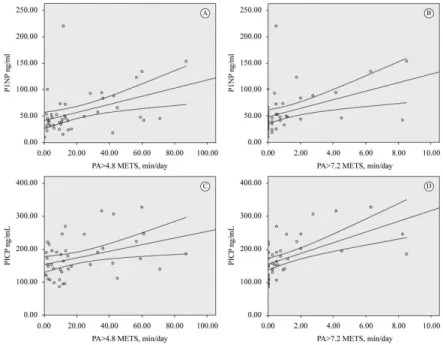
a higher coeficient in vigorous activity and formation markers. The relationship of bone formation markers with 4.8 METs and 7.2 METs is presented in Figure 1.
Discussion
The foundations for lifetime bone health are
established from infancy to adolescence and require adequate nutrition, body mass, PA, and hormone
production18. CF compromises the gains during puberty when growth and mineral accrual are most rapid19. The values of BMD found in our CF patients
are consistent with previous indings2. Our results show that patients diagnosed with CF with high levels
Table 1. General characteristics of patients with cystic ibrosis stratiied according to sex.
Total Cohort (N=50) Males (n=23) Females (n=27) p
Demographic data
Age years (IQR) 24.5 (19-28) 25 (19-30) 24 (20-27) 0.59 Corticosteroids 48 (96) 22 (95.6) 26 (96.3) 1.00 BMI Kg/m2 (IQR) 20.5 (18.8-21.9) 20.6 (18.9-22.8) 19.9 (18.7-21.6) 0.30
ΔF508 mutation 33 (66.0) 16 (69.6) 17 (63.0) 0.62 ΔF508 homocigosis 15 (30.0) 8 (34.8) 7 (25.9) 0.50 ΔF508 heterocigosis 18 (36.0) 8 (34.8) 10 (37.0) 0.87 R334W mutation 10 (20.0) 6 (26.1) 4 (14.8) 0.32 Exocrine pancreatic insuficiency 36 (72.0) 15 (65.2) 21 (77.8) 0.32
Exercise tolerance
FEV1% predicted (IQR) 60.3 (35.2-80.9) 47.4 (35.2-80.8) 60.7 (32.6-82.6) 1.00
FVC % predicted (IQR) 78.0 (59.3-95.3) 77 (59.3-93.5) 78.6 (58.1-99.9) 0.90 VO2max% (IQR) 66.5 (58.0-81.5) 65 (58-75) 72 (60.9-89.0) 0.22
Wmax (IQR) 110 (80-130) 130 (110-150) 100 (80-110) 0.001
6MWT distance meters (IQR) 640 (582-679) 647 (620-695) 615 (562-675) 0.05 Daily physical activity
Device wearing min/day (IQR) 618 (598-637) 614 (598-643) 622 (589-634) 0.63 PA >3 METs min/day (IQR) 152 (107-231) 154 (117-239) 121 (97-230) 0.37 PA >4.8 METs min/day (IQR) 11.6 (5.5-35.4) 24 (6.25-56.55) 10.2 (3.5-16.6) 0.04
PA >7.2METs min/day (IQR) 0.5 (0-2.2) 0.75 (0.25-2.0) 0.16 (0-1.12) 0.05 Steps/day (IQR) 8,361 (5,662-10,322) 8,859 (5,676-12,472) 7,972 (5,294-9,660) 0.22
Osteoporosis
Z score <1 SD (%) 25 (51.0) 12 (52.2) 13 (48.1) 0.45 Z score <2 SD (%) 2 (4) 2 (8.7) 0 (0) 0.22
IQR: interquartile range; BMI: body mass index; FEV1: forced expiratory volume in one second; FVC: forced vital capacity; VO2max% theoretical:
maximal oxygen consumption; Wmax (watts): maximal power output; PA: physical activity; METs: metabolic equivalents (minutes/day). Data given as median (IQR).
Figure 1. Correlation beetween bone formation markers and daily physical activity. (A) PA>4.8 METs min/day versus P1NP ng/mL,
serum levels of bone formation markers. The bone remodeling markers in our sample were signiicantly
different from the reference values of the normal
adult population.
Routine screening for reduced BMD using
dual energy X-ray absorptiometry (DXA) scans is recommended as detailed in published guidelines2,20. The recommendation is for this procedure to be
performed every 5 years, 2 years, or 1 year, depending on Z-score values in young adults. Bone loss has been
observed in several longitudinal studies in young
adults with CF21,22 reporting annualized losses in BMD of 0.5 to 2.1%. This signiicant loss requires a more
thorough investigation. The positive correlation found
between BMI and the BMD values was in agreement with several previous studies9,23 and suggests that improving vigilance in the periods between DXA
scans could result in an alternative marker.
Concerning vitamins, the normal rate of 25OHD in these patients was 20-40 ng/mL. We only found a weak positive relationship between 25OHD and
BMD values, although all patients had vitamin D
supplementation. Recent meta-analyses show that
vitamin D supplementation increases the levels of vitamin D in the blood but does not improve BMD values24. This suggests that recommended vitamin
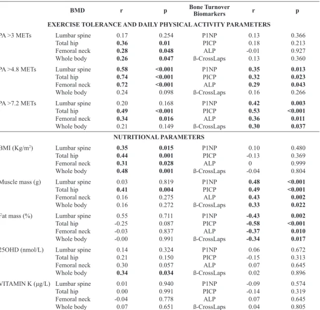
Table 2. Spearman’s correlations of exercise tolerance parameters, daily physical activity, and nutritional parameters with bone mineral
density and bone turnover biomarkers in adult CF patients.
BMD r p Bone TurnoverBiomarkers r p
EXERCISE TOLERANCE AND DAILY PHYSICAL ACTIVITY PARAMETERS
PA >3 METs Lumbar spine
Total hip Femoral neck Whole body 0.17 0.36 0.28 0.26 0.254 0.01 0.048 0.047 P1NP PICP ALP ß-CrossLaps 0.13 0.18 -0.01 0.13 0.366 0.213 0.927 0.360 PA >4.8 METs Lumbar spine
Total hip Femoral neck Whole body 0.58 0.74 0.72 0.24 <0.001 <0.001 <0.001 0.098 P1NP PICP ALP ß-CrossLaps 0.35 0.32 0.29 0.16 0.013 0.023 0.043 0.266 PA >7.2 METs Lumbar spine
Total hip Femoral neck Whole body 0.20 0.49 0.34 0.21 0.168 <0.001 0.016 0.149 P1NP PICP ALP ß-CrossLaps 0.42 0.53 0.36 0.30 0.003 <0.001 0.011 0.037 NUTRITIONAL PARAMETERS
BMI (Kg/m2) Lumbar spine
Total hip Femoral neck Whole body 0.35 0.44 0.31 0.48 0.015 0.001 0.028 0.001 P1NP PICP ALP ß-CrossLaps 0.10 -0.13 0 -0.04 0.480 0.369 0.999 0.804
Muscle mass (g) Lumbar spine Total hip Femoral neck Whole body 0.03 0.41 0.16 0.16 0.819 0.004 0.275 0.272 P1NP PICP ALP ß-CrossLaps 0.48 0.49 0.43 0.33 <0.001 <0.001 0.002 0.022
Fat mass (%) Lumbar spine
Total hip Femoral neck Whole body 0.55 -0.25 -0.03 -0.00 0.711 0.087 0.837 0.991 P1NP PICP ALP ß-CrossLaps -0.43 -0.58 -0.37 -0.34 0.002 <0.001 0.010 0.017
25OHD (nmol/L) Lumbar spine
Total hip Femoral neck Whole body 0.14 0.21 0.30 0.34 0.324 0.150 0.057 0.034 P1NP PICP ALP ß-CrossLaps 0.06 -0.15 0.07 0.02 0.672 0.313 0.645 0.896 VITAMIN K (μg/L) Lumbar spine
Total hip Femoral neck Whole body 0.01 0.00 -0.04 0.07 0.940 0.991 0.778 0.651 P1NP PICP ALP ß-CrossLaps -0.09 -0.14 0.07 0.04 0.574 0.319 0.645 0.805
supplementation is useful but insuficient for the
maintenance of bone health in these patients2. PA and exercise training play an important role in the clinical management of patients with cystic
ibrosis (CF). Exercise training is more common and recognized as an essential part of rehabilitation programs and overall CF care2,20. However, exercise remains underutilized and not always incorporated into the CF management routine.
Our results suggest that there is no correlation of
turnover markers in PA with low intensity. However, when daily physical activity is moderate (>4.8 METS) or vigorous (>7.2 METS), they are signiicantly
correlated with circulating levels of bone formation
markers. The present study is the irst to demonstrate
that vigorous daily PA may have a correlation with the rate of bone remodeling by favoring bone formation in
adolescent and young adult CF patients. These indings
suggest that the intensity of the exercise can have
an inluence on the bone turnover of CF patients
and therefore should be controlled in the exercise
prescriptions to improve bone health.
This is suggested in a review of weight-bearing exercise25, which states that exercise prescriptions should generate bone strains that are not only of
a suficiently high magnitude, but also unusually distributed and high rate.
Published data on the amount of time spent on
PA among patients with CF are limited. Although Troosters et al.12 have previously described the use of accelerometers to monitor daily PA in this population group, they did not focus the possible effects on BMD
nor on bone remodeling markers. In contrast, other authors have established models in which a patient’s maximal oxygen consumption (VO2max) could be used as a BMD predictor26,27.
There are some limitations to the present study. First, results on bone turnover markers in CF patients should be interpreted with caution. In particular, levels of bone turnover markers may be altered in a speciic subset of CF patients with impaired liver function.
In addition, alterations in BMI, use of steroids, changes in lifestyle, and other variables may have
confounded the results of bone turnover markers.
The cross-sectional nature of our study means that the effect of exercise on bone metabolism was collected retrospectively and there was no control of vitamin
supplementation adherence. Nevertheless, it should be noted that exercise is unlikely to result in short-term changes in bone turnover markers28. Future longitudinal
studies with long follow-up periods are needed to
conirm and expand our indings.
Despite these limitations, BMI and daily time spent on moderate physical activity were found to be
correlated with femoral neck BMD in CF patients.
The correlation between daily PA and biochemical
markers of bone formation suggests that exercise is linked to bone health in this patient group. Further longitudinal studies are needed in order to conirm these indings.
Conclusion
BMI and daily time spent on moderate PA were
found to be correlated with femoral neck BMD in CF patients. The correlation between daily PA and biochemical bone formation markers suggests that
moderate and vigorous levels of PA may improve
bone health in this population. Further research is needed in order to conirm these indings.
Acknowledgments
We would like to thank the cystic ibrosis patients who took the time out of their busy lives to participate in the study. The authors are grateful to Juan Manuel Praena Fernández from the Methodology and Research Evaluation Unit at the Virgen del Rocío University Hospital for his valuable statistical advice. We thank the Andalusian Cystic Fibrosis Association for their unconditional support. The authors also wish to acknowledge the cooperation of Dr. Luis Jiménez Jiménez and Inmaculada Domínguez from the Biochemistry Laboratory Department, Virgen del Rocío University Hospital, Seville, Spain. This study was supported
by a grant from the Association of Pulmonologists
of Southern Spain (Neumosur).
References
1. Boyle MP. Adult cystic fibrosis.JAMA. 2007;298(15):1787-93.
http://dx.doi.org/10.1001/jama.298.15.1787. PMid:17940235.
2. Aris RM, Merkel PA, Bachrach LK, BorowitzDS, Boyle MP, ElkinSL, et al. Guide to bone health and disease in
cystic fibrosis.J Clin Endocrinol Metab. 2005;90(3):1888
-96. http://dx.doi.org/10.1210/jc.2004-1629. PMid:15613415. 3. CastellaniC, Malerba G, Sangalli A, Delmarco A, Petrelli E,
Rossini M, et al. The genetic background of osteoporosis in
cystic fibrosis: association analysis with polymorphic markers in four candidate genes.J Cyst Fibros. 2006;5(4):229-35.
http://dx.doi.org/10.1016/j.jcf.2006.03.008. PMid:16713399. 4. Paccou J, Zeboulon N, CombescureC, Gossec L, Cortet B.
among adults with cystic fibrosis: a systematic literature review with meta-analysis.Calcif Tissue Int. 2010;86(1):1-7.
http://dx.doi.org/10.1007/s00223-009-9316-9. PMid:19949942. 5. Grey V, AtkinsonS, Drury D, CaseyL, Ferland G, Gundberg
C, et al. Prevalence of low bone mass and deficiencies of
vitamins D and K in pediatric patients with cystic fibrosis from 3 Canadian centers. Pediatrics. 2008;122(5):1014-20.
http://dx.doi.org/10.1542/peds.2007-2336. PMid:18977981.
6. Aris RM, StephensAR, Ontjes DA, Denene Blackwood A,
LarkRK, Hensler MB, et al. Adverse alterations in bone metabolism are associated with lung infection in adults with
cystic fibrosis.Am J Respir Crit Care Med. 2000;162(5):1674-8.
http://dx.doi.org/10.1164/ajrccm.162.5.2002100. PMid:11069795. 7. SheadEF, HaworthCS, BarkerH, Bilton D, CompstonJE.
Osteoclast function, bone turnover and inflammatory
cytokines during infective exacerbations of cystic fibrosis. J Cyst Fibros. 2010;9(2):93-8. http://dx.doi.org/10.1016/j.
jcf.2009.11.007. PMid:20006563.
8. Penafortes JT, Guimarães FS, Moço VJ, Almeida VP, Menezes
SL, LopesAJ. Relationship between body balance, lung function, nutritional status and functional capacity in adults
with cystic fibrosis.Braz J Phys Ther. 2013;17(5):450-7.
http://dx.doi.org/10.1590/S1413-35552012005000111. PMid:24037240.
9. Buntain HM, Greer RM, SchluterPJ, Wong JC, Batch JA, Potter JM, et al. Bone mineral density in Australian children,
adolescents and adults with cystic fibrosis: a controlled cross sectional study. Thorax. 2004;59(2):149-55. http://
dx.doi.org/10.1136/thorax.2003.006726. PMid:14760157.
10. Tejero GarcíaS, Giráldez Sánchez MA, Cejudo P, Quintana Gallego E, Dapena J, García JiménezR, et al. Bone health, daily physical activity, and exercise tolerance in patients
with cystic fibrosis.Chest. 2011;140(2):475-81. http://dx.doi.
org/10.1378/chest.10-1508. PMid:21292759.
11. American Thoracic Society, American College of Chest Physicians. ATS/ACCP statement on cardiopulmonary
exercise testing.Am J Respir Crit Care Med. 2003;167:11-227. 12. Troosters T, Langer D, Vrijsen B, SegersJ, Wouters K,
Janssens W, et al. Skeletal muscle weakness, exercise tolerance and physical activity in Adults with cystic
fibrosis.Eur Respir J. 2009;33(1):99-106. http://dx.doi.
org/10.1183/09031936.00091607. PMid:18715878. 13. St-Onge M, Mignault D, Allison DB, Rabasa-Lhoret
R. Evaluation of a portable device to measure daily energy expenditure in free-living adults.Am J Clin Nutr. 2007;85(3):742-49. PMid:17344495.
14. US Department of Health and Human Services. Physical
Activity and Health: a report of the surgeon general. Atlanta:
US Department of Health and Human Services, Public Health Service, CDC, National Center for Chronic Disease Prevention and Health Promotion; 1996.
15. Sheahan NF, Dowling A, O’Reilly G, Malone JF.
Commissioning and quality assurance protocol for dual energy X-ray absorptiometry systems.Radiat Prot Dosimetry. 2005;117(1-3):288-90. http://dx.doi.org/10.1093/rpd/nci741.
PMid:16461504.
16. KanisJA, Melton LJ3rd, ChristiansenC, JohnstonCC,
Khaltaev N. The diagnosis of osteoporosis.J Bone Miner Res.
1994;9(8):1137-41. http://dx.doi.org/10.1002/jbmr.5650090802.
PMid:7976495.
17. Dixon WJ, Massey FJJr, editors. Introduction to statistical analysis. 4th ed. New York: McGrawHill; 1983. 224 p.
18. Bachrach LK. Acquisition of optimal bone mass in childhood
and adolescence.Trends Endocrinol Metab. 2001;12(1):
22-8. http://dx.doi.org/10.1016/S1043-2760(00)00336-2. PMid:11137037.
19. Bailey DA, McKayHA, Mirwald RL, CrockerPR, Faulkner
RA. A six-year longitudinal study of the relationship of physical activity to bone mineral accrual in growing
children: the university of Saskatchewan bone mineral accrual study.J Bone Miner Res. 1999;14(10):1672-9. http://
dx.doi.org/10.1359/jbmr.1999.14.10.1672. PMid:10491214.
20. Sermet-Gaudelus I, Bianchi ML, Garabédian M, Aris RM, Morton A, HardinDS, et al. European cystic fibrosis bone
mineralisation guidelines.J Cyst Fibros. 2011;10(Suppl 2):S16-23. http://dx.doi.org/10.1016/S1569-1993(11)60004-0.
PMid:21658635.
21. HaworthCS, SelbyPL, Horrocks AW, Mawer EB, Adams JE, Webb AK. A prospective study of change in bone mineral
density over one year in adults with cystic fibrosis. Thorax. 2002;57(8):719-23. http://dx.doi.org/10.1136/thorax.57.8.719.
PMid:12149534.
22. Aris RM, LesterGE, Caminiti M, Blackwood AD, Hensler M, LarkRK, et al. Efficacy of alendronate in adults with
cystic fibrosis with low bone density.Am J Respir Crit Care Med. 2004;169(1):77-82. http://dx.doi.org/10.1164/
rccm.200307-1049OC. PMid:14563654.
23. Legroux-Gérot I, LeroyS, Prudhomme C, Perez T, FlipoRM, Wallaert B, et al. Bone loss in adults with cystic fibrosis: prevalence, associated factors, and usefulness of biological
markers.Joint Bone Spine. 2012;79(1):73-7. http://dx.doi.
org/10.1016/j.jbspin.2011.05.009. PMid:21733729. 24. FergusonJH, Chang AB. Vitamin D supplementation for cystic
fibrosis.Cochrane Database Syst Rev. 2014;5:CD007298. PMid:24823922.
25. HindK, Truscott JG, ConwaySP. Exercise during childhood
and adolescence: a prophylaxis against cystic fibrosis-related bone mineral density? Exercise for bone health in children with cystic fibrosis.J Cyst Fibros. 2008;7(4):270-6. http://
dx.doi.org/10.1016/j.jcf.2008.02.001. PMid:18378195.
26. Frangolias DD, Paré PD, KendlerDL, Davidson AGF, Wong L, RaboudJ, et al. Role of exercise and nutrition
status on bone mineral density in cystic fibrosis.J Cyst Fibros. 2003;2(4):163-70.
http://dx.doi.org/10.1016/S1569-1993(03)00087-0. PMid:15463868.
27. Dodd JD, Barry SC, Barry RBM, CawoodTJ, McKennaMJ, Gallagher CG. Bone mineral density in cystic fibrosis: benefit
of exercise capacity.J Clin Densitom. 2008;11(4):537-42.
http://dx.doi.org/10.1016/j.jocd.2008.05.095. PMid:18619882. 28. Maïmoun L, SultanC. Effects of physical activity on bone
remodeling. Metabolism. 2011;60(3):373-88. http://dx.doi.
org/10.1016/j.metabol.2010.03.001. PMid:20359721.
Correspondence Sergio Tejero García
Department of Trauma and Orthopedic Surgery Hospital Universitario Virgen del Rocío Av. Manuel Siurot, s/n
41013, Sevilla, Spain