UNIVERSIDADE DE TRÁS-OS-MONTES E ALTO DOURO
Functional Neurorehabilitation and Stem Cell Therapy in Canine
Degenerative Myelopathy
Dissertation of the Integrated Master in Veterinary Medicine
Inês da Silva Ferreira Dias
Supervisor: Professor Doutor Artur Severo Proença Varejão Co-supervisor: Mestre Ângela Paula Neves Rocha Martins
Vila Real, 2020
III
UNIVERSIDADE DE TRÁS-OS-MONTES E ALTO DOURO
Functional Neurorehabilitation and Stem Cell Therapy in Canine
Degenerative Myelopathy
Dissertation of the Integrated Master in Veterinary Medicine
Inês da Silva Ferreira Dias
Supervisor: Professor Doutor Artur Severo Proença Varejão Co-supervisor: Mestre Ângela Paula Neves Rocha Martins
Jury’s composition:
Professor Doutor Celso Alexandre de Sá Santos Professor Doutor João Filipe Freire Requicha Professor Doutor José Eduardo Teixeira Pereira
Vila Real, 2020
V DECLARATION
NAME: Inês da Silva Ferreira Dias
IDENTIFICATION NUMBER: 14003226 MOBILE NUMBER: (+351) 918827543 ELETRONIC MAIL: ines.sfd@hotmail.com
MASTER DESIGNATION: Dissertation of the Integrated Master in Veterinary Medicine
TITLE: Functional Neurorehabilitation and Stem Cell Therapy in Canine Degenerative Myelopathy
SUPERVISOR: Professor Doutor Artur Severo Proença Varejão
CONCLUSION YEAR: 2020
I declare that this master’s dissertation is the result of my research and personal work and the guidance of my supervisors. Its content is original, and all sources consulted are duly mentioned in the text and in the references. I further declare that this work was not presented in any other institution to obtain any academic degree.
Vila Real, February 2020,
Inês da Silva Ferreira Dias
VII Dedication To my parents…
IX
Acknowledgements
First of all, I would like to express my deepest gratitude to Dr. Ângela Martins for being an incredible inspiration, for the endless encouragement and support. I am very grateful for the opportunity and for everything I learned from you.
I would also like to express my grateful appreciation to my supervisor, Professor Artur Varejão, for believing in me, for your helpful advices and incessant availability. Your professionalism and enthusiasm in teaching exerted a huge and positive influence on my academic path and future career.
My special thanks to Professor António Ferreira, for the excellence work performed in the Neurology Department of Veterinary Medicine School of Lisbon University, for the great opportunity to learn from you and for being always available to embrace new projects.
I would like to thank the team from Hospital Veterinário da Arrábida and Centro de Reabilitação Animal da Arrábida for the kind integration. Your guidance and patience were essential during my internship. A special appreciation to Rita Cruz, who helped me gather all the information necessary for the initiation of this work.
I am so very grateful to my family for the love and devotion, for being there for me all the time.
Finally, I would like to thank all my dearest friends, for the lightness and color you brought to this journey of my life.
Inês Dias | Abstract
XI
Abstract
Canine degenerative myelopathy is a progressive and fatal neurodegenerative disease with an adult-onset, is described in several breeds and currently doesn’t have an effective treatment. Some forms of amyotrophic lateral sclerosis in humans have homology to degenerative myelopathy, sharing a mutation in the superoxide dismutase 1 (SOD1), which is considered a risk factor to the development of the disease.
Functional neurorehabilitation is a modality of restorative neurology and has a fundamental role in neurologic patients. Furthermore, mesenchymal stem cells, due to its differentiation, immunomodulation and regeneration capacity, are an interesting therapeutic option for diverse neurodegenerative conditions.
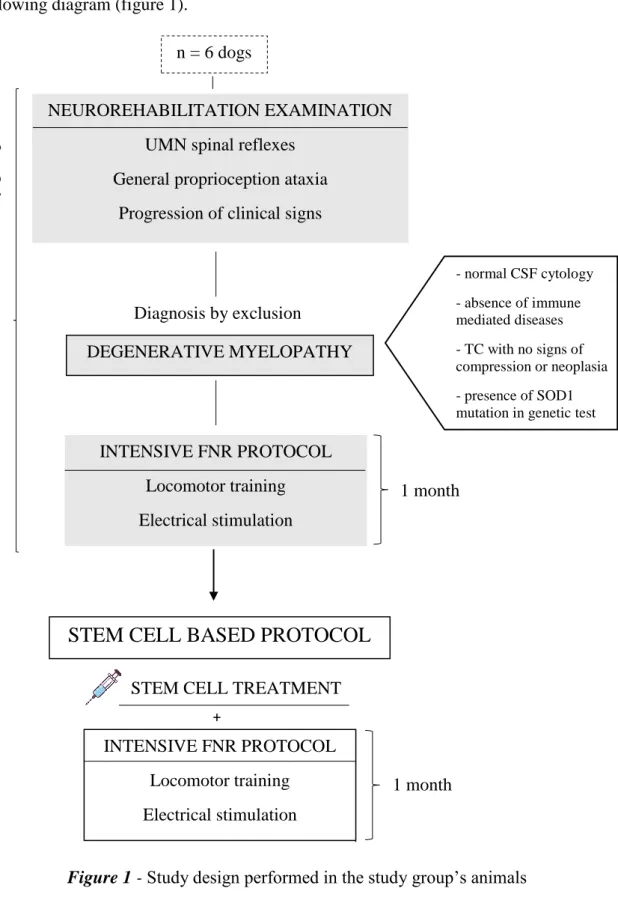
The present study was carried out in Hospital Veterinário da Arrábida, Centro de Reabilitação Animal da Arrábida and Veterinary Medicine School of Lisbon University. The main aim was to evaluate the therapeutic potential of adipose-derived mesenchymal stem cells (MSC) associated with an intensive protocol of functional neurorehabilitation (FNR) in degenerative myelopathy affected dogs. For such, a sample of ten dogs with a diagnosis of exclusion of degenerative myelopathy was selected, four constituted the control group and performed a physical therapy protocol and the other six comprised the study group and were submitted to a stem cell based protocol (MSC and FNR).
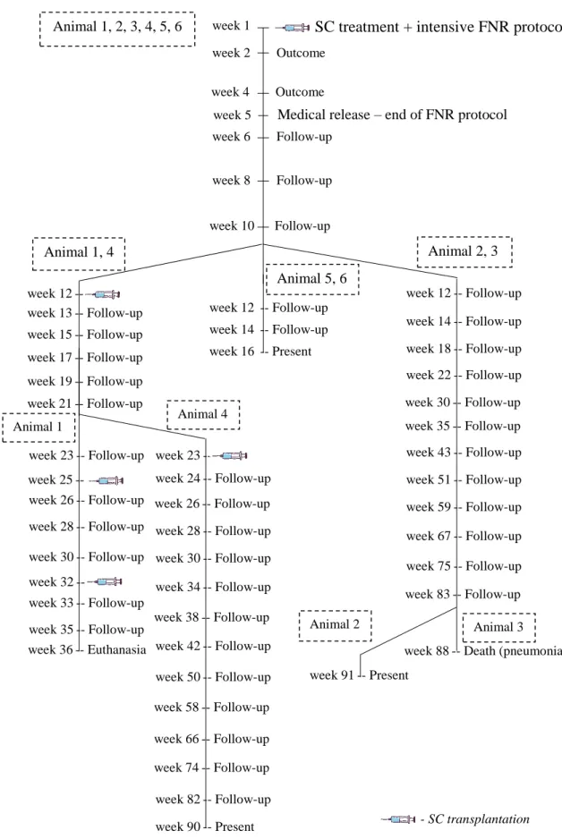
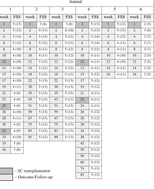
The results revealed an increase in survival time of the study group’s animals comparing to the ones in the control group. The average survival time since the beginning of physical therapy protocol for the control group was 15 weeks, in contrast with the study group, in which a dog survived 36 weeks and another 88 weeks (death from clinical occurrence) since the initiation of stem cell based protocol. Additionally, two animals in the study group had follow-ups for 88 and 91 weeks, still with acquired motor functionality. It was also possible to verify that 67% of study group’s animals improved according to a functional scoring system (FSS), after the stem cells transplantation.
In conclusion, the current study reveals a synergetic effect of the stem cell based protocol in canine degenerative myelopathy.
Keywords: Degenerative myelopathy, functional neurorehabilitation, mesenchymal stem cells, amyotrophic lateral sclerosis, dog.
Inês Dias | Resumo
XIII
Resumo
A mielopatia degenerativa é uma doença neurodegenerativa de caracter progressivo e fatal, que afeta cães adultos descrita em diversas raças, não tendo atualmente um tratamento eficaz. Algumas formas de esclerose lateral amiotrófica em humanos apresentam características homólogas com a mielopatia degenerativa, partilhando uma mutação no gene superóxido dismutase 1 (SOD1), sendo este um fator de risco para o desenvolvimento da doença.
A neurorreabilitação funcional é uma medida de neurologia restaurativa com um papel fundamental nos doentes neurológicos. Por outro lado, as células estaminais mesenquimatosas, devido às suas capacidades de diferenciação, de imunomodulação e regeneração, são consideradas uma fascinante opção terapêutica para diversas doenças neurodegenerativas.
O presente trabalho foi realizado no Hospital Veterinário da Arrábida, no Centro de Reabilitação Animal da Arrábida e na Faculdade de Medicina Veterinária da Universidade de Lisboa. Teve como objetivo avaliar o potencial terapêutico das células estaminais mesenquimatosas (MSC) derivadas de tecido adiposo juntamente com protocolos intensivos de neurorreabilitação funcional (PINRF) no tratamento de cães com mielopatia degenerativa. Foram incluídos dez cães com diagnóstico de exclusão de mielopatia degenerativa, quatro representaram o grupo de controlo e realizaram um protocolo de fisioterapia e os restantes seis constituíram o grupo de estudo, tendo estes sido sujeitos a um protocolo baseado em células estaminais (MSC e PINRF).
Os resultados obtidos revelaram um aumento do tempo de sobrevivência dos animais do grupo de estudo em relação aos do grupo de controlo. O tempo médio de vida desde o início do protocolo de fisioterapia no grupo de controlo foi de 15 semanas, contrastando com um animal do grupo de estudo que sobreviveu 36 semanas e outro que sobreviveu 88 semanas (morte por ocorrência clínica) após o início do protocolo baseado em células estaminais. Para além disso, dois animais do grupo de estudo têm um seguimento de 88 e 91 semanas, continuando com funcionalidade motora adquirida. Foi ainda possível verificar que 67% dos animais do grupo de estudo tiveram uma melhoria segundo uma escala de pontuação funcional (FSS) depois da transplantação das células estaminais.
Em conclusão, o presente estudo evidencia o papel sinergético do protocolo baseado em células estaminais na mielopatia degenerativa.
Palavras-chave: Mielopatia degenerativa, neurorreabilitação funcional, células estaminais mesenquimatosas, esclerose lateral amiotrófica, cão.
Inês Dias | List of abbreviations and acronyms
XV
List of abbreviations and acronyms
DM – Degenerative Myelopathy CSF – Cerebrospinal Fluid
ALS – Amyotrophic Lateral Sclerosis PWC - Pembroke Welsh Corgi LMN – Lower Motor Neuron UMN – Upper Motor Neuron PL – Pelvic Limbs
SOD1 – Superoxide Dismutase 1 MRI – Magnetic Resonance Imaging EMG - Electromyography
CT – Computed Tomography MSC – Mesenchymal stem cells CNS - Central Nervous System FNR – Functional Neurorehabilitation CPG – Central Pattern Generator SCI – Spinal Cord Injury
TENS - Transcutaneous Electrical Nerve Stimulation NMES - Neuromuscular Electrical Stimulation FES - Functional Electrical Stimulation
LT - Locomotor Training SC – Stem Cell
Inês Dias | List of abbreviations and acronyms
XVI
HVA/CRAA - Hospital Veterinário da Arrábida/Centro de Reabilitação Animal da Arrábida
PTP - Physical Therapy Protocol SCBP – Stem Cell Based Protocol
CCRP - Certified Canine Rehabilitation Practitioner MFS – Modified Frankel Scale
Inês Dias | General Index
XVII
General Index
Abstract ... XI Resumo ... XIII List of abbreviations and acronyms ... XV List of graphs ... XIX List of figures ... XXI List of tables ... XXIII
1. Introduction ... 1
2. Spinal Cord Histopathology ... 3
2.1 Brain Pathology ... 5
2.2 Nerve and Muscle Pathology ... 5
3. Clinical Spectrum ... 7 4. Etiopathogenesis ... 11 4.1 Mutation ... 13 5. Diagnosis ... 15 5.1 Genetic testing ... 17 6. Treatment ... 19 6.1 Functional Neurorehabilitation ... 21
6.1.1 Properties of Central Nervous System ... 22
6.1.2 Locomotor Training ... 23
6.1.3 Electrical Stimulation ... 25
6.1.4 Laser therapy ... 26
6.2 Stem Cell Therapy ... 26
6.2.1 Mesenchymal Stem Cells ... 29
6.2.2 Amyotrophic Lateral Sclerosis ... 34
6.2.3 Advancements for the future ... 37
Inês Dias | General Index
XVIII
8. Materials and Methods ... 41
8.1 Participants ... 41
8.2 Study Design ... 42
8.3 Functional Neurorehabilitation Examination ... 45
8.4 Functional Neurorehabilitation Protocol... 46
8.5 Physical Therapy Protocol ... 47
8.6 Stem Cell Protocol ... 47
8.7 Outcomes and Follow-ups ... 49
8.8 Home Rehabilitation Program ... 51
8.9 Statistical analysis ... 51 9. Results ... 53 10. Discussion ... 65 11. Conclusion ... 69 References ... 71 Appendix I ... I
Inês Dias | List of graphs
XIX
List of graphs
Graph 1 - Distribution of the control group by age ... 53
Graph 2 - Distribution of the study group by age ... 53
Graph 3 - Distribution of the control group by weight ... 54
Graph 4 - Distribution of the study group by weight ... 54
Graph 5 - Distribution of the control group by gender ... 55
Graph 6 - Distribution of the study group by gender ... 55
Graph 7 - Distribution of the control group by breed ... 56
Graph 8 - Distribution of the study group by breed ... 56
Graph 9 - Distribution of the control group by the genetic test result ... 57
Inês Dias | List of figures
XXI
List of figures
Figure 1 - Study design performed in the study group’s animals ... 42
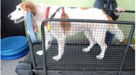
Figure 2 – Animal 1 from study group performing locomotor training ... 46
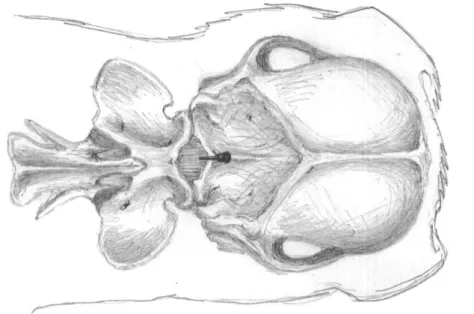
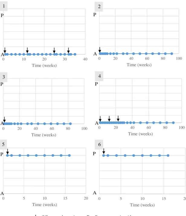
Figure 3 – Anatomical landmarks for cerebellomedullary cisternal stem cell transplantation in the dog, dorsoventral view of the cerebellomedullary cistern region 48 Figure 4 - Representation of outcomes and follow-ups performed in the study group’s animals ... 49
Figure 5 - Evolution of the patellar reflex in the study group’s animals ... 59
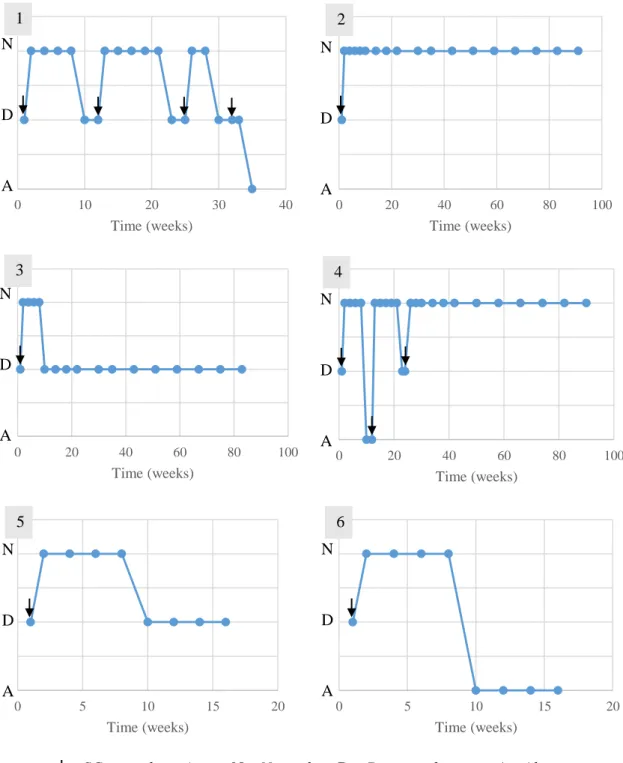
Figure 6 - Evolution of the withdrawal reflex in the study group’s animals ... 60
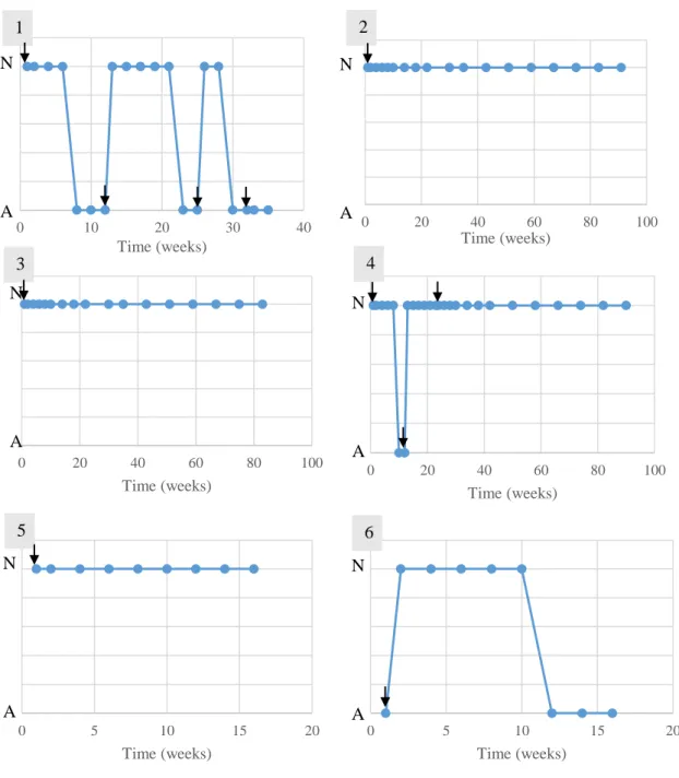
Figure 7 - Evolution of the cutaneous trunci reflex in the study group’s animals... 61
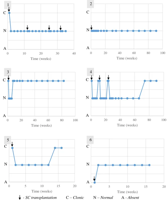
Figure 8 - Evolution of the crossed extensor reflex in the study group’s animals ... 62
Inês Dias | List of tables
XXIII
List of tables
Table 1 - Classification scheme for clinical presentation of DM categorized into four stages. Adapted from (Coates & Wininger, 2010; Toedebusch et al., 2017) ... 9 Table 2 - Classification of patients according to MFS and FSS, at admission and
medical release of PTP (control group) or intensive FNR protocol (study group) ... 45 Table 3 - Evolution of the study group patients’ classifications according to the FSS (outcomes and follow-ups) ... 58 Table 4 - Adapted Modified Frankel Spinal Cord Injury Scale for Dogs ... I Table 5 - Functional Scoring System in Dogs with Acute Spinal Cord Injuries ... II
Inês Dias | Introduction
1
1. Introduction
Canine degenerative myelopathy (DM) is a progressive and fatal neurodegenerative disorder that affects the spinal cord of adult dogs (Yokota et al., 2018).
DM was first described by Averill in 1973, when it was recognized as a specific neurological disease (Nardone et al., 2016). Other terms used were chronic degenerative radiculomyelopathy, German Shepherd Dog myelopathy, and progressive myelopathy (Kathmann et al., 2006).
German Shepherd dog is the most commonly affected breed and the mainly represented in the initial reports of DM. Nowadays this disease is identified in numerous breeds and mixed breeds (Granger & Neeves, 2015; March et al., 2009; Shelton et al., 2012). Predominantly it occurs in large size dogs and the overall prevalence rate is 0,19% (Capucchio et al., 2014; Steven & Coates, 2018).
DM was histopathologically confirmed in the following breeds: German shepherd Dog, Siberian Husky, Miniature Poodle, Boxer, Pembroke Welsh Corgi, Chesapeake Bai Retriever, Rhodesian Ridgeback, Bernese Mountain Dog, Standarf Poodle, Kerry Blue Terrier, Cardigan Welsh Corgi, Golden Retriever, Wire Fox Terrier, American Eskimo Dog, Pug, Soft-coated Wheaten Terrier and mixed breeds. However, based on a presumptive diagnosis without histopathologic confirmation, many other dog breeds were previously reported to be affected with this disease (Coates & Wininger, 2010; Granger & Neeves, 2015).
There is no sex predilection in DM, although one study revealed a predominance of females in a clinical characterization in Pembroke Welsh Corgi dogs and older studies indicated a bigger incidence in males in samples of large size breeds (Averill, 1973; Griffiths & Duncan, 1975; Coates et al., 2007).
Even though DM is considered an adult onset disease, this condition has been described in dogs aged between 6 months and 15 years old (Cherubini et al., 2010; Longhofer, et
al., 1990).
Most dogs affected are at least 8 years old before the onset of neurologic signs (Coates & Wininger, 2010; Kobatake et al., 2016; Zeng et al., 2014) and the mean age of onset of clinical signs in large dog breeds is 9 years old, although study in Pembroke Welsh Corgis (PWC) refer a mean age of 11 years (Coates et al., 2007)
Inês Dias | Spinal Cord Histopathology
3
2. Spinal Cord Histopathology
The spinal cord histopathological changes of DM affected dogs are consistent with a noninflammatory axonal degeneration and demyelination. The lesions are identified in all funiculi and involves the somatic sensory, general proprioceptive sensory and motor tracts of the spinal cord (Coates & Wininger, 2010; Holder et al., 2014; Nakamae et al., 2015).
The degenerative changes in the white matter funiculi are characterized by dilation of the myelin sheath, axon cylinder vacuolization and concurrent fragmentation and phagocytosis of axonal and myelin debris. Regional axonal loss is severe in many dogs, as evidenced by complete loss of recognizable axonal and myelin profiles and replacement by large areas of astrogliosis, with proliferation of fibrous or reactive astrocytes. It is described the occasional presence of macrophages and a study demonstrated that there is an increase of CD18-positive macrophages in severe lesions (Crisp et al., 2013; March et al., 2009).
The most severely affected region of white matter is the dorsolateral portion of the lateral funiculus and the dorsal funiculi in some dogs (Averill, 1973; Braund & Vandevelde, 1978; Bichsel et al., 1983; Matthews & de Lahunta, 1985; Johnston et al., 2000; Coates et al., 2007; Nardone et al., 2016).
Location of lesions coincide with the anatomic distribution of descending and ascending tracts in the spinal cord, with no apparent predilection for a specific proprioceptive or upper motor neuron (UMN) system. The dorsal portion of the lateral funiculus comprises the spinocerebellar, reticulospinal, rubrospinal and corticospinal tracts. Within the dorsal funiculus, lesions tend to localize medially, corresponding to the location of the fasciculus gracilis (Coates & Wininger, 2010; Nardone et al., 2016). Neuronal degeneration and loss were identified in the red nucleus, the lateral vestibular nucleus, the fastigeal nucleus, and the dentate nucleus of German Shepherd Dogs with DM (Johnston et al., 2000).
Neuropathologic lesions occur at all levels of the spinal cord, however are characteristically more severe and extensive in the mid to caudal thoracic segments, where degeneration and loss of large-diameter myelinated fibers are consistently
Inês Dias | Spinal Cord Histopathology
4
greater, with mild to moderate degeneration found in cervical and lumbar segments (Wininger et al., 2011; Bichsel et al., 1983; Matthews & de Lahunta, 1985).
It was proposed that predilection for lesion severity in the thoracic spinal cord may be a result of decreased perfusion from smaller diameter radicular artery and paucity of vessels when compared with other spinal cord regions. This fact may predispose neural tissue to ischemic processes associated with oxidative stress and excitotoxicity (Matthews & de Lahunta, 1985; Coates et al., 2007; Caulkins et al., 1989).
Descriptions of histopathological changes in DM vary within and across different breeds. Yet with different degrees of lesion severity and funicular distribution, they present similar patterns of axonal and myelin degeneration (March et al., 2009). In affected German Shepherd dogs, degenerative lesions are described as discontinuous, multifocal, bilateral, and asymmetric. Pathologic studies in this breed rarely report extensive areas of complete fiber loss in large regions of spinal cord white matter. In PWC and in an adult Miniature Poodle with degenerative myelopathy, severe and continuous regions of axonal and myelin degeneration were described (Coates et al., 2007; Miller et al., 2009; de Lahunta A., 1985).
Severity of lesions in individual dogs positively correlated with the duration of clinical signs. In longer clinical course cases it is possible to observe increased neural degeneration within funicular tracts across a longitudinally greater number of spinal cord segments (Johnston et al., 2001; March et al., 2009).
Lesions in the ascending and descending white matter pathways can explain the proprioception loss and paraparesis. The extension of lesions to the UMN pathways in the cranial thoracic and cervical spinal cord explain the clinical progression from paraparesis to tetraparesis. Moreover, fecal and urinary incontinence present in some dogs may be associated with histopathologic changes in the sensory pathways of the dorsal funiculus of the spinal cord, which signaling colorectal and urinary bladder distension. Involvement of these pathways may also contribute to a lack of visceral sensory feedback to the brain centers, altering recognition of visceral distention and involuntary evacuation. Nevertheless, urinary incontinence may also be caused by lower motor neuron (LMN) abnormality in the micturition pathway (Coates & Wininger, 2010; March et al., 2009).
Inês Dias | Spinal Cord Histopathology
5
2.1 Brain Pathology
The major hallmark histopathology of DM is the degenerative lesions in the white matter of the spinal cord, as described before. There is very limited information about the brain pathology in this condition, and some studies who examined brains from DM-affected dogs by light microscopy did not find lesions (March et al., 2009). However, one study revealed that the main differences between controls and affected German Shepherd dogs were the more marked gliosis and neuronal loss in the red nucleous, lateral vestibular nucleous and, occasionally, in the dentate nucleous and fastigial nucleous of the cerebellum, and the presence of Wallerian degeneration in the ventral tegmental decussation of the dogs with DM (Johnston et al., 2000).
2.2 Nerve and Muscle Pathology
Initial studies in DM described pathologic changes in the lumbar dorsal nerve roots, including severe degeneration. It has been reported chromatolysis in the intermediate gray matter, however motor neuron loss has not been recognized as a pathologic feature of this disease (Griffiths & Duncan, 1975; Shelton et al., 2012).
Spinal nerve root axons have been characterized as normal or exhibiting axonal loss comparing with age matched controls (Katz et al., 2017; Morgan et al., 2014; Zeng et
al., 2014).
Concerning skeletal muscles, histopathologic findings includes substantial change in the ratio of the two main muscle fiber types and in late stage disease can involve denervation atrophy and excessive variability in myofiber size (Coates & Wininger, 2010; Zeng et al., 2014). Regarding intercostal muscle atrophy describe DM-affected dogs, it is not preceded by physical loss of the motor neurons innervating these muscles, nor of their axons wich indicate that intercostal muscle atrophy is not secondary to loss of physical contact with motor neurons (Morgan et al., 2014). Similar to what is observed in the thoracic intercostal muscles, extensor carpi radialis muscle analysis shows that its acetylcholine recept complexes retained contact with motor nerve terminals, demonstrating that impairment of forelimb motor function is not secondary to denervation by lower motor neurons (Katz et al., 2017).
Thus, some findings suggest that changes in muscle fiber type and size in DM appear to be due to primary alterations in the muscles (Katz et al., 2017).
Inês Dias | Spinal Cord Histopathology
6
Other histopathologic lesions in dogs with DM can include nerve fiber loss in peripheral nerves resulting from axonal degeneration, endoneural fibrosis, numerous inappropriately thinly myelinated fibers, and secondary demyelination (Awano et al., 2009; Coates & Wininger, 2010; Zeng et al., 2014; Matthews & de Lahunta A., 1985). Studies of the common fibular (peroneal) nerve reveal that these changes are more severe as the disease progresses. (Awano et al., 2009; Coates & Wininger, 2010)
There is evidence that Pembroke Welsh Corgis and Boxers with chronic DM develop peripheral nerve pathology consistent with an axonopathy, and to a lesser degree demyelination. Reported pathologic changes within common peroneal nerve are consistent with axonal degeneration and to a lesser extent demyelination, which results in a neurogenic pattern of muscle fiber atrophy in longstanding DM. These dogs developed peripheral nerve pathology as the disease progresses, supported by the worsening of LMN weakness (Shelton et al., 2012).
In contrast, mild histologic changes in peripheral nerves reported in other studies appeared to be compatible with age-related changes and did not support an obvious peripheral nervous system component of this disorder (March et al., 2009).
Inês Dias | Clinical Spectrum
7
3. Clinical Spectrum
The clinical presentation of DM comprises an insidious, progressive UMN spastic paraparesis and a general proprioceptive ataxia of the pelvic limbs (PL), usually asymmetric, which progresses to LMN paraparesis and then spreads to involve the thoracic limbs and brainstem (Coates & Wininger, 2010; March et al., 2009; Steven & Coates, 2018).
One of the key features of DM is the lack of paraspinal hyperesthesia, in any of the disease’s phases (Coates et al., 2007).
Early disease
Initially, affected dog owners may report abnormal gait and falling. In the physical examination it is usually observable asymmetric lameness of the pelvic limbs, knuckling over of the toes, wearing of the nails, and stumbling. Thereby, in most dogs, clinical signs are consistent with a lesion in the UMN system that is characterized by proprioceptive deficits, mild paresis and increased muscle tone in the pelvic limbs (Braund & Valdevelde, 1978; Griffiths & Duncan, 1975; Kathmann et al., 2006; Okada
et al., 2009). Spinal reflex abnormalities are consistent with a lesion between the 3rd thoracic and 3rd lumbar spinal cord segments. Patellar reflexes may be normal or exaggerated to clonus and flexor (withdrawal) reflexes may also be normal or show crossed extension (suggestive of chronic UMN dysfunction) (Coates et al., 2007; Averill, 1973).
However, at the disease onset LMN signs can be observed in 10-15% of affected dogs, involving decreased patellar reflex, which can be attributed to normal age-related changes. Other possible explanations are the involvement of dorsal spinal nerve roots or peripheral nerves (Coates et al., 2007; Okada et al., 2009; Griffiths & Duncan, 1975).
As the disease evolves, affected dogs lose the ability to ambulance in the pelvic limbs, often within 12 months from the time of detection of first clinical signs (Coates & Wininger, 2010; Nardone et al., 2016; Kanazono et al., 2013). At this point, large dogs are usually elected for euthanasia. Owners can care for smaller dog breeds over a longer time (Coates et al., 2007; Matthwes & de Lahunta, 1985). The median disease duration in the Pembroke Welsh Corgi until euthanasia is 19 months (Coates et al., 2007).
Inês Dias | Clinical Spectrum
8
Late disease
In late disease, clinical signs progress to LMN paraplegia and ascend to affect the thoracic limbs within 18–24 months (Awano et al., 2009). The paresis becomes more symmetrical and progresses to flaccid tetraplegia, and severe muscle atrophy occurs in the axial and appendicular musculature (Awano et al., 2009; Matthwes & de Lahunta, 1985; Averill, 1973). Hereupon, neurologic examination includes hyporeflexia of the patellar and withdrawal reflexes. Although it is not an usual clinical sign on this condition, it has been described urinary and fecal incontinence that appears near the development of nonambulatory paraparesis (Steven & Coates, 2018).
If the disease is allowed to progress, DM affected dog can exhibit cranial nerve signs such as dysphagia and dysphonia (Steven & Coates, 2018).
Scientific evidence demonstrates that, in the later stages, DM leads to a decline in respiratory function that correlates with an alteration in respiratory movements and atrophic changes in the intercostal muscles. This hypoventilation results in hypoxemia and respiratory movement is consequently changed to the abdominal breathing pattern (Oyake et al., 2016). Hence many affected dogs are able to live with extensive care until respiratory failure develops, which is commonly approximately 3 years or later after the disease onset (Nakamae et al., 2015).
Inês Dias | Clinical Spectrum
9 Table 1 - Classification scheme for clinical presentation of DM categorized into four
stages. Adapted from (Coates & Wininger, 2010; Toedebusch et al., 2017) - Disease severity +
Stage 1 Stage 2 Stage 3 Stage 4
6 – 12 months 9 – 18 months 14 – 24 months >36 months
- Asymmetric and spastic paresis in PL - General proprioceptive ataxia in PL - Normal spinal reflexes - Nonambulatory paraparesis to paraplegia in PL - Mild to moderate PL muscle atrophy - Decreased to absent PL spinal reflexes - Urinary/fecal incontinence (+/-) - Flaccid paraplegia in PL - TL paresis - Severe PL muscle atrophy - Absent spinal reflexes - Urinary/fecal incontinence - Flaccid tetraplegia - Severe widespread muscle atrophy - Absent spinal reflexes - Urinary/fecal incontinence - Dysphagia, dysphonia - Respiratory distress
Inês Dias | Etiopathogenesis
11
4. Etiopathogenesis
Several studies have been considering possible causes underlying DM, however, conclusive evidence for a specific pathogenic mechanism is lacking (Cherubini et al., 2010; March et al., 2009). Genetic, metabolic, nutritional, vascular, and immune-mediated etiologies have been proposed (March et al., 2009).
In 2009, a study identified a mutation in superoxide dismutase 1 (SOD1) gene, which encodes Cu/Zn superoxide dismutase. A G to A transition in the SOD1 gene predicted a missense underlying DM, which is a genetic change that results in the substitution of one amino acid for another, and there was a highly significant association between homozygosity for the mutant allele and the DM phenotype in Pembroke Welsh Corgis, Boxer, Chesapeake Bay Retriever, German Shepherd Dog, and Rhodesian Ridgeback. In humans, mutations in the SOD1 gene are known to cause some forms of familial amyotrophic lateral sclerosis (ALS) (Rothstein, 2009; Steven & Coates, 2018).
The SOD1 protein is involved in antioxidant mechanisms to protect the cell from reactive oxygen species toxicity and his mutations do not lead to disease by loss of enzyme function. Instead, the mutation results in a misfolded protein, which accumulates in spinal neurons and astrocytes (Nakamae et al., 2015; Yokota et al., 2018).
To date, two SOD1 mutations were described in affected dogs, E40K and T18S. These two enzymatically active dimers possess an increased aggregation propensity in cell culture and support the proposed toxic role of SOD1 in DM (Crisp et al., 2013; Shelton
et al., 2012).
Through medical genomics it is possible to use individual’s genomic information to assist in diagnosis and to help assess the risk of developing a disease, in other words, to show the susceptibility to suffer from a determined illness.
Despite the connection between the SOD1 mutation and the development of the disease, mechanisms underlying his effect in neurodegeneration are still uncertain (Awano et al., 2009).
It appears to be probable that mutations in SOD1 determine MN death due to multifactorial and complex pathogenesis which are still not fully understood, as it happens in ALS. These mechanisms include a possible dysregulation of energy supply
Inês Dias | Etiopathogenesis
12
to neurons by oligodendrocytes, supporting the thesis that glial components are affected (Golubczyk et al., 2019; Nardone et al., 2016). Moreover, selective vulnerability of MN with SOD1 mutation can be related to mitochondrial dysfunction, since motor neurons are cells that require for a high level of mitochondrial activity and axons with the mutation are less resistant to activity-induced changes in ion concentrations (Alvarez et
al., 2013; Shaw & Eggett, 2002).
In another study, it was identified a modifier gene, SP110 nuclear body protein, involved in the regulation of gene transcription, which variations may affect overall disease risk and age of onset in Pembroke Welsh Corgis at risk for DM (Ivansson et al., 2016).
Based on what happens in SOD1-associated familial ALS, reports also suggest an association between endoplasmic reticulum stress and DM pathogenesis, demonstrated by the up-regulated expression of an endoplasmic reticulum stress marker in the spinal cords of affected dogs (Yokota et al., 2018).
Vitamin E deficiency was suggested to be involved in DM pathogenesis, although recent data indicate that it is unlikely (Johnston et al., 2001; Fechner et al., 2003). Pro-inflammatory mediators are known to play a role in the pathogenesis of ALS and it is possible to occurs a pro-inflammatory state in DM, with future studies needed to clarify its significance (Lovett et al., 2014).
Other potential etiologies indicated are abnormalities of autophagy, that have recently been reported to occur in various neurodegenerative diseases (Nardone et al., 2016; Ogawa et al., 2015); disturbed immune response, with immunohistochemical evidence for immunoglobulin and complement deposition in spinal cord lesions of German Shepherd dogs suffering from DM (Barclay & Haines, 1994); excitotoxicity via deficiency of the glutamine/glutamate cycle, that may contribute to the striking neuron loss in the ventral horn of DM dogs during disease progression (Ogawa et al., 2014); and lastly, oxidative stress and accumulation of denatured ubiquitinated proteins in spinal cord lesions, revealed in a study of immunohistochemical features of DM in two PWC dogs with a homozygous mutation in SOD1 (Ogawa et al., 2011).
Inês Dias | Etiopathogenesis
13
4.1 Mutation
Mutations in the SOD1 gene are known to cause some forms of familial ALS in humans. That fact plus the clinical similarities between DM and ALS, made this a viable candidate gene to investigate and subsequently a mutation in affected dogs was identified. In a study realized by Awano and colleagues, the sequencing of SOD1 gene revealed a G to A transition in exon 2 (SOD1:c.118A), which corresponds to nucleotide 118, resulting in a glutamic acid to lysine missense mutation at amino acid 40 (E40K) (Awano et al., 2009; Coates & Wininger, 2010).
Significant associations between the DM phenotype and homozygosity for the A allele were observed in 5 dog breeds: Pembroke Welsh corgi, Boxer, Rhodesian ridgeback, German Shepherd dog, and Chesapeake Bay retriever (Awano et al., 2009).
SOD1 is one of the most abundant proteins in the Central Nervous System (CNS), possesses 153 amino acids and functions as a homodimer, which converts superoxide radicals to hydrogen peroxide and molecular oxygen.
A second SOD1 mutation was detected in a Bernese Mountain dog suffering from DM. It predicts a T18S missense mutation that was homozygous (Wininger et al., 2011). Later, another report associated the first discovered mutant SOD1 allele with DM in cross-bred dogs and 124 different canine breeds (Zeng et al., 2014). The great majority of histopathologic confirmed affected dogs are SOD1:c.118A homozygous but a small quantity are heterozygous, being homozygosity a major risk for this condition (Kobatake et al., 2017; Pfahler et al., 2014).
Still, dogs that only carry a single copy of the SOD1 mutation rarely develop the clinical signs of DM. It is possible that asymptomatic heterozygotes represent the subclinical state of this disease and therefore they can constitute one line of research regarding early pathologic events (Kobatake et al., 2017).
There has been no conclusive evidence, however, as a considerable number of homozygous dogs for the mutant allele didn’t develop clinical signs, it was suggested that DM is an autosomal recessive disease with incomplete penetrance (Awano et al., 2009; Steven & Coates, 2018).
Inês Dias | Etiopathogenesis
14
Through immunohistochemical analysis, studies oftentimes detected accumulation and aggregate formation of SOD1 in the spinal neurons of affected dogs (Crisp et al., 2013; Kohyama et al., 2016; Nakamae et al., 2015).
Inês Dias | Diagnosis
15
5. Diagnosis
Diagnosis of DM can be challenging because its early signs can mimic several spinal cord diseases that also compromises UMN pathways. As this condition affects mainly older dogs, it is not uncommon for co-morbidities to exist and complicate the interpretation of neurologic examination (Crisp et al., 2013; Steven & Coates, 2018). A complete history must be taken to establish the onset and recognize the insidious progression pattern of clinical signs (Cherubini et al., 2010). It is imperative to perform a thorough general clinical examination to rule out the presence of concomitant diseases, such as metabolic and systemic disorders and cardiovascular conditions (Cherubini et al., 2010). An orthopedic examination is particularly important in older dogs and degenerative joint disease, bilateral cranial cruciate disease and hip dysplasia must be considered in the list of differential diagnosis (Cherubini et al., 2010).
Regardless of age, assessment of any animal with suspected neurologic disease involves a complete and accurate neurologic examination (Bagley, 1997). Common differentials include intervertebral disk disease, spinal cord trauma, lumbosacral stenosis, inflammatory disease and spinal cord neoplasia (Coates et al., 2007; Wahl et al., 2008). Lack of paraspinal hyperesthesia is a key clinical feature of DM that differentiates it from compressive myelopathies (Steven & Coates, 2018). Furthermore, in contradiction to DM, orthopedic conditions are not associated with proprioceptive deficits, and lumbosacral disease is associated with LMN rather than UMN signs (Bagley, 1997). Presently, there is no specific antemortem diagnostic test available and because SOD1 mutations are incompletely penetrant, genetic screening is insufficient for diagnosis (Bagley, 1997; Toedebusch et al., 2017). A definitive diagnosis of DM can only be made postmortem through histologic examination of the spinal cord and detection of axonal degeneration, demyelination along with astrogliosis (Nardone et al., 2016). As such, having ruled out non-neurological causes for the clinical presentation and after a neurological examination is performed, tentative antemortem diagnosis is based on the anamnesis, the clinical signs and its progression pattern, SOD1 mutation and a process of elimination of other causes supported by complementary tests (Cherubini et al., 2010; Coates & Wininger, 2010).
Inês Dias | Diagnosis
16
Neurodiagnostic techniques for evaluation of spinal cord disease include cerebrospinal fluid (CSF) analysis, electrodiagnostic testing and spinal cord imaging procedures as myelography, computed tomography (CT), and magnetic resonance (Coates et al., 2007; Coates & Wininger, 2010).
A presumptive diagnosis of DM is, thereby, based on ruling out spinal cord compressive diseases (Steven & Coates, 2018).
If there are no other concurrent spinal diseases, radiographic and myelographic studies are normal and CT and magnetic resonance imaging (MRI) don’t reveal significant abnormalities (Bagley, 1997; Okada et al., 2009). However, if another spinal disease is found with diagnostic testing, it is not possible to exclude degenerative myelopathy as a contributive factor to the clinical signs (Bagley, 1997).
Survey spinal radiographs can help rule out bone diseases such as diskospondylitis, bone neoplasia and spinal fractures. A lumbar myelography can be performed to detect spinal cord compression, nevertheless CT myelography is more sensitive for characterizing morphology of the spine (Cherubini et al., 2010; Jones et al., 2005). MRI is the modality of choice to investigate spinal cord diseases and is especially helpful for identifying early spinal cord neoplasia and evidence of extradural compressive myelopathy (Coates & Wininger, 2010).
Frequently, imaging reveals disc protrusions that can confound a diagnosis of DM. Ultimately, the clinician must be guided by clinical experience and evaluate the disease progression, paraspinal hyperesthesia, and amount of spinal cord compression to estimate the severity of the compressive myelopathy (Coates & Wininger, 2010; Steven & Coates, 2018).
In DM-affected dogs, results of CSF analysis are normal or, more commonly, indicate elevated total protein without a concurrent pleocytosis (Bagley, 1997; Cherubini et al., 2010). CSF analysis provides information to the existence of inflammation or infection of CNS and therefore can help rule out meningitis. It may also be a promising source for diagnostic biomarker of DM, and a possible help in establishing prognosis and mechanism of disease (Coates & Wininger, 2010).
Myelin basic protein is a protein restricted to the nervous system and in humans, its concentration in CSF is used as a biochemical marker to evaluate active demyelinating
Inês Dias | Diagnosis
17 disorders. One study reported an increased concentration of myelin basic protein in CSF collected from the lumbar cistern of dogs with DM, but more investigation is needed to have valid conclusions (Oji et al., 2007). CSF of affected dogs has also been evaluated for markers of immune responses with intrathecal formation of immunoglobulins by detecting the presence of oligoclonal banding, but results were not significantly different from control dogs and large-scale study including cases with other spinal cord disorders is warranted (Kamishina et al., 2008). Another study verified that chaperon protein clusterin, that is protective against oxidative stress, is elevated in the CSF of chronic spinal cord disorders compared to meningitis and idiopathic epilepsy, indicating that additional markers are required to differentiate DM from a concurrent condition (Shafie et al., 2014). More recently, CSF levels of phosphorylated neurofilament heavy (pNF-H) were investigated in all stages of DM. pNF-H is an abundant structural protein of myelinated motor axons and a promising biomarker for nervous system diseases and its concentrations in blood and CSF have shown high association with disease progression in humans diagnosed with ALS. The results from this report indicate that pNF-H is increased in the CSF but not in serum of affected dogs relative to control groups and that quantification of this protein can be used as an antemortem diagnostic tool for DM, however further studies are necessary (Toedebusch et al., 2017).
Electrodiagnostic testing is helpful for detecting evidence of neuromuscular disease, but the results vary in the various disease stages. Early in DM, electromyography (EMG) and nerve conduction velocity studies don’t detect any change from normal limits, suggesting that peripheral nerves and motor fibers are not affected in this stage. Later in the disease course, EMG reveals multifocal spontaneous activity, fibrillation potentials, and positive sharp waves in the appendicular musculature. The proximal and distal motor neuron conduction velocities in stimulated tibial and ulnar nerves were decreased than normal, providing evidence of the presence of axonopathy and demyelination in disease’s late stages (Awano et al., 2009; Steven & Coates, 2018).
5.1 Genetic testing
DM is associated until now with two single nucleotide variations in exon 1 (specific to Bernese Mountain dog) and exon 2 (not specific to the breed) of the SOD1 gene (Awano et al., 2009; Wininger et al., 2011). A genetic test based on the SOD1 mutation is commercially available and can assist in the diagnosis (Steven & Coates, 2018).
Inês Dias | Diagnosis
18
Due to the late onset and insidious nature of the disease process and because dogs homozygous for the mutation are likely as fertile as other genotypes, the natural selection is insignificant and they will contribute one chromosome with the mutant allele to all of their offspring. The heterozygotes are DM carriers and are less likely to develop clinical signs but could pass on a chromosome with the mutant allele to half of their offspring (Capucchio et al., 2014; Steven & Coates, 2018). Thus, the mutant allele may reach high frequencies in dog populations and an easy to implement diagnostic test may contribute to control the gene diffusion (Capucchio et al., 2014).
The SOD1 DNA test for breeding strategies is strongly recommended in an effort to reduce the incidence of DM and ultimately leading to an eventual eradication of the disease in some breeds (Kohyama et al., 2016; Turba et al., 2013). Working dogs can also be submitted to genetic testing to identify at-risk genotypes prior to buying or training (Shaffer et al., 2018).
However, an overly aggressive breeding program to eliminate the mutant allele may create a ―bottleneck‖ effect and possibly select for other diseases or alter desirable qualities of the breed. (Coates & Wininger, 2010)
Inês Dias | Treatment
19
6. Treatment
Currently, there is no prophylactic or curative treatment for DM. Therefore, management strategies have been empiric and aim to relieve clinical signs and maintain the quality of life of the affected dogs. However, long-term prognosis of DM is poor (Coates & Wininger, 2010; Oyake et al., 2016; Steven & Coates, 2018).
In 2008, a study evaluated a therapeutic protocol including the administration of ε-aminocaproic acid (a fibrinolysin inhibitor) and N-acetylcysteine (a glutathione precursor and free radical scavenger) along with supplementation of vitamins B, C and E and controlled daily exercise. The progression of the disease was not affected in any animal and the rate of neurological worsening did not change (Clemmons, 1992; Polizopoulou et al., 2008).Therapy with corticosteroids didn’t delay the progression of DM, as well. Another report studied the long-term parenteral cobalamin or oral tocopherol administration and found no beneficial effects. Furthermore, there was no significant differences between serum concentrations of α-tocopherol in affected German Sheppard dogs and controls (Johnston et al., 2001; Polizopoulou et al., 2008). Physiotherapy and principles of physical rehabilitation have shown to be crucial in DM management, because it may improve the quality of life and prolong the survival time. When affected animals are left untreated, non-ambulatory paraplegia is usually established within 6 months after the initial diagnosis (Clemmons, 1992; Polizopoulou
et al., 2008).
Kathmann and colleagues reported survival data from 22 dogs with DM that received varying degrees of physiotherapy. Dogs that received intensive physiotherapy had significantly longer survival times (mean 255 days) compared with that for animals with moderate (mean 130 days) or no (mean 55 days) physiotherapy. The physiotherapy routine consisted of active and passive exercises regardless the disease stage (Kathmann
et al., 2006).
As already mentioned, some of the earliest pathological changes in DM occur in the muscle fibers and upper motor and sensory neuron tracts in the spinal cord. A more thorough characterization of this cascade of pathological events will provide targeting therapeutic interventions most likely to be effective in slowing disease progression (Katz et al., 2017).
Inês Dias | Treatment
20
Recent studies examined therapeutic potential of the endocannabinoid system for the treatment of chronic neurodegenerative diseases of the CNS, such as Alzheimer’s disease, Parkinson’s disease, Huntington’s disease, and ALS that has many similar features with DM. As it is known, the endocannabinoid system exerts a modulatory effect of important functions such as neurotransmission, glial activation, oxidative stress, or protein homeostasis and dysregulation of this processes is a common neuropathological hallmark in neurodegenerative diseases (Aymerich et al., 2018; Fernández-Trapero et al., 2017).
Reports observed that CB2 receptors become strongly up-regulated in activated astrocytes recruited at the damaged spinal cord in mutant SOD1 mice, an experimental model of ALS. These results were also found in the spinal cord of DM affected dogs, a natural occurring model of some forms of ALS. These findings support that the elevation of CB2 receptors in activated glial elements is an endogenous response of the endocannabinoid to neuronal damage. Such up-regulation occurred in absence of changes in other endocannabinoid elements. Targeting the CB2 receptor afforded neuroprotection in transgenic rodent ALS models. Thereby, more research in cannabinoid-based therapies must be conducted and selectively targeting this receptor can be used to enhance the protective effects exerted by these glial cells to improve neuronal homeostasis and integrity and slow disease progression (Aymerich et al., 2018; Fernández-Trapero et al., 2017).
Perspectives for the future include targeted stem cell (SC) therapy and delivery of small molecule therapies to the spinal cord with viral vectors (Coatti et al., 2015; Nayak et al., 2006).
A promising adjunctive therapy for a variety of surgical and medical disorders is hyperbaric oxygen therapy and its use is increasing in both human and veterinary medicine (Birnie et al., 2018; Edwards, 2010). It is based on the therapeutic use of intermittent inhalation of 100% oxygen inside a chamber pressurized above atmospheric pressure, which increases the dissolved oxygen concentration in blood and can alter tissue responses to disease and injury (Birnie et al., 2018; Edwards, 2010). It has already been demonstrated that this treatment is well tolerated and safe in dogs and cats and that hyperoxia has many potential benefits (Birnie et al., 2018). Physiologic effects of hyperbaric oxygen may include a relief of oxidative stress, antimicrobial effects, inflammation decrease, enhanced neuroprotective mechanisms and even
Inês Dias | Treatment
21 immunomodulation, making this a promising adjuvant therapy for neurologic patients (Patel & Huang, 2017; Zhou et al., 2019).
6.1 Functional Neurorehabilitation
Physical Rehabilitation of companion animals has become increasingly common and it proves to be very important to attain recovery in many orthopedic and neurologic conditions (Drum, 2010; Riegger-Krugh et al., 2014).
Functional neurorehabilitation (FNR) is a practice dedicated to the restauration of function to a body impaired by injury or disease, in order to improve the quality of life of the animal (Millis & Ciuperca, 2015; Weigel et al., 2005).
After a neurologic injury, FNR intend to recover animal’s postural control, balance, locomotion capacity and ultimately functional independence (Harkema et al., 2012). A rehabilitation plan of care, created to each individual case, should be intense and applied as soon as possible, within the limitation of the disease, in order to achieve success (Martins, 2015; Norrie, 2005; Goncalves et al., 2016).
Rehabilitation of dogs with neurologic disease can involve a combination of many therapeutic strategies as passive and reflexive exercises, active exercises and therapeutic modalities such as electrical stimulation, ultrasound, cryotherapy and heat therapy (Olby et al., 2005). An important attention must be given to the possible consequences secondary to the condition in order to prevent and treat them like pain, decubital ulcers and muscle contractures (Riegger-Krugh et al., 2014).
FNR is based on neuroanatomical key principles and CNS properties as neuroplasticity and neuromodulation, presents in both human being and domestic animals (Martins, 2015; Goncalves et al., 2016; Rossignol & Frigon, 2011).
One of the goals of this growing branch of veterinary medicine is the translation of these principles into rational strategies to promote recovery of function (Celnik & Cohen, 2004).
Inês Dias | Treatment
22
6.1.1 Properties of Central Nervous System
6.1.1.1 Central Pattern Generators
Neuronal circuits within the spinal cord interacts with specific sensory information and are capable of generate self-sustained patterns of locomotor-like neural activity, independently of supraspinal and afferent input (Dietz & Harkema, 2004). These networks of interneurons are defined as Central Pattern Generators (CPG) and are located in the lumbar segments of the spinal cord (Dietz & Harkema, 2004; Guertin, 2014) After a spinal cord injury (SCI), different changes in cellular and circuit properties occur spontaneously and can be promoted by rehabilitation approaches based on these functional networks (Rossignol et al., 2006; Rossignol & Frigon, 2011).
6.1.1.2 Neuroplasticity
Neuroscience has demonstrated that plasticity occurs throughout the CNS and throughout life (Thompson & Wolpaw, 2014; Yue et al., 2017). In physiologic conditions the spinal cord is continuously interacting with brain and peripheral sensory input during postural and locomotor activities and the activity-dependent plasticity plays an important role in the acquisition and maintenance of motor skills and in the effects of SCI or a progressive disease of the spinal cord (Edgerton & Roy, 2009; Wolpaw & Tennissen, 2006). Thus, CNS has the potential to reorganize connections through synaptic plasticity of pre-existing circuits and anatomical plasticity whereupon new circuits might develop through anatomical reorganization (Edgerton et al. 2006; Grasso et al., 2004).
After a SCI, motor training can provide sufficient stimulation of specific neural pathways to facilitate functional reorganization within the spinal cord and improve motor output. The most successful type of training includes repetitive pattern and variability in the performed task (Dietz & Harkema, 2004; Edgerton & Roy, 2009). Thus, training-induced plasticity might be responsible for changes in the locomotor network, that are probably adaptive and learnt (being specific to the trained task) (Dietz, 2012; Grasso et al., 2004). Furthermore, appropriate sensory input during training is of high importance to achieve an optimal motor output of the spinal neuronal circuitry (Dietz & Harkema, 2004).
Inês Dias | Treatment
23 The use of robotic devices for training specific motor tasks has become more prevalent recently and can provide a high probability of successful rehabilitation (Edgerton & Roy, 2009).
Further work should aim to identify the timing, type, and quantity of exercise and pharmacological interventions that can be used in new therapeutic approaches to maximizing function after spinal cord injury or restoring function to a newly regenerated spinal cord (Jakeman et al., 2011; Wolpaw & Tennissen, 2006).
6.1.1.3 Neuromodulation
Neuroplasticity that occurs after a SCI doesn’t always have beneficial effects, that is why FNR must modulate the spinal cord excitability and define specific protocols with multimodal approach (Edgerton et al., 2006).
Interventions that may contribute to improve sensorimotor function include practice of the specific motor task that needs to be improved; combining with modulation of the excitability of the spinal circuitry through pharmacological modulation and/or via epidural stimulation (Celnik & Cohen, 2004; Edgerton et al., 2006; Zbogar et al., 2017). Moreover, peripherally or centrally applied electrical stimulation is a valuable tool to promote functional improvement related to modulation of specific areas of CNS (Yue et
al., 2017).
Several reports have shown the involvement of neurotrophic factors in mechanisms related to neuroplasticity, as their actions are seen in a wide range of neuronal events and are also factors for axonal growth and synaptic plasticity (Côté et al., 2010; Yue et
al., 2017). Also, molecules involved in axon guidance during CNS development may
have an important role in the process of neurorehabilitation, as well as some neurotransmitters that have a modulator function in locomotor patterns (Edgerton et al., 2004; Yue et al., 2017).
6.1.2 Locomotor Training
Locomotor training (LT) is a rehabilitation strategy and an effective method for improving the recovery of postural control, balance, postural standing, gait, independence of function and quality of life after a neurological injury (Dietz & Harkema, 2004; Martins, 2015). Intensive activity-based rehabilitation interventions
Inês Dias | Treatment
24
such as step-training on a treadmill can result in significant functional improvements in individuals with chronic incomplete SCI (Harkema et al., 2012). It was also shown to improve gait kinematics, recover of phase-dependent modulation of spinal reflexes, and prevent loss of muscle mass (Côté et al., 2010).
LT can activate and modulate spinal locomotor centers, the already mentioned central pattern generators, and so by exploiting plasticity of CNS, it is possible to improve recovery of hindlimb function (Dietz & Harkema, 2004). In the spinal cord, exercise can also improve microenvironment and regulate physiological and metabolic function of motor neurons (Fu et al., 2016). However, besides the improvement of spinal cord function, LT can improve the function in different levels from end-effector organ to cerebral cortex through reshaping skeletal muscle structure and muscle fiber type and remodeling function of the cerebral cortex, thereby promoting functional recovery by improving neural and muscular function (Fu et al., 2016).
In humans affected by multiple sclerosis, rehabilitation interventions including LT can reduce sequels of the disease and should be considered early for maintaining functional capacity and reducing risk for losing important abilities, to achieve the highest possible quality of life (Beer et al., 2012). Furthermore, regular exercise may promote anti-oxidant defenses and neurotrophic support that could reduce CNS vulnerability to neuronal degeneration (White & Castellano, 2008). In poststroke subjects, short-term intensive rehabilitation using body weight support treadmill training has also shown to be useful for improving gait capacity (Takao et al., 2015).
A study concerning individuals with ALS determined that repetitive rhythmic exercise of supported treadmill ambulation training is feasible, tolerated and safe and it is consistent with improved gait function (Sanjak et al., 2010).
Chronic spinal cord conditions, such as Hansen type II intervertebral disc disease and degenerative myelopathy, are typically associated with progressive neurologic deficits and warrant a therapeutic approach with less focus on aggressive management and a greater emphasis on frequent repetition of low-intensity activities. Thus, preserving neuromuscular and musculoskeletal function, and manage the pain in some animals (Sims et al., 2015).
Inês Dias | Treatment
25 Hydrotherapy is another rehabilitation strategy that results in improvement in motor functions. There is evidence that an aquatic training program is appropriate and beneficial for individuals with multiple sclerosis and SCI and can be an effective method in reducing spasticity severity (Kesiktas et al., 2004; Salem et al., 2011).
6.1.3 Electrical Stimulation
Therapeutic exercise in rehabilitation is often complemented by physical agent modalities that can assist to limit impairments and disability and to maximize function. Electrical stimulation, ultrasound, cryotherapy, and heat therapy are the most commonly used modalities in neurologic rehabilitation and they have been used to reduce swelling, relieve pain, enhance healing, increase muscle strength, improve muscle tone, affect the elasticity of connective tissue and promote of soft-tissue and fracture healing (Drum, 2010; Hanks et al., 2015).
Electrical stimulation (ES) is most commonly used to reduce acute and chronic pain and spasticity and increase muscle strength and it is broadly categorized transcutaneous electrical nerve stimulation (TENS) or neuromuscular electrical stimulation (NMES) (Drum, 2010; Hanks et al., 2015; Peckham & Knutson, 2005). Generally, the intensity must always be adapted to the needs, comfort, and response of the animal and varies with the objective of treatment and toleration (Hanks et al., 2015).
TENS is used primarily for pain, however fine muscle fasciculations can be attained. It can also be used in the acute phase around incision lines to reduce pain and edema (Drum, 2010). In combination with active therapy, TENS can be an adjunct therapy for management of limb spasticity and improve voluntary motor control (Hofstoetter et al., 2014; Mills & Dossa, 2016). Moreover, a study tested the effects of TENS on spasticity in rats with SCI and suggested that high frequency TENS alleviates spasticity by inhibiting activated microglia (Hahm et al., 2015).
NMES is used to stimulate a muscle contraction, slowing muscle atrophy but there is also a component of pain control. It can help simulate walking motion if applied in an alternating contracting fashion and can be combined with active exercise to maximize muscle contraction (Drum, 2010; Hanks et al., 2015).
Inês Dias | Treatment
26
Therefore, through electrical activation of intact lower motor neurons using electrodes placed on or near the innervating nerve fibers, paralyzed or paretic muscles can be made to contract in a coordinated manner. Functional electrical stimulation (FES) is the application of electrical current to excitable tissue to provide functional restoration in neurologically impaired individuals (Peckham & Knutson, 2005). By means of FES, it is possible to increase strength (muscle force) and endurance (fatigue resistance) and improve upper or lower limb mobility (Gordon & Mao, 1994; Hamid & Hayek, 2008). In addition to motor system, FES can also be applied to restoring function in bladder and bowel, and respiratory system (Hamid & Hayek, 2008; Peckham & Knutson, 2005).
6.1.4 Laser therapy
Among the various modalities of FNR, laser therapy is applied to several conditions in dogs, including neurologic disorders. Therapeutic lasers have become increasingly popular for rehabilitation purposes and although its mechanism of action has yet to be fully characterized, it may have direct beneficial effects on nerve cells and their supporting structures. Most studies of laser use in rehabilitation are focused on wound healing and pain management, however, it may help to modulate cellular functions, reduce degenerative changes in neurons, induce proliferation of astrocytes and oligodendrocytes, and may also have anti-inflammatory effects (Riegger-Krugh et al., 2014; Sims et al., 2015).
6.2 Stem Cell Therapy
Regenerative medicine is an emerging field of research in tissue engineering, biomaterials and cellular therapies that aims to restore structure and function of diseased, nonfunctional or malfunctional tissues or organs (Gugjoo et al., 2019). Cell therapy had his first steps in the 1950s with the first bone marrow transplant involving identical twins. For several decades now, in human medicine, the application of stem cells represents already a well-established clinical procedure (Ciervo et al., 2017).
Thus, stem cell sciences are a promising biologic tool for a better understanding of several diseases as well as a potential therapy in many conditions (Glicksman, 2018).
Inês Dias | Treatment
27 Stem cell term refers to an unspecialized and precursor cell characterized by the properties of self-renewal, multiplication and multi lineage differentiation potential (Bonafede & Mariotti, 2017; Haidet-Phillip et al., 2015; Quimby, 2019). Self-renewal reflect their capacity to undergo multiple/limitless divisions (Los et al.,2019).
According to their regenerative potential, SC can be classified as totipotent, pluripotent, multipotent, oligopotent and unipotent (Los et al., 2019). Totipotency is defined as the ability to differentiate into cell lineages from all three germ layers: mesoderm, endoderm, and ectoderm including placental cell. In a human organism, only zygote and first blastomere have this capability. Pluripotency is the ability to differentiate into several but not all cell types, which is a characteristic of cells composing blastocyst’s inner cell mass, embryonic stem cells. Multipotent cells differentiate into many types of cells that originate from one germ layer. Oligopotent cells are capable of differentiate into a few types of cells and, finally, unipotency is the ability to produce cells of their own type (Los et al., 2019; Nayak et al., 2006).
Stem cells can also be classified according to their origin and divided into two major subtypes: embryonic stem cells that are able to differentiate to the more than 200 cell types present in the body, if provided the right conditions for differentiation, and adult stem cells, which are located in organs of mature body and can usually only form the cell types present in the organ from which they were derived, being multipotent (Gugjoo et al., 2019; Mummery et al., 2010).
The use of embryonic stem cells raises ethical concerns, as most methods of their isolation involve the destruction of embryo. Also, its use carries a risk of teratoma formation and immune rejection (Cashman et al., 2008; Los et al., 2019).
Adult stem cells are found in niches which are microenvironments in specific anatomic locations that influence and regulate stem cell fate (Los et al., 2019; Rossi & Salvetti, 2019). Examples are mesenchymal stem cells (MSC), hematopoietic SC, skeletal SC, pancreatic SC and neural SC. Neural stem cell are found in the brain of both growing and mature mammals and are defined based on their ability to differentiate into three major CNS cell types: neurons, astrocytes and oligodendrocytes. They may enable a better understanding of diseases pathology and in the future may become a treatment