Mitochondrial Dysfunction Induced by
N-
Butyl-1-(4-Dimethylamino)Phenyl-1,2,3,4-Tetrahydro-
β
-Carboline-3-Carboxamide Is
Required for Cell Death of
Trypanosoma cruzi
Hélito Volpato1, Vânia Cristina Desoti2, Rodrigo Hinojosa Valdez3, Tânia Ueda-Nakamura2,
Sueli de Oliveira Silva2, Maria Helena Sarragiotto4, Celso Vataru Nakamura1,2*
1Programa de Pós-Graduação em Ciências Biológicas—Biologia Celular e Molecular, Universidade Estadual de Maringá, Maringá, Paraná, Brazil,2Programa de Pós-Graduação em Ciências Farmacêuticas, Universidade Estadual de Maringá, Paraná, Brazil,3Departamento de Farmácia, Instituto Federal do Paraná, Palmas, Paraná, Brazil,4Departamento de Química, Universidade Estadual de Maringá, Maringá, Paraná, Brazil
Abstract
Background
Chagas’disease is caused by the protozoanTrypanosoma cruziand affects thousands of
people worldwide. The available treatments are unsatisfactory, and new drugs must be developed. Our group recently reported the trypanocidal activity of the synthetic compound N-butyl-1-(4-dimethylamino)phenyl-1,2,3,4-tetrahydro-β-carboline-3-carboxamide (C4), but
the mechanism of action of this compound was unclear.
Methodology/Principal Findings
We investigated the mechanism of action ofC4against epimastigote and trypomastigote forms ofT.cruzi. The results showed alterations in mitochondrial membrane potential, alter-ations in cell membrane integrity, an increase in the formation of reactive oxygen species, phosphatidylserine exposure, a reduction of cell volume, DNA fragmentation, and the for-mation of lipid inclusions.
Conclusion/Significance
These finding suggest that mitochondria are a target ofC4, the dysfunction of which can
lead to different pathways of cell death. OPEN ACCESS
Citation:Volpato H, Desoti VC, Valdez RH, Ueda-Nakamura T, Silva SdO, Sarragiotto MH, et al. (2015) Mitochondrial Dysfunction Induced byN -Butyl-1-(4-Dimethylamino)Phenyl-1,2,3,4-Tetrahydro-β -Carboline-3-Carboxamide Is Required for Cell Death ofTrypanosoma cruzi. PLoS ONE 10(6): e0130652. doi:10.1371/journal.pone.0130652
Academic Editor:Herbert B. Tanowitz, Albert Einstein College of Medicine, UNITED STATES
Received:January 22, 2015
Accepted:May 22, 2015
Published:June 18, 2015
Copyright:© 2015 Volpato et al. This is an open access article distributed under the terms of the
Creative Commons Attribution License, which permits unrestricted use, distribution, and reproduction in any medium, provided the original author and source are credited.
Data Availability Statement:All relevant data are within the paper.
Introduction
Chagas’disease is a tropical infection caused byTrypanosoma cruzi. Approximately 7–8 mil-lion people worldwide are infected by this protozoan, mostly in Latin America. Up to 30% of chronically infected individuals develop cardiac complications [1]. It is found endemically in 21 Latin American countries, and 28 million people are at risk of acquiring this infection around the world [2].
The available treatment for Chagas’disease is based on only two drugs, nifurtimox and benznidazole, which were discovered approximately 40 years ago. Both drugs are only partially effective and have many side effects [3,4]. The search for new drugs must be intensified. Differ-ent research groups are investigating the effectiveness of possible trypanocidal agDiffer-ents [5]. Our group demonstrated thein vitroandin vivoeffects onT.cruziof someβ-carboline compounds, especiallyN-butyl-1-(4-dimethylamino)phenyl-1,2,3,4-tetrahydro-β-carboline-3-carboxamide (C4)(Fig 1) [6,7]. This compound was effective against the three evolutive forms ofT.cruzi. Furthermore, transmission electron microscopy indicated that the mitochondrion is the major organelle affected by this compound in trypanosomatids, such asT.cruziandLeishmania
ama-zonensis[6,8]. This compound has also been shown to have low toxicity in mammalian cellsin
vitroand other animal models [6,7].
The present study evaluated biochemical alterations in epimastigote and trypomastigote forms ofT.cruzitreated withC4. Flow cytometry, fluorimetry, and fluorescence microscopy were used to investigate cellular and subcellular structures and identify organelles that are affected byC4treatment. We found that mitochondrial damage may be a possible target for
Fig 1. Chemical structure ofN-butyl-1-(4-dimethylamino)phenyl-1,2,3,4-tetrahydro-β-carboline-3-carboxamide (C4).
doi:10.1371/journal.pone.0130652.g001 data collection and analysis, decision to publish, or preparation of the manuscript.
C4in these parasites, thus providing a better understanding of the mechanism of action of this compound. Based on our results, we suggest that mitochondrial dysfunction induced byC4
can lead to different pathways of cell death inT.cruzi.
Materials and Methods
2.1. Chemicals
Actinomycin D, antimicyn A (AA), carbonyl cyanidem-chlorophenylhydrazone (CCCP), 2’,7’-dichlorodihydrofluorescein diacetate (H2DCFDA), digitonin, dimethylsulfoxide (DMSO), rhodamine 123 (Rh123), and Nile red were purchased from Sigma-Aldrich (St. Louis, MO, USA). Dulbecco’s modified Eagle’s medium (DMEM) and fetal bovine serum (FBS) were obtained from Invitrogen (Grand Island, NY, USA). Annexin-V FITC, the MitoSOX kit, propi-dium iodide (PI), and the TUNEL kit were obtained from Invitrogen (Eugene, OR, USA). All of the other reagents were of analytical grade.
2.2. Synthesis of C4
C4was synthesized as previously described [6].
2.3. Substance preparation
C4was prepared in DMSO. All of the groups, including the controls were tested at final con-centrations of less than 1% DMSO, a concentration that was found not to affect the parasite.
2.4. Parasites
The experiments were performed with the Y strain ofT.cruzi. Epimastigote forms were grown in Tryptose Liver Infusion (LIT) supplemented with 10% FBS at 28°C for 96 h. Trypomastigote forms were obtained from the supernatant of an infected LLCMK2cells monolayer (epithelial cell of monkey kidney;Macaca mulatta) in DMEM supplemented with 2 mM L-glutamine, 10% FBS, 50 units/mL penicillin, and 0.05 mg/mL streptomycin and buffered with sodium bicarbonate in a 5% CO2air mixture at 37°C. Sub-confluent cultures of LLCMK2cells were infected with 1 × 10⁶trypomastigotes/mL. Extracellular parasites were removed after 24 h. The cells were washed, and these cultures were maintained in DMEM that contained 10% FBS until trypomastigotes emerged from the infected cells.
2.5. Mitochondrial membrane potential
Epimastigotes (5 × 10⁶cells/mL treated with 18.0 and 77.0μM ofC4) and trypomastigotes
(1 × 10⁷cells/mL treated with 45.0 and 230.0μM ofC4) ofT.cruziwere incubated at 28°C and
37°C, respectively, for 3 h. Afterward, the parasites were washed and incubated with 5μg/mL
Rh123 for 15 min to verify mitochondrial membrane potential (ΔCm). CCCP (100.0μM) was
used as a positive control. The data acquisition and analysis were performed using a FACSCali-bur flow cytometer (Becton-Dickinson, Rutherford, NJ, USA) equipped with CellQuest soft-ware (Joseph Trotter, The Scripps Research Institute, La Jolla, CA, USA). A total of 10,000 events were acquired in the region that was previously established as the one that corresponded to the parasites.
2.6. Fluorimetric detection of mitochondrial-derived O
2•−KCl, 120 mM NaCl, 0.7 mM Na2HPO4, and 1.5 mM NaH2PO4(pH 7.3). The cells were loaded with 5μM MitoSOX reagent and incubated for 10 min at room temperature while protected
from light. After incubation with MitoSOX reagent, the parasites were washed twice with KH buffer and untreated or treated with 18.0 and 77.0μM ofC4(for epimastigotes) and 45.0 and
230.0μM ofC4(for trypomastigotes). Antimycin A (10μM), which is known to induce
super-oxide anion (O2•−) production by mitochondria, was used as a positive control. MitoSOX
detection was performed using black 96-well plates for 3 h. Fluorescence was measured in a fluorescence microplate reader (Victor X3, PerkinElmer) at an excitation wavelength of 510 nm and emission wavelength of 580 nm [9].
2.7. Fluorimetric detection of reactive oxygen species
Epimastigotes (1 × 10⁶cells/mL treated with 18.0 and 77.0μM ofC4) and trypomastigotes
(1 × 10⁷cells/mL treated with 45.0 and 230.0μM ofC4) ofT.cruziwere incubated at 28°C and
37°C, respectively, for 24 h. Afterward, the parasites were washed and resuspended in PBS (pH 7.4). Hydrogen peroxide (20μM) was used as a positive control. Afterward, these parasites
were loaded with 10μM of the cell-permeable probe H2DCFDA in the dark for 45 min.
Reac-tive oxygen species (ROS) were measured as an increase in fluorescence caused by the conver-sion of nonfluorescent dye to highly fluorescent 20,70-dichlorofluorescein, with an excitation wavelength of 488 nm and emission wavelength of 530 nm, in a fluorescence microplate reader (Victor X3, PerkinElmer).
2.8. Evaluation of Nile red accumulation
Epimastigotes (1 × 10⁶cells/mL treated with 18.0 and 77.0μM ofC4) and trypomastigotes
(1 × 10⁷cells/mL treated with 45.0 and 230.0μM ofC4) ofT.cruziwere incubated at 28°C and
37°C, respectively, for 24 h. After treatment, the parasites were washed twice in PBS, pH 7.4, and incubated with 10μg/mL of Nile red in the dark for 30 min. Fluorescence was measured in
a fluorescence microplate reader (Victor X3, PerkinElmer) and analyzed using an Olympus BX51 fluorescence microscope at an excitation wavelength of 485 nm and emission wavelength of 535 nm. The images were captured using an Olympus UC30 camera.
2.9. Exposure of phosphatidylserine
Phosphatidylserine exposure was detected using annexin-V FITC, a calcium-dependent phos-pholipid binding protein. Epimastigotes (5 × 10⁶cells/mL treated with 18.0 and 77.0μM ofC4)
and trypomastigotes (1 × 10⁷cells/mL treated with 45.0 and 230.0μM ofC4) ofT.cruziwere
incubated at 28°C and 37°C, respectively, for 3 h. Afterward, the cells were washed and resus-pended in 100μL of binding buffer (140 mM NaCl, 5 mM CaCl2, and 10 mM HEPES-Na, pH 7.4), followed by the addition of 5μL annexin-V FITC for 15 min at room temperature.
Bind-ing buffer (400μL) and 0.2μg/mL PI were then added. Data acquisition and analysis were
per-formed using a FACSCalibur flow cytometer equipped with CellQuest software. A total of 10,000 events were acquired in the region that was previously established as the one that corre-sponded to the parasites. The following analyzes were performed: cells apoptotic (annexin V-positive—FL1, but negative—FL2), late apoptotic cells (annexin V-V-positive—FL1, but PI-positive—FL2) and cells in necrosis (annexin V-negative—FL1, but PI-PI-positive—FL2) [10].
2.10. Cell volume determination
Epimastigotes (5 × 10⁶cells/mL treated with 18.0 and 77.0μM ofC4) and trypomastigotes
37°C, respectively, for 3 h. Afterward, the protozoa were collected by centrifugation, washed twice in PBS, and resuspended in PBS. Data acquisition and analysis were performed using a FACSCalibur flow cytometer equipped with CellQuest software. A total of 10,000 events were acquired in the region that was previously established as the one that corresponded to the para-sites. Histograms and analyses were performed using CellQuest software. Forward light scatter (FSC-H) was considered to represent cell volume.
2.11. Evaluation of DNA fragmentation
DNA double-strand ruptures were analyzedin situusing a TUNEL kit. Epimastigotes (1 × 10⁶ cells/mL) were treated with 18.0 and 77.0μM ofC4for 24 h at 28°C, after the cells were
sub-jected to the TUNEL assay according to the manufacturer’s instructions. Actinomycin D (10.0μg/mL) was used as a positive control. Fluorescence was observed in an Olympus BX51
fluorescence microscope, and pictures were captured with an Olympus UC30 camera.
2.12. Cell membrane integrity
Epimastigotes (5 × 10⁶cells/mL treated with 18.0 and 77.0μM ofC4) and trypomastigotes
(1 × 10⁷cells/mL treated with 45.0 and 230.0μM ofC4) ofT.cruziwere incubated at 28°C and
37°C, respectively, for 3 h. Afterward, the parasites were washed with PBS and marked with 0.2μg/mL PI for 10 min to verify possible alterations in cell membrane integrity. Digitonin
(40μM) was used as a positive control for cell membrane alterations. The material was kept on
ice until analysis. Data acquisition and analysis were performed using a FACSCalibur flow cytometer equipped with CellQuest software. A total of 10,000 events were acquired in the region that was previously established as the one that corresponded to the parasites.
2.13. Statistical analysis
The data that are shown in the graphs are expressed as mean ± standard error (SE) of at least three independent experiments. The data were analyzed using two-way and one-way analysis of variance (ANOVA), with significant differences among means identified using the Bonfer-roni and Tukeypost hoctests. Values ofp0.05 were considered statistically significant. The statistical analysis was performed using GraphPad software.
Results
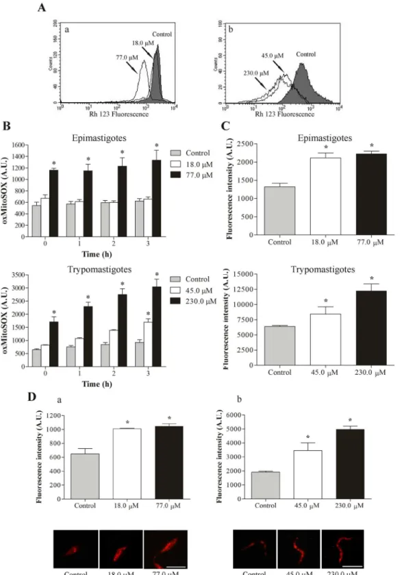
3.1. C4 induces mitochondrial depolarization
Based on previous studies that reported the effect ofC4onT.cruzimitochondria [6], we
evalu-atedΔCm inC4-treated cells by flow cytometry. The histograms showed a noticeably pro-nounced loss ofΔCm in both the epimastigote and trypomastigote forms ofT.cruziat the highest concentrations assayed after 3 h of treatment, with>60.0% reductions ofΔCm com-pared with the control group (Fig 2A). The positive control CCCP decreasedΔCm by 50.7% and 76.3% in epimastigotes and trypomastigotes, respectively (data not shown).
3.2. C4 increases mitochondrial O
2•−production
Based on ourΔCm results, we evaluated mitochondrial superoxide anion (O2•−) production.
Fig 2Bshows a significant increase in mitochondrial O2•−production at the highest
concentra-tions assayed for epimastigote and trypomastigote forms compared with the control group at all times tested. In epimastigotes that were treated with 77.0μM ofC4, we observed a 115.0%
Fig 2. Evaluation of mitochondrial membrane potential, ROS production and lipid inclusions inT.cruzitreated with C4. (A)Mitochondrial depolarization inT.cruzitreated withC4for 3 h and stained with the fluorescence probe Rh 123.(a)Epimastigote forms.(b)Trypomastigote forms. Arrows correspond to the concentrations tested. The control group (untreated parasites) is also shown.(B)Mitochondrial O2•−production in epimastigote and
trypomastigote forms ofT.cruzitreated withC4for up to 3 h. Mitochondrial O2•−production was evaluated using the fluorescence probe MitoSOX. At the indicated times, oxidized MitoSOX (oxMitoSOX) was fluorimetrically measured in the parasites.(C)Total ROS production in epimastigote and trypomastigote
C4, this increase was 84.0% and 230.0%, respectively, with 3 h of incubation. The positive con-trol (AA) also increased mitochondrial O2•−production (data not shown).
3.3. C4 increases total reactive oxygen species
In addition to mitochondrial O2•−production, we evaluated the production of reactive oxygen
species (ROS) inC4-treated parasites.Fig 2Cshows thatC4significantly increased total ROS production at both forms ofT.cruziafter 24 h of treatment compared with the control group. In epimastigotes that were treated with 18.0 and 77.0μM ofC4, the increase in total ROS was
60.0% and 68.0%, respectively. In trypomastigotes that were treated with 45.0 and 230.0μM of
C4, the increase was 32.0% and 92.0%, respectively. The positive control (H2O2) also increased total ROS production (data not shown).
3.4. C4 induces lipid body formation
Epimastigotes and trypomastigotes ofT.cruzithat were treated for 24 h withC4exhibited the presence of many lipid bodies marked with Nile red. Two assays showed this alteration: (i) fluorescence microscopy revealed the presence of lipid bodies, and (ii) the fluorimetric assay quantified this accumulation. These assays showed a concentration-dependent increase in the number of lipid bodies (Fig 2D), with an increase>50% for epimastigotes and trypomastigotes at both concentrations tested.
3.5. C4 induces phosphatidylserine exposure
Increases in ROS can lead to apoptosis-like cell death. Apoptosis is characterized by biochemi-cal alterations, including phosphatidylserine exposure [11,12]. We evaluated whetherC4
induces phosphatidylserine exposure. As shown inFig 3A, epimastigote and trypomastigote forms that were treated withC4exhibited an increase in annexin-V fluorescence intensity after 3 h of treatment compared with the untreated parasites, indicating phosphatidylserine expo-sure. The histograms showed a>30.0% increase in the intensity of annexin-V fluorescence at both concentrations tested for trypomastigote forms (Fig 3: e and f). For epimastigote forms, at the higher concentration, annexin-V fluorescence was observed in approximately 40.0% of the parasites (Fig 3: b and c).
3.6. C4 decreases cell volume
The present results indicate thatC4induced phosphatidylserine exposure, and we explored the action of this compound on the apoptosis cell death pathway. We performed additional experi-ments to evaluate cell shrinkage, a hallmark of apoptotic death [12,13]. As shown inFig 3B, a decrease in cell volume was observed in trypomastigotes at both concentrations ofC4tested after 3 h, with reductions of approximately 90.0% (Fig 3B: b). For epimastigotes, at the higher concentration, we observed a decrease in cell volume in approximately 20.0% of the parasites (Fig 3B: a).
3.7. C4 induces DNA fragmentation
Continuing the same line of reasoning, we then evaluated possible cell death by apoptosis, reflected by DNA fragmentation, using the TUNEL assay.Fig 3Cillustrates the analysis of
stained with the fluorescence probe Nile red.(a)Epimastigote forms.(b)Trypomastigote forms. The images suggest a random distribution of lipid-storage bodies in the parasites. Scale bars = 10μm. The data (B,CandD) are expressed as the mean fluorescence (in arbitrary units)±SE.*p0.05, significant
difference compared with the control group (untreated parasites).
Fig 3. Evaluation of phosphatidylserine exposure, cell volume, DNA fragmentation and cell membrane integrity inT.cruzitreated with C4. (A) Phosphatidylserine exposure in epimastigote and trypomastigote forms ofT.cruzitreated withC4for 3 h and stained with the fluorescence probes annexin V-FITC and PI.(a)Untreated epimastigote.(b, c)Epimastigotes treated with 18.0 and 77.0μM ofC4.(d)Untreated trypomastigotes.(e, f)Trypomastigotes treated with 45.0 and 230μM ofC4.(B)Reduction of cell volume ofT.cruzitreated withC4for 3 h and analyzed by flow cytometry.(a)Epimastigote forms.
(b)Trypomastigote forms. Forward light scatter (FSC-H) was considered to represent cell volume. Arrows correspond to the concentrations tested. The control group (untreated parasites) is also shown.(C)DNA fragmentation in epimastigote forms ofT.cruzitreated withC4for 24 h and stained with TUNEL. The gray column indicates differential interference contrast, and the black column indicates fluorescence.(a-b)Untreated epimastigotes.(c, d)
Epimastigotes treated with 18.0μM.(e, f)Epimastigotes treated with 77.0μM. Green fluorescence indicates DNA fragmentation. Scale bars = 10μm.(D)
Alteration of the cell membrane in epimastigote and trypomastigote forms ofT.cruzitreated withC4for 3 h and stained with the fluorescence probe PI. (a) Untreated epimastigotes. (b, c) Epimastigotes treated with 18.0 and 77.0μM ofC4. (d) Untreated trypomastigotes. (e, f) Trypomastigotes treated with 45.0 and 230.0μM ofC4. The numbers show the percentage of PI-positive cells in the upper right and left quadrants.
DNA fragmentation, which was performed following treatment of the parasites with different concentrations ofC4for 24 h. Epimastigotes that were treated with 18.0 and 77.0μM ofC4
(Fig 3C: d and f, respectively) exhibited bright fluorescence, indicating DNA double-strand ruptures compared with untreated parasites. Bright fluorescence was also observed with the positive control (actinomycin D; data not shown).
3.8. C4 induces alterations in cell membrane integrity
Previous work also demonstrated the effect ofC4on the cell membrane [6]. We further
evalu-ated the effect ofC4on membrane integrity in epimastigote and trypomastigote forms ofT.
cruzi.C4affected membrane integrity in both forms ofT.cruziafter 3 h of treatment compared
with untreated parasites. The histograms showed an increase in the intensity of PI fluorescence at both concentrations tested (49.34% and 73.64% PI-positive parasites inFig 3D: e and f), mainly for trypomastigotes, indicating alterations in cell membrane integrity. In epimastigotes at the higherC4concentrations, approximately 27% of the parasites were PI-positive (Fig 3D:
c). The positive control (digitonin) increased fluorescence by 41.02% and 93.82% in epimasti-gotes and trypomastiepimasti-gotes, respectively (data not shown).
Discussion
β-carbolines have presented numerous biological properties, such as antimicrobial [14], antitu-moral [15], antiviral [16] and antiparasitic [6–8] effects. In our recent studies, we demonstrated
thein vitroandin vivoactivity ofC4againstT.cruzi[6,7]. Additionally,C4induced low
cyto-toxicity, with a selective index higher to the parasites than for mammalian cells [6]. In the present study, we focused on elucidating the mechanism of action ofC4in the cell death of epi-mastigotes and trypoepi-mastigotes ofT.cruzi.
Our previous study reported ultrastructural alterations, especially in the mitochondria, in parasites that were treated withC4[6]. The present results confirmed that mitochondria are a target ofC4, reflected by the depolarization ofΔCm and increase in the production of mito-chondrial ROS and formation of lipid droplets in parasites treated withC4. Changes inΔCm are associated with opening of the permeability transition pore (PTP) in the mitochondrial membrane [17,18]. Thus,C4might induce the opening of PTP in the mitochondrial mem-brane, leading to activation of the apoptotic pathway [19]. This programmed cell death is commonly characterized by different morphological characteristics, such as exposure of phos-phatidylserine residues on the external leaflet of the cell membrane, decrease of cell volume and DNA fragmentation [20]. In addition, analysis of red Nile showed an increase of lipid bod-ies in the cytoplasm, which may indicate that theC4changes the content of phospholipids and sterols ofT.cruzi, and is strongly related to mitochondrial dysfunction [21]. Previous studies with an alkyl phosphocholine-dinitroaniline hybrid molecule [22] and antifungal azoles [23] showed similar results.
Our results demonstrated thatC4can induce several changes in the parasites that lead to cell death either by apoptosis or necrosis [12,27–29]. Our results suggest that mitochondria are one of the target organelles that may be involved in the increase in ROS production through the electron transport chain, which affects cellular structures and induces parasite death. Alto-gether, the present results suggest that new chemotherapeutic agents can be developed for the treatment of Chagas’disease.
Acknowledgments
We thank all the staff of the‘‘Laboratório de Inovação Tenológica no Desenvolvimento de Fármacos e Cosméticos”for their collaboration.
Author Contributions
Conceived and designed the experiments: HV VCD RHV CVN. Performed the experiments: HV VCD RHV. Analyzed the data: HV VCD RHV TUN MHS CVN. Contributed reagents/ materials/analysis tools: TUN SOS MHS CVN. Wrote the paper: HV VCD RHV TUN SOS MHS CVN.
References
1. World Health Organization (WHO).“Chagas disease (American trypanosomiasis)”. Fact sheet N° 340 Updated March 2015. World Health Organization, Geneva, Switzerland, 2015. Available:http://www. who.int/mediacentre/factsheets/fs340/en/
2. World Health Organization,“First WHO report on neglected tropical diseases: working to overcome the global impact of neglected tropical diseases”, Geneva, Switzerland, 2010. Available:http://whqlibdoc. who.int/publications/2010/9789241564090eng.pdf.
3. Coura JR, Castro SL. A critical review on Chagas disease chemotherapy. Mem Inst Oswaldo Cruz. 2002; 97: 3–24. PMID:11992141
4. Urbina JA, Docampo R. Specific chemotherapy of Chagas disease: controversies and advances. Trends Parasitol. 2003; 19 (11): 495–501. PMID:14580960
5. Coura JR. Present situation and new strategies for Chagas disease chemotherapy—a proposal. Mem Inst Oswaldo Cruz. 2009; 104: 549–554. PMID:19722074
6. Valdez RH, Dusman-Tonin LT, Ueda-Nakamura T, Dias Filho BP, Morgado-Diaz JÁ, Sarragiotto MH, et al. Biological Activity of 1,2,3,4-tetrahydro-β-carboline-3-carboxamide againstTrypanosoma cruzi.
Acta Trop. 2009; 110: 7–14. doi:10.1016/j.actatropica.2008.11.008PMID:19063858
7. Valdez RH, Dusman-Tonin LT, Ueda-Nakamura T, Silva SO, Dias Filho BP, Kaneshima PT, et al.In vitroandin vivotrypanocidal synergistic activity ofN
-butyl-1-(4-dimethylamino)phenyl-1,2,3,4-tetrahy-dro-β-carboline-3-carboxamide associated with benznidazole. Antimicrob Agents Chemother. 2012; 56 (1): 507–512. doi:10.1128/AAC.05575-11PMID:22037851
8. Volpato H, Desoti VC, Cogo J, Panice MR, Sarragiotto MH, Silva SO, et al. The effects of
N-Butyl-1-(4-dimethylamino)phenyl-1,2,3,4-tetrahydro-β-carboline-3-carboxamide againstLeishmania amazonensis
are mediated by mitochondrial dysfunction. Evid Based Complementary Altern Med. 2013; article ID 874367, 7 pages, 2013. doi:10.155/2013/874367
9. Piacenza L, Irigoin F, Alvarez MN, Pellufo G, Taylor MC, Kelly JM, et al. Mitochondrial superoxide radi-cals mediate programmed cell death inTrypanosoma cruzi: cytoprotective action of mitochondrial iron superoxide dismutase overexpression. Biochem J. 2007; 403 (2): 323–334. PMID:17168856
10. Yahya M, Fatemeh G, Abdolhosein D, Zohreh S, and Zuhair H. Effect of cantharidin on apoptosis of the
Leishmania majorand on parasite load in BALB/c mice. Res J Parasitol. 2013; 8 (1): 14–25.
11. Arambage SC, Grant KM, Pardo I, Ranford-Cartwright L, Hurd H. Malaria ookinetes exhibit multiple markers for apoptosis-like programmed cell deathin vitro. Parasit Vectors. 2009; 2 (32). doi:10.1186/ 1756-3305-2-32PMID:19604379
12. Duszenko M, Figarella K, Macleod ET, Welburn SC. Death of a trypanosome: a selfish altruism. Trends
Parasitol. 2006; 22: 536–542. PMID:16942915
14. Cao R, Chen H, Peng W. Design, synthesis and in vivo anti-tumor activities of novelβ-carboline deriva-tives. Eur J Med Chem. 2005: 40: 991–1001. PMID:15950325
15. Formagio ASN, Tonin LTD, Foglio MA, Madjarof C, de Carvalho JE, da Costa WF, et al. Synthesis and antitumoral activity of novel 3-(2-substituted-1,3,4-oxadiazol-5-yl) and 3-(5-substituted-1,2,4-triazol-3-yl)β-carboline derivatives. Bioorg Med Chem. 2008; 15 (6): 9660–9667.
16. Formagio ASN, Santos PR, Zanoli K, Ueda-Nakamura T, Dusman-Tonin LT, Nakamura CV, et al.
Syn-thesis and antiviral activity of 389β-carboline derivatives bearing a substituted carbohydrazide at C-3 against poliovirus and 390 herpes simplex virus (HSV-1). Eur J Med Chem. 2009; 44 (391): 4695– 4701.
17. Skulachev VP. Why are mitochondria involved in apoptosis? Permeability transition pores and apopto-sis as selective mechanisms to eliminate superoxide-producing mitochondria and cell. FEBS Lett. 1996; 397 (1): 7–10. PMID:8941703
18. Mandelker L. Introduction to oxidative stress and mitochondrial dysfunction. Vet Clin N Am-Small. 2008; 38 (1): 1–30. doi:10.1016/j.cvsm.2007.10.005PMID:18249243
19. Luo Y, Chang CK, Kessel D. Rapid initiation of apoptosis by photodynamic therapy. Photochem Photo-biol. 1996; 63: 528–534. PMID:8934765
20. Jiménez-Ruiz A, Alzate JF, MacLeod ET, Luder CG, Fasel N, Hurd H. Apoptotic markers in protozoan parasites. Parasit Vectors. 2010; 3: 1–15. doi:10.1186/1756-3305-3-1PMID:20051120
21. Lee SJ, Zhang J, Augustine MKC, Hong PK. Mitochondrial dysfunction induces formation of lipid drop-lets as a generalized response to stress. Oxid Med Cell Longev. 2013, article ID 327167, 10 pages. doi: 10.1155/2013/327167
22. Godinho JLP, Georgikopoulou K, Calogeropoulou T, De Souza W, Rodrigues JCF. A novel alkyl phos-phocholine-dinitroaniline hybrid molecule exhibits biological activity in vitro againstLeishmania amazo-nensis. Exp Parasitol. 2013; 135: 153–165. doi:10.1016/j.exppara.2013.06.015PMID:23845259
23. Macedo-Silva ST, Urbina JA, Souza W, Rodrigues JCF.In vitroactivity of the antifungal azoles itraco-nazole and posacoitraco-nazole againstLeishmania amazonensis. PloS ONE. 2013; 8 (12): e83247. doi:10.
1371/journal.pone.0083247PMID:24376670
24. Fernandes MP, Inada NM, Chiaratti MR, Araujo FFB, Meireles FV, Correia MT, et al. Mechanism of Try-panosoma cruzideath induced byCratyliamollisseed lectin. J Bionerg Biomembr. 2010; 42 (1): 69–78. doi:10.1007/s10863-010-9268-9PMID:20155390
25. Lazarin-Bidóia D, Desoti VC, Ueda-Nakamura T, Dias Filho BP, Nakamura CV, Silva SO. Further evi-dences for the mechanism of action of eupomatenoid-5: confirmation of ROS involvement owing mito-chondrial dysfunction inTrypanosoma cruzi. Free Radical Biol Med. 2013; 60: 17–28. doi:10.1016/j. freeradbiomed.2013.01.008PMID:23376033
26. Britta EA, Scariot DB, Falzirolli H, Ueda-Nakamura T, Silva CC, Filho BP, et al. Cell death and ultrastru-tural alterations inLeishmania amazonensiscaused by new compound 4-Nitrobenzaldehyde thiosemi-carbazone derived from S-limonene. BMC Microbiology. 2014; 14:236. doi: 10.1186/s12866-014-0236-0PMID:25253283
27. Matsuo AL, Silva LS, Torrecilhas AC, Pascoalino BS, Ramos TC, Rodrigues EG, et al.In VitroandIn Vivotrypanocidal effects of the cyclopalladated compound 7a, a drug candidate for treatment of Cha-gas' disease. Antimicrob Agents Chemother. 2010; 54 (8): 3318–3325. doi:10.1128/AAC.00323-10 PMID:20479201
28. Kroemer G, Galluzzi L, Vandenabeele P, Abrams J, Alnemri ES, Baehrecke EH, et al. Classification of cell death: recommendations of the nomenclature committee on cell death 2009. Cell Death Differ. 2009; 16: 3–11. doi:10.1038/cdd.2008.150PMID:18846107