746
Revista da Sociedade Brasileira de Medicina Tropical 43(6):746-748, nov-dez, 2010
Case Report/Relato de Caso
1. Oswaldo Cruz Foundation, Brasília, DF, Brazil. 2. School of Medicine, Catholic University of Brasília, Brasília, DF, Brazil. 3. Fernandes Figueira Institute, Oswaldo Cruz Foundation, Rio de Janeiro, RJ, Brazil. 4. Evandro Chagas Clinical Research Institute, Oswaldo Cruz Foundation, Rio de Janeiro, RJ, Brazil.
Address to: Dr. Vitor Laerte Pinto Junior. FIOCRUZ Brasília. PO Box 04311, ZIP 70904-970 Brasília, DF, Brazil.
Phone: 55 61 3329-4600 e-mail: [email protected] Received in 16/06/2010 Accepted in 20/08/2010
INTRODUCTION
CASE REPORT
Cryptococcus gatii
molecular type VGII as agent of meningitis in a healthy
child in Rio de Janeiro, Brazil: report of an autochthonous case
Cryptococcus gatii
tipo molecular VGII como agente causador de meningoencefalite em criança
saudável no Rio de Janeiro, Brasil: relato de um caso autóctone
Vitor Laerte Pinto Junior
1,2, Marcos Vinicius da Silva Pone
3, Sheila Moura Pone
3, João Maurício Scarpellini Campos
3,
José Roberto Pereira Garrido
3, Ana Cláudia Mamede Wiering de Barros
3, Luciana Trilles
4, Gláucia Gonçalves Barbosa
4,
Bernardina Penarrieta Morales
4, Cláudia de Carvalho Falci Bezerra
4and Márcia dos Santos Lazéra
4ABSTACT
Cryptococcus gatii causes meningoencephalitis in immunocompetent hosts, occurring endemically in some tropical and subtropical regions. Recently, this fungus was involved in an outbreak in Vancouver Island and British Columbia (Canada). In this temperate region, the VGII type is predominant. he paper describes an autochthonous case of meningoencephalitis by
C. gatii VGII in a previously health child in Rio de Janeiro, considered nonendemic region of Brazil. he fungus was identiied by biochemical tests and the molecular type was determined by UA5-RFLP. he present report highlights the need for clinical vigilance for primary cryptococcal meningitis in nonendemic areas.
Key-words: Meningoencephalitis. Immunocompetent. Cryptococcus gatii.
RESUMO
Cryptococcus gattii é causa de meningoencefalite em hospedeiros imunocompetentes, ocorrendo endemicamente em regiões tropicais e subtropicais. Recentemente foi causador de surtos na Ilha de Vancouver e na Columbia Britânica (Canadá). Nesta região de clima temperado, o tipo VGII é predominante. Relatamos um caso de meningoencefalite pelo C.gatii tipo VGII acometendo criança previamente saudável autóctone do Rio de Janeiro, região não endêmica do Brasil. O agente foi identiicado por testes bioquímicos e o tipo molecular determinado através de UA5-RFLP. O presente relato enfatiza a necessidade de vigilância clínica para a meningite criptocóccica primária em áreas não endêmicas.
Palavras-chaves: Meningoencefalite. Imunocompetente. Cryptococcus gatii.
Cryptococcus gatii is an agent of life-threatening disseminated infections in healthy, immunocompetent hosts. he most common clinical manifestations are meningoencephalitis and pulmonary disease, occurring mainly as endemic mycosis in tropical and subtropical regions. his primary emerging pathogen has atracted special atention during an outbreak of pulmonary and disseminated infection in Vancouver Island since 19991.
Using speciic primers for the minisatellite-speciic core sequence of the wild-type phage M13 and/or UA5-RFLP analysis2, four
molecular types VGI-VGIV are identiied and used for epidemiological studies for this species. VGII type is the principal agent, which caused human cases in Vancouver1. Infantile cryptococcosis is a rare event,
but it has been frequently diagnosed in healthy children in the north and northeast regions of Brazil3,4.
Considering the unexpected occurrence of meningitis caused by
C. gatii type VGII in an immunocompetent child born and resident in the State of Rio de Janeiro, the clinical-epidemiological features of this case are discussed.
Clinical and epidemiological data were obtained from analysis of the patient`s medical records, outpatient follow-up, domiciliary visits and interviews with family members. he family of the patient signed a consent form authorizing this report.
he primary isolate obtained from cerebrospinal luid (CSF) seeded on Sabouraud dextrose agar 2% medium was identiied by morphological and physiological tests, including phenol oxidase production on niger seed agar medium (NSA), cycloheximide sensitivity, assimilation of C and N sources (Vitek ICB, bioMerieux, Durham, USA), and the canavanine-glycine-bromothymol blue medium (CGB test) to identify the species.
High molecular weight DNA was extracted, according to Ferrer et al5, and the molecular type was identiied by UA5-RFLP, according
to Meyer et al2. The RFLP patterns were assigned visually by
comparison with the paterns obtained from the reference strains.
A ive years-old boy, born and resident in the Metropolitan area of Rio de Janeiro, was admited to a medical facility near his residence on January 3rd 2005 complaining of an abrupt onset of
fever, malaise, frontal headache, abdominal pain and post alimentary vomiting for two days. hese unspeciic manifestations were treated with oral amoxicillin and symptomatic medication. Ater four days, with improvement of his symptoms, he was discharged to continue treatment at home.
747
Pinto Junior VL et al - C. gatii VGII meningitis in a healthy child in Rio de Janeiro, Brazil
DISCUSSION
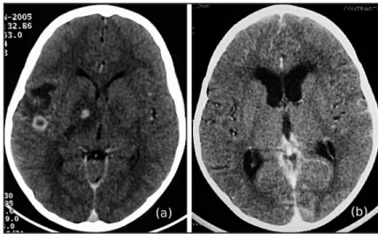
consciousness disturbance or meningeal signs during clinical examination. Computerized tomography (CT) scans of the brain revealed hypodense areas in the right basal ganglia and subcortical region, with ring enhancement ater iodine contrast infusion, strongly suggestive of neurotoxoplasmosis (Figure 1). Sulfadiazine and pyrimethamine administration were initiated. Ater a short stay, the patient was transferred to the Fernandes Figueira Institute (IFF).
Upon admission to IFF, laboratorial tests were negative for HIV (ELISA) and IgG and IgM serology for Toxoplasma gondii. A spinal tap was performed and showed 62 leukocytes/mm³ with 92% of mononuclear cells, protein levels of 70mg/dl and glucose levels of 63mg/dl. All bacteriological tests were negative, including tuberculosis investigation (direct bacterioscopy and culture). Antimicrobial drugs for neurotoxoplasmosis were discontinued and vancomycin, ceftriaxone and metronidazole were initiated to treat a possible cerebral abscess. Dexamethasone was also added to ameliorate the cerebral edema. Improvement in the patient’s clinical condition was observed.
FIGURE 1 - A: Initial contrasted CT scan of the brain showing hypodense nodular lesions with ring enhancement in the right basal ganglia and subcortical region with marked perilesional edema afecting the ipsilateral internal capsule. B: CT scan of the brain, ten days later (a), showing cerebral ventricle dilatations.
On day 10 of admission to IFF, the patient presented worsening of intracranial hypertension (ICH) along with meningeal irritation and tonic-clonic seizures. CSF culture was positive for Cryptococcus sp and following this result, alatex agglutination test also proved positive. As a consequence, all antibiotics were interrupted and 1mg/kg/d of amphotericin B deoxycholate was initiated. A new CT scan of the brain presented dilatations of the ventricular system (Figure 1) and another spinal tap was performed. The CSF revealed 19 leukocytes, 97% mononuclear cells, protein of 45mg/dL and glucose of 46mg/dL. The patient evolved with fluctuation of mental status. After failure of ICH control with serial lumbar punctures, a peritoneal shunt was implanted.
On day 18 of admission the patient presented with bilateral blindness and severe motor deficits, mainly limbs. Negative results of CSF cultures for fungus and of direct examination for
Cryptococcus sp occurred on day 22 following the onset of treatment. Other complications observed during inpatient stay were arterial hypertension, salt waste syndrome and low potassium levels as a consequence of amphotericin B administration.
The patient was discharged after 95 days with accumulated amphotericin B dosage of 400mg, over a six week period. The remainder of treatment was achieved with luconazole at a daily dose of 12mg/kg for four weeks.
Outpatient follow-up continues, showing the persistence of neurological sequelae, mainly hypotonus of the leg muscles, two seizure episodes (controlled with anticonvulsant drugs) and bilateral blindness, during the initial visits. At present, the patient presents expressive improvement of motor disability and atends a school for the visually handicapped.
Cr y ptococcosi s meningoencephaliti s af fecting an immunocompetent child represents a diagnostic puzzle, mainly because C. gatii is not a typical etiological agent of meningitis, but also because of its nonspeciic clinical manifestations. Consequently, delay in the diagnosis and in the onset of specific treatment is frequently observed4.
Studies involving infantile cryptococcosis in Brazil demonstrated that the most common manifestations are fever, headache, neck stifness and vomiting3; in this case, these suggestive manifestations
occurred ater the second week of onset.
Cranial CT scans can also cause confusion, since the aspect of the lesion is very similar to other diseases, such as neurotoxoplasmosis and cerebral abscess. In a study of tomographic alterations in 11 children with meningitis by C. gatii in the State of Pará, Brazil, all cases presented hypodense nodules, most of them located in the basal ganglia6. he same study also described hydrocephalus and
difuse cortical atrophy.
In the present case, the cryptococcal isolate from CSF was only identiied at a species level by a reference laboratory. he fact that the commercially available kits for yeast identiication do not discriminate C. neoformans from C. Gatii, must be taken into account.
his fact, together with the absence of case surveillance, limit current knowledge regarding C. gatii epidemiology in Brazil.
In the north and northeast regions of Brazil, encompassing the Brazilian Amazon forest and the semiarid savanna areas, the high proportion of HIV-negative children with cryptococcal meningitis conigure an unique epidemiological picture in the world. In the State of Pará, it an infantile cryptococcal meningitis frequency of 18.6% was veriied for 43 cases diagnosed from 2003 to 20076 and
24.3% in 78 cases diagnosed from 1992 to 1998 in the same state3.
A frequency of 33% of cases in children was observed in the State of Amazonas7 and 21% in the State of Piaui8. Epidemiological studies
in these regions show that the molecular type VGII is the main agent for cryptococcal meningitis in young adults and children9,10.
he previous few VGII cases identiied in the State of Rio de Janeiro were from patients who came from the northeast of the country9.
748
Rev Soc Bras Med Trop 43(6):746-748, nov-dez, 2010
REFERENCES
those from other regions are also necessary to verify possible variants in the molecular type VGII.
An autochthonous case in Rio de Janeiro caused by C. gatii
VGII type demands atention. he same molecular type is endemic in the north and northeast regions and seems to be spreading to Southeastern Brazil. he adaptive potential and expansive behavior of VGII has been observed in North America, where this agent is responsible for the ongoing outbreak of meningoencephalitis in Vancouver and is spreading into Paciic Northwest Region of the United States1.
It is necessary to include this agent as a potential etiological agent of meningitis in clinical practice in all regions of Brazil, to increment laboratorial conditions for storage and identiication of cryptococcal isolates and to establish surveillance concerning cryptococcal infection in our country.
FAPERJ, Rio de Janeiro, Brazil, for the inancial support provided (grant E-26/110.486/2007).
FINANCIAL SUPPORT
1. Kidd SE, Hagen F, Tscharke RL, Huynh M, Bartlett KH, Fyfe M, et al. A rare genotype of Cryptococcus gatii caused the cryptococcosis outbreak on Vancouver Island (British Columbia, Canada). Proc Natl Acad Sci USA 2004; 101:17258-17263.
2. Meyer W, Castañeda A, Jackson S, Huynh M, Castañeda E. Molecular typing of Ibero American Cryptococcus neoformans isolates. Emerging Infect Dis 2003; 9:189-195.
3. Corrêa MDP, Oliveira EC, Duarte RR, Pardal PP, Oliveira FDM, Severo LC. Cryptococcosis in children in the State of Pará, Brazil. Rev Soc Bras Med Trop 1999; 32:505-508.
4. Severo CB, Xavier MO, Gazzoni AF, Severo LC. Cryptococcosis in children. Paediatr Respir Rev 2009;10:166-171.
5. Ferrer C, Colom F, Frasés S, Mulet E, Abad JL, Alió JL. Detection and identiication of fungal pathogens by PCR and by ITS2 and 5.8S ribosomal DNA typing in ocular infections. J Clin Microbiol 2001; 39:2873-2879.
6. Correa MDPSC, Severo LC, Oliveira FDM, Irion K, Londero AT. he spectrum of computerized tomography (CT) indings in central nervous system (CNS) infection due to Cryptococcus neoformans var. gatii in immunocompetent children. Rev Inst Med Trop Sao Paulo 2002; 44:283-287.
7. Santos L. Criptococose no estado do Amazonas: estudo de 75 casos diagnosticados na Fundação de Medicina Tropical (1988-1998) [dissertation]. [Rio de Janeiro]: Instituto Oswaldo Cruz; 2000. 154p.
8. Martins L. Epidemiologia da criptococose em crianças e adultos jovens e diversidade de Cryptococcus neoformans no meio Norte do Brasil [dissertation]. [Rio de Janeiro]: Instituto Oswaldo Cruz; 2003. 78p.
9. Trilles L, Lazéra MDS, Wanke B, Oliveira RV, Barbosa GG, Nishikawa MM, et al. Regional patern of the molecular types of Cryptococcus neoformans and Cryptococcus gatii in Brazil. Mem Inst Oswaldo Cruz 2008; 103:455-462. 10. Santos WAD, Meyer W, Wanke B, Costa SPSE, Trilles L, Nascimento JLMD,