132
Wajnberg E. Takotsubo cardiomyopathy following subarachnoid hemorrhage
Radiol Bras. 2012 Mar/Abr;45(2):132–134
Takotsubo cardiomyopathy following subarachnoid
hemorrhage
*
Síndrome de takotsubo após hemorragia subaracnoidea
Eduardo Wajnberg1
Takotsubo cardiomyopathy corresponds to a syndrome characterized by a transient myocardial dysfunction affecting the left ventricular apex that classically occurs after major physical or emotional stress (also called “broken heart syn-drome” or “stress-induced cardiomyopathy”). The author describes the case of a patient with takotsubo cardiomyopa-thy induced by subarachnoid hemorrhage.
Keywords: Subarachnoid hemorrhage; Aneurysm; Takotsubo cardiomyopathy.
A síndrome de takotsubo corresponde a uma cardiomiopatia caracterizada por uma disfunção miocárdica transitória afetando ápice do ventrículo esquerdo, que classicamente ocorre após grande estresse físico ou emocional (também chamada “síndrome do coração partido” ou “cardiomiopatia induzida por estresse”). Descrevemos a ocorrência de síndrome de takotsubo em uma paciente induzida por hemorragia subaracnoidea.
Unitermos: Hemorragia subaracnoidea; Aneurisma; Cadiomiopatia de takotsubo. Abstract
Resumo
* Study developed at Hospital Universitário Clementino Fraga Filho – Universidade Federal do Rio de Janeiro (HUCFF-UFRJ), Rio de Janeiro, RJ, Brazil.
1. MD, MSc, Hospital Universitário Clementino Fraga Filho – Universidade Federal do Rio de Janeiro (HUCFF-UFRJ), Rio de Janeiro, RJ, Brazil.
Mailing Address: Dr. Eduardo Wajnberg. Rua Nina Rodrigues, 72/102, Jardim Botânico. Rio de Janeiro, RJ, Brazil, 22461-010. E-mail: [email protected]
Received July 19, 2011. Accepted after revision September 22, 2011.
Wajnberg E. Takotsubo cardiomyopathy following subarachnoid hemorrhage. Radiol Bras. 2012 Mar/Abr;45(2):132–134.
0100-3984 © Colégio Brasileiro de Radiologia e Diagnóstico por Imagem
CASE REPORT
pic and diuretic drugs. Electrocardiogram (ECG) revealed lateral ST segment eleva-tion and long QT interval. Cardiac enzyme tests demonstrated slightly high troponin and CPK levels, and echocardiogram showed decreased left ventricular ejection fraction associated with apical akinesia and to endovascular treatment by embolization
with platinum microcoils. In the postopera-tive period, the patient progressed with hemodynamic instability associated with left-sided heart failure and cardiogenic shock, requiring orotracheal intubation, mechanical ventilation, and use of inotro-INTRODUCTION
Takotsubo syndrome corresponds to a cardiomyopathy characterized by a tran-sient myocardial dysfunction affecting the left ventricular apex that classically occurs after major physical or emotional stress, and that is also called “broken heart syn-drome” or stress-induced cardiomyopa-thy”(1).
The authors report the case of a patient with takotsubo syndrome induced by sub-arachnoid hemorrhage.
CASE REPORT
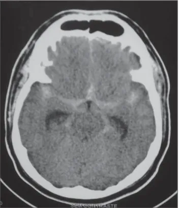
A 55-year-old woman presented an epi-sode of sudden and intense migraine with signs of Fisher grade 3 subarachnoid hem-orrhage (Figure 1). After digital angiogra-phy, which revealed the presence of an anterior communicating aneurysm with 3 mm in diameter, the patient was submitted
133
Wajnberg E. Takotsubo cardiomyopathy following subarachnoid hemorrhage
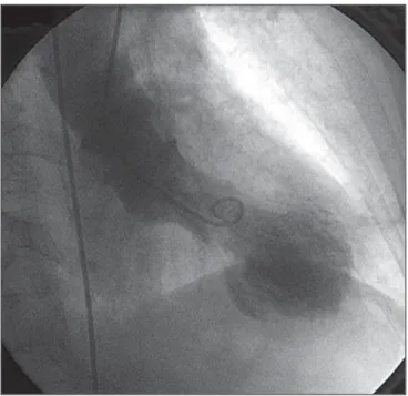
Radiol Bras. 2012 Mar/Abr;45(2):132–134 basal hypercontractility (Figure 2). The patient was referred for catheterization which did not revealed any significant ath-erosclerotic lesion. Ventriculography con-firmed the ballooned appearance of the left ventricle, resembling the shape of an am-phora characteristic of the syndrome (Fig-ure 3). In about one week, the clinical con-dition of the patient progressively im-proved, the isotropic drugs dose was de-creased, and the left ventricular function was recovered.
DISCUSSION
Takotsubo syndrome, also called “tran-sient left ventricular apical ballooning”, was described in 1990 by Sato et al.(2). The name of this syndrome refers to the appear-ance of the left ventricle during end systole at ventriculography and its resemblance with the amphora-shaped octopus trap (cor-responding to the Japanese term tako =
oc-topus, tsubo = trap – Figure 4). A
multifac-torial etiology is probable, but certainly there is involvement of a failure in auto-regulation of the myocardial microvascu-lature, transient coronary vasospasm and an abnormal response to catecholamines re-leased as a reaction against stress(3). There is a marked predominance (82% to 100%) of this syndrome in women, particularly
among those in the premenopausal period, with mean age between 62 and 75 years.
Diagnostic criteria include the balloon-ing appearance of the ventricle at echocar-diography or ventriculography, besides basal segments hypercontraction and alter-ations of the ST segment and T wave at ECG. In terms of clinical, biological and ECG features(4), the syndrome many times resembles acute myocardial infarction, but myocarditis and drug abuse, such as co-caine are also included in the differential diagnosis. The absence of significant coro-nary artery disease at cardiac catheteriza-Figure 2. Echocardiography, coronal section, four-chamber view
demonstrat-ing the ballooned appearance of left ventricle.
Figure 3. Ventriculography revealing the characteristic appearance of takotsubo syndrome: the amphora shape of the left ventricle at end systole.
tion must suggest the diagnosis of this syn-drome and, if suspected, left ventriculog-raphy should be performed(5).
The excessive release of catechola-mines triggered by physical or emotional stress is proposed as the main underlying mechanism in the pathogenesis of this phe-nomenon, which in the present case is sec-ondary to subarachnoid hemorrhage. Mag-netic resonance imaging is a promising method for the diagnosis and assessment of this new entity, allowing the differentiation between irreversible lesion characterized by late gadolinium enhancement and myo-Figure 4. Drawing of the
134
Wajnberg E. Takotsubo cardiomyopathy following subarachnoid hemorrhage
Radiol Bras. 2012 Mar/Abr;45(2):132–134 cardial edema(6). Thus, cardiac MRI can
assess myocardial viability, constituting a relevant prognostic tool(7). Generally, it may be said that the prognosis of this dis-ease is good, with complete myocardial contractility recovery after some days/ weeks(8,9).
CONCLUSION
Takotsubo syndrome is an increasingly recognized and diagnosed disease and, with the present case report, the authors highlight that it should be considered in the differential diagnosis of myocardial dys-function occurring in the context of intrac-ranial hemorrhages. Different imaging methods play a critical role in the
diagno-sis, and MRI plays a promising role in the assessment of this disease.
REFERENCES
1. Gianni M, Dentali F, Grandi AM, et al. Apical bal-looning syndrome or takotsubo cardiomyopathy: a systematic review. Eur Heart J. 2006;27:1523–9. 2. Sato H, Tateishi H, Uchida T, et al. Takotsubo type
cardiomyopathy due to multivessel spasm. In: Kodama K, Haze K, Hon M, editors. Clinical as-pect of myocardial injury: from ischemia to heart failure. Tokyo: Kagaku Hyoronsha; 1990. p. 56– 64. [in Japanese].
3. Wittstein IS, Thiemann DR, Lima JA, et al. Neu-rohumoral features of myocardial stunning due to sudden emotional stress. N Engl J Med. 2005;352: 539–48.
4. Bybee KA, Kara T, Prasad A, et al. Systematic re-view: transient left ventricular apical ballooning: a syndrome that mimics ST-segment elevation myocardial infarction. Ann Intern Med. 2004;141: 858–65.
5. Azzarelli S, Galassi AR, Amico F, et al. Clinical features of transient left ventricular apical balloon-ing. Am J Cardiol. 2006;98:1273–6.
6. Fernández-Pérez GC, Aguilar-Arjona JA, de la Fuente GT, et al. Takotsubo cardiomyopathy: as-sessment with cardiac MRI. AJR Am J Roentgenol. 2010;195:W139–45.
7. Eitel I, Behrendt F, Sareban M, et al. The utility of cardiovascular magnetic resonance imaging in Takotsubo cardiomyopathy (apical ballooning) for differential diagnosis, pathophysiological insights and additional findings. J Cardiovasc Magn Res. 2009;11(Suppl 1):O23.
8. Sharkey SW, Windenburg DC, Lesser JR, et al. Natural history and expansive clinical profile of stress (tako-tsubo) cardiomyopathy. J Am Coll Cardiol. 2010;55:333–41.