Immunohistochemical Expression of Cell Differentiation and Growth
in Neonate Cardiomyocytes
Tarcísio Fulgêncio Alves da Silva
1, Greyce Kelly de Souza
1, Mona Adalgisa Simões
1, Francisco Cesar Pabis
1, Lucia
de Noronha
1,2Pontifícia Universidade Católica do Paraná - PUC-PR1, Universidade Federal do Paraná - UFPR2, PR - Brazil
Mailing Address: Mona Adalgisa Simões
Itaiopolis, 254, apto 302, America. Postal Code 89204-100, Joinville, SC – Brazil
E-mail: [email protected] , [email protected]
Manuscript received February 21, 2012; manuscript revised February 21, 2012; accepted April 16, 2012.
Abstract
Background: The cardiac alterations during the fetal heart transition to extrauterine life have been explored by several animal studies and the cell mechanisms responsible for these modifications are not well documented in humans.
Objective: To evaluate the mechanism of cell differentiation into cardiomyocytes that occur in the first days of life, through immunohistochemical analysis of proteins involved in proliferation and muscle contraction processes, in samples of human neonate myocardium.
Methods: Cross-sectional study of paraffin-sample sections of myocardium from an autopsy database of human
neonates, divided into two sample groups: full-term neonates who died after a maximum of two days of life (NEO1) with 10 cases, and full-term infants who died between 3 and 10 days of life (NEO2) with 14 cases, in order to follow a temporal line that would contemplate the transition from fetal circulation to extrauterine life. The samples were studied in tissue microarray and the antibodies used were Ki67, PCNA, PTEN, Bcl2 (proliferation), HHF35 and sarcomeric actin (contractile proteins).
Results: Difference was observed regarding Ki67, p = 0.02; HHF35, p <0.01 and sarcomeric actin, p = 0.02, with Ki67 expression being higher in NEO1 group, whereas HHF35 and sarcomeric actin expression was higher in the NEO2 group.
Conclusion:The results suggest that cardiomyocytes have a proliferation characteristic (Ki67) in NEO1 which, following a temporal line, will be replaced by a differentiation characteristic (HHF35 and sarcomeric actin) in NEO2 (Arq Bras Cardiol 2012;99(3):797-801)
Keywords: Infant, newborn; myocytes, cardiac / classification; immunohistochemistry; phase transition;
placental circulation.
decrease in DNA synthesis with a consequent decrease in cell proliferation.
The cell mechanisms that are responsible for triggering and regulating this transition are not fully understood4.
Porrello et al. performed partial resection of the heart in rats aged one and seven days of life. They observed regeneration only in one-day-old rats, thus demonstrating that, for a brief period after birth, the mammalian heart seems to have capacity for regeneration5.
The design of these mechanisms is particularly important to understand cardiomyocyte regeneration processes, through undifferentiated primitive cells, after myocardial lesions in adults. The knowledge of intrinsic proliferation and cardiomyogenic potential of newborns can lead to a better understanding of these processes in adults, resulting in future clinical benefits in patients with severe myocardial lesions5.
The hypothesis of the present study is based on the fact that cardiomyocytes can undergo a gradual transition from the hyperplastic to the hypertrophic phenotype, following the temporal evolution of neonates, involving alterations in the immunohistochemical expression of some proteins that act
Introduction
During cardiac development, in animal studies, cardiomyocytes undergo numerous phenotypic alterations, going from an intrauterine proliferative phenotype (hyperplastic) to a cell growth and differentiation pattern in extrauterine life (hypertrophic), according to MacLellan and Schneider and Cortius et al.1,2.
Anversa and Nadal-Ginard reported that the transition of cardiomyocytes from a hyperplastic to a hypertrophic phenotype would provide a significant increase in cardiac contractile force3.
on cell growth and differentiation. Therefore, the objective of this study is to evaluate, by immunohistochemical analysis of some proteins involved in the processes of muscle proliferation and contraction, the mechanism of cell differentiation of cardiomyocytes in humans that can demonstrate the transition from fetal circulation to extrauterine life.
Methods
This study analyzed samples of left ventricular myocardium fixed in formalin and embedded in paraffin, from autopsies of full-term newborns (gestational age between 38 and 42 weeks), who died within 10 days of post-natal life (n = 106 cases) at the Pediatric and Perinatal Pathology Unit, Department of Pathology, Hospital de Clinicas, Universidade Federal do Parana (UFPR), between the years 1985 and 2005. This study was approved by the UFPR Ethics Committee under protocol # 1319.167/2006-11.
The cases were divided into two groups: NEO1, full-term newborns that died no more than two days after birth, and NEO2, who died between three and 10 days of post-natal life.
The study consisted initially of 56 cases in NEO1 and 50 cases in NEO2. After exclusion criteria were applied, 10 cases were selected for the NEO1 group and 14 cases for the NEO2 group.
The exclusion criteria included all cases of malformed, syndromic fetuses, fetal erythroblastosis cases, placental alterations or those with a gestational history of chronic arterial or specific gestational hypertension, pre-pregnancy or gestational diabetes, maternal infections, intrauterine growth retardation, oligohydramnios and polyhydramnios.
All cases were classified according to sex, gestational age, age and cause of death. Samples of myocardium were fixed in formalin and preserved in paraffin blocks, which led us to adopt the immunohistochemistry technique as the method of analysis of protein expression.
Immunohistochemistry seems to be the most appropriate technique for materials fixed in formalin and embedded in paraffin. It has been described over the years, as an important tool in the study of protein expression, in addition to being an inexpensive and easy-to-perform technique.
The results, however, are influenced by factors such as fixation time and processing of the material, the quality of the fixation agent and the chosen antibodies, the reactions themselves and the subjectivity when interpreting the slides. Nevertheless, immunohistochemistry has been widely used in material fixed in formalin and processed in paraffin due to its expression capacity even when using poor-quality tissues6.
Myocardium samples from each group were then mounted in tissue microarrays (TMA), using three fragments of LV myocardium per case, and each fragment measured 3 mm in diameter.
The immunohistochemical technique was used to demonstrate the specific antigens in myocardial samples fixed in formalin and embedded in paraffin and all antibodies were diluted with antibody diluent (Dakocytomation ®). The EnVision ® + Dual Link / Peroxidase (Dakocytomation ®) was used as the secondary antibody. As chromogen, used to reveal
the reaction, 3,3’-diaminobenzidine or DAB, chromogen-substrate system (Dakocytomation ®) was used. The following primary antibodies and dilution ratios were used: anti-PTEN (1:400; ® Novocastra), anti-Ki67 (1:150, Dako ®), anti-PCNA (1:400, Dako ®) and anti-Bcl2 (1:200, Dako ®) to assess cell proliferation, and HHF35 (1:400, Dako ®) and sarcomeric anti-actin (1:400, Dako ®) to assess contractile proteins. The reactions were controlled reactions with positive and negative controls.
The reactions of antibodies Ki67, PCNA, Bcl2 and PTEN for cell proliferation were analyzed by simple quantitative method by counting the nuclei of positive cardiomyocytes in the entire sample, under 400 X magnification.
The reactions of HHF35 and sarcomeric actin antibodies for contractile proteins were analyzed by the morphometric quantitative method, and this method reads the color intensity of the region with positive immunohistochemistry reaction and converts it into an area measurement, using the micrometer as unit. For that purpose an optical microscope was used with a 200 X magnification, in a binocular microscope (Olympus BX50 ®) coupled to a computer in which the Image Pro Plus ® software had been installed to perform the analysis. To compare the groups in terms of quantitative variables, the ANOVA model was considered, with a significance level of 0.05.
Results
A total of 24 newborn hearts were analyzed, showing, regarding gender, mean gestational age or days of life and cause of death, the following profile shown in Table 17.
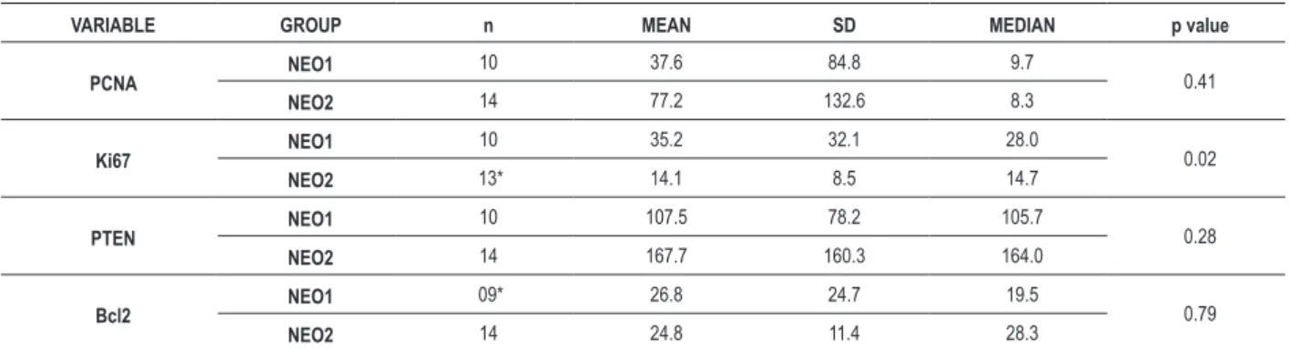
Among the immunohistochemical markers that suggest cell proliferation, Ki67 with p = 0.02 showed significant difference between NEO1 and NEO2 groups. Table 2 shows the descriptive statistics and p values for antibodies PCNA, Ki67, Bcl2 and PTEN that evaluate cell proliferation. The sarcomeric actin and HHF35 antibodies that evaluate contractile proteins showed significant differences between the two groups, with higher values in the NEO2 group, with their results shown in Table 3.
Discussion
PCNA and Ki67 have been used to study the cell cycle as important markers of proliferation and have been used in studies on cardiomyocyte cell growth1,2,8.
In the mammalian embryo, proliferation and differentiation of cardiomyocytes occur concomitantly during embryonic and fetal periods and continue to occur until a few days after birth2.
Recent studies of the immunohistochemical expression of Ki67 in infarcted human hearts have shown, however, that 4% of the cardiomyocytes showed positivity for this proliferative protein in infarcted regions. Furthermore, the phenotypic alterations found in the transition from fetal circulation to extrauterine life could include a gradual decrease in PCNA and Ki67 expression1,2,8-12.
In the present study, Ki67 showed a decrease in expression when groups NEO1 and NEO2 were compared (p = 0.02). This is in agreement with the idea of a decrease in the proliferative characteristics of cardiomyocytes in this period.
Another antibody that has an important role in cell cycle regulation is PTEN, which is involved in cell cycle progression,
cell migration and growth, as well as apoptosis13. Samples in
this study showed a difference in PTEN expression between the two groups; however, the differences were not statistically significant. PTEN, when activated, could be involved in blocking the cell capacity to move from G1 to S phase through cyclin inhibition, consequently blocking the proliferative capacity of cardiomyocytes in the extrauterine pathway13.
Increased expression of Bcl2 was observed, which followed the temporal evolution of the sample groups, although with no statistically significant difference. This fact might indicate greater resistance to apoptosis in the group of newborns who died after two days of life and therefore, a greater tendency to cell growth and differentiation2,9.
Table 2 – Results of the quantitative analysis of PCNA, Ki67, PTEN and Bcl2 antibodies, in number of positive cells per ield
VARIABLE GROUP n MEAN SD MEDIAN p value
PCNA NEO1 10 37.6 84.8 9.7 0.41
NEO2 14 77.2 132.6 8.3
Ki67 NEO1
10 35.2 32.1 28.0
0.02
NEO2 13* 14.1 8.5 14.7
PTEN NEO1
10 107.5 78.2 105.7
0.28
NEO2 14 167.7 160.3 164.0
Bcl2 NEO1
09* 26.8 24.7 19.5
0.79
NEO2 14 24.8 11.4 28.3
ANOVA with p < 0.05; * One case excluded due to low quality of the immunohistochemistry reaction.
Table 3 – Results of the morphometric analysis of positive areas of antibodies HHF35 and sarcomeric actin, in square micrometers per field
VARIABLE GROUP n MEAN SD MEDIAN p value
HHF35 Area NEO1 10 36876.8 13369.0 36353.5 < 0.01
NEO2 14 56540.0 10605.3 57433.1
Sarcomeric Actin Area
NEO1 10 22299.6 13698.3 23326.0
0.02
NEO2 14 12314.4 7221.7 12023.8
ANOVA com p < 0.05.
Table 1 – Proile of sample groups according to sex, mean age and cause of death
PROFIL
GROUP
SEX AGE
CAUSE OF DEATH %
MALE FEMALE MEAN SD
NEO1 30% 70% 16.3 hours 10.7 hours Perinatal Hypoxia 100%
NEO2 64.3% 35.7% 5.4 days 2.8 days
Hemorrhage Perinatal Hypoxia Bronchopneumonia
Other
The HHF35 antibodies and sarcomeric actin, which are used for the study of contractile proteins, showed statistically significant difference between the two sample groups, and the Neo2 group showed higher expression of both proteins (p < 0.01 and p = 0.02, respectively). This fact possibly demonstrates that the differentiation of cardiomyocytes occurs by increasing the volume of cardiac cells, through changes in the expression of sarcomeric proteins, which would result in changes in the functional properties of cardiomyocytes with an increase in capacity of power generation by the heart muscles during the postnatal development14.
Conclusions
We observed a decrease in Ki67 expression in relation to sample temporal evolution, showing a reduction in cardiomyocyte proliferative phenotype. The antibodies that marked contractile proteins (sarcomeric anti-actin and HHF35) showed increased expression following the temporal
evolution of the sample groups. These data are consistent with the substitution of the hyperplasic proliferative characteristic of this tissue during intrauterine life by a cell differentiation characteristic in extrauterine life.
Potential Conflict of Interest
No potential conflict of interest relevant to this article was reported.
Sources of Funding
There were no external funding sources for this study.
Study Association
This article is part of the thesis of doctoral submitted by Tarcísio Fulgêncio Alves da Silva, from Pontífica Universidade Católica do Paraná.
1. MacLellan RW, Schneider MD. Genetic dissection of cardiac growth control pathways. Annu RevPhysiol. 2000;62:289-319.
2. Corstius HB, Zimanyi MA, Maka N, Herath T, Thomas W, van der Laarse A, et al. Effect of intrauterine growth restriction on the number of cardiomyocytes in rat hearts. Pediatr Res. 2005;57(6):796-800.
3. Anversa P, Nadal-Ginard B. Myocyte renewal and ventricular remodelling. Nature. 2002;415(6868):240-3.
4. Evans HJ, Sweet JK, Price RL, Yost M, Goodwin RL. Novel 3D culture system for study of cardiac myocyte development. Am J Phisiol Heart Circ Physiol. 2003;285(2):H570-8.
5. Porrello ER, Mahmoud AI, Simpson E, Hill JA, Richardson JA, Olson EN, et al. Transient regenerative potential of the neonatal mouse heart. Science. 2011;331(6020):1078-80.
6. Cronin M, Pho M, Dutta D, Stephans JC, Shak S, Kiefer MC, et al. Measurement of gene expression in archival paraffin-embedded tissues: development and performance of a 92-gene reverse transcriptase-polymerase chain reaction assay. Am J Pathol. 2004;164(1):35-42.
7. Lahmers S, Wu Y, Call DR, Labeit S, Granzier H. Developmental control of titin isoform expression and passive stiffness in fetal and neonatal myocardium. Circ Res. 2004;94(4):505-13.
8. Field LJ. Modulation of the cardiomyocyte cell cycle in genetically altered animals. Ann N Y Acad Sci. 2004;1015:160-70.
9. Pasumarthi KB, Field LJ. Cardiomyocyte cell cycle regulation. Circ Res. 2002;90(10):1044-54.
10. McGill CJ, Brooks G. Cell cycle control mechanisms and their role in cardiac growth. Cardiovasc Res. 1995;30(4):557-69.
11. Li F, Wang X, Capasso JM, Gerdes AM. Rapid transition of cardiac myocytes from hyperplasia to hypertrophy during postnatal development. J Mol Cell Cardiol. 1996;28(8):1737-46.
12. Tang MK, Kindler PM, Cai DQ, Chow PH, Li M, Lee KK. Heart-type fatty acid binding proteins are upregulated during terminal differentiation of mouse cardiomyocytes, as revealed by proteomic analysis. Cell Tissue Res. 2004;316(3):339-47.
13. Simpson L, Parsons R. PTEN: life as a tumor suppressor. Exp Cell Res. 2001;264(1):29-41.
14. Siedner S, Kruger M, Schoeter M, Metzler D, Roell W, Fleischmann BK, et al. Developmental changes in contractility and sarcomeric proteins from the early embryonic to the adult stage in the mouse heart. J Physiol. 2003;548(Pt 2):493-505.