J. Braz. Chem. Soc., Vol. 18, No. 2, 437-439, 2007. Printed in Brazil - ©2007 Sociedade Brasileira de Química 0103 - 5053 $6.00+0.00
Short Report
*e-mail: [email protected]
Xeractinol – A New Flavanonol C-glucoside from
Paepalanthus
argenteus
var.
argenteus
(Bongard) Hensold
(Eriocaulaceae)
Anne Lígia Dokkedal,*,a Francisco Lavarda,a Lourdes Campaner dos Santosb and Wagner Vilegasb
a
Faculdade de Ciências, Universidade Estadual Paulista, CP 473, 17033-360 Bauru - SP, Brazil
b
Instituto de Química, Universidade Estadual Paulista, CP 355, 14801-970 Araraquara - SP, Brazil
Um novo composto, isolado a partir do extrato metanólico das folhas de Paepalanthus argenteus var. argenteus (Bongard) Hensold foi caracterizado como xeractinol, um novo diidroflavonol C-glucosilado. Sua estrutura foi elucidada com base em extensiva análise espectroscópica (RMN uni- e bidimensionais, EM, HREIMS, IV, UV). Cálculos ab initio de estrutura eletrônica corroboram nossa proposição para a estrutura molecular. O diidroflavonol aqui isolado pode servir como marcador taxonômico de Paepalanthus subg. Xeractis, uma vez que este flavonóide não foi encontrado em nenhum outro taxon de Eriocaulaceae.
New compound isolated from methanolic extract from the leaves of Paepalanthus argenteus
var. argenteus (Bongard) Hensold was characterized as xeractinol, a new dihydroflavonol C-glucoside. The structure was elucidated on the basis of extensive spectroscopic analysis (1D and 2D NMR, MS, HREIMS, IR and UV). Ab initio electronic structure calculations support our proposal to the molecular structure. The dihydroflavonol herein isolated may serve as taxonomic marker of Paepalanthus subgenus Xeractis, because this flavonoid have not been reported in any other taxon of Eriocaulaceae.
Keywords: Paepalanthus argenteus var. argenteus, Eriocaulaceae, dihydroflavonol, xeractinol, ab initio calculations
Introduction
Eriocaulaceae is a pantropical, predominantly herbaceous monocotyledonous family, comprising around 1200 species included in 10 genera.1 They are frequent
components of the vegetation in montane shallow pools or swamps, especially on sandy ground. This family is characterized as a monophyletic taxon with the following synapomorphies: very small, unisexual, white flowers, in dense capitula, with only one ovule per locule, and spiraperturate pollen.1
Eriocaulaceae is a rich source of phenolic compounds, like naphthopyranones and glycosilated flavonóides.2-5 We have already reportedthat flavonoids
and naphthopyranones are of taxonomic significance to the systematic of Eriocaulaceae.6,7 We report here
the isolation and characterization of xeractinol, a new flavanonol from the leaves of Paepalanthus argenteus
var. argenteus (Bongard) Hensold.
Results and Discussion
The flavanonol C-glucoside 1 (xeractinol) was obtained directly from the fractionation of the methanolic extract of leaves of P. argenteus var. argenteus by column chromatography on Sephadex LH-20 (Pharmacia) eluted with MeOH. It gave a yellow spot on TLC under UV light. The IR spectrum exhibited bands at 3425 (OH) and at 1656 (C=O) and 1604 cm-1 (C=C). The UV spectra showed
absorptions bands at 287 and 330 nm. The ES-MS spectrum of 1 showed a pseudo molecular ion [M+H]+ at m/z 467, consistent with a molecular formula C21H22O12. The [(M+H)-120]+ ion was formed by an A-type cleavage,
which is consistent with a C-6 glucoside substituent.9 The
peak at m/z 177 is due to the ion with formula C9H15O5, resulting from the retro-Diels-Alder fragmentation of 1
in which part of the sugar moiety is bounded to the A-ring, thus evidencing that the sugar moiety is linked to this part of the molecule. The [(M+H)-90]+ ion corresponds
to the cleavage of the B-ring. The negative HREIMS [M-H]- at m/z 465.1039 suggest C
438 Xeractinol – A New Flavanonol C-glucoside J. Braz. Chem. Soc.
formula. The 1H NMR of 1 showed two doublets with J
11 Hz at δ 4.95 and at δ 4.46, related to the H-2 and H-3, respectively, of a 3-hydroxy substituted flavanone.9 The
singlet at δ 5.92 was assigned to H-8. The doublet at δ
6.74 (2H, J 2.0) is related to H-2’ and H-6’ (magnetically equivalent) and the doublet at δ 6.87 (J 2.0) is related to H-4’. The glucose moiety was confirmed in C-linkage by the presence of an anomeric proton at δ 4.48 (J 10.0) as well as from six other signals between δ 3.67 and δ 3.98. The 13C NMR (DMSO d
6) spectrum showed 21 signals,
six of which could be assigned to a β-D-glucopyranosyl moiety and fifteen other signals were similar to those of a flavanonol moiety.8,12 Aglycone moiety 197.9 (C-4), 166.2
7), 162.7 5), 161.4 9), 145.9 3’), 145.1 5’), 128.1 1’), 119.5 6’), 115.4 4’), 115.3 (C-2’), 106.1 (C-6), 100.3 (C-10), 94.9 (C-8), 83.1 (C-2), 71.7 (C-3); sugar moiety 81.6 (C-5”), 79.2 (C-3”), 73.1 (C-1”), 70.8 (C-4”), 70.5 (C-2”), 61.7 (C-6a”). DEPT experiment allowed differentiating between C, CH and CH2. The C-2 signal appeared at δ 83.1 and the C-3 signal at δ 71.7. According to these values, aryl and hydroxyl substituents at C-2 and C-3 are equatorially oriented
(2,3-trans).8 The HMQC spectrum showed all the direct
correlations between C and H. The HMBC experiment showed the coupling between H-1”(δ 4.48) and C-6 (δ
106.1), C-7 (δ 166.2) and C-5 (δ 162.7). The HMBC experiment also showed correlations between the low field OH-5 signal (δ 12.41) and C-5 (δ 162.7), C-6 (δ 106.1) and C-10 (δ 100.3), confirming that the glucose moiety is bonded to C-6; between H-4’ (δ 6.87) andC-3’ (δ 145.5), C-5’ (δ 145.1), C-2’ (δ 115.3), C-6’ (δ 119.5); between H-2 (δ 4.95) and C-6’ (δ 119.5), C-2’ (δ 115.3), C-1’ (δ
128.1), C=O (δ 197.9); between H-3 (δ 4.46) and C-1’ (δ
128.1), C=O (δ 197.9); between H-2’ (δ 6.74) and C-1’ (δ
128.1), C-4’ (δ 115.4); between H-6’ (δ 6.74) and C-1’ (δ
128.1), C-4’ (δ 115.4); between H-8 (δ 5.92) and C-6 (δ
106.1), C-9 (δ 161.4) and C-10 (δ 100.3).
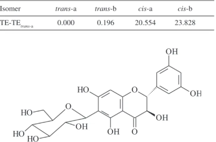
Considering the two hydrogen atoms bonded to carbon 2 and 3, there are four possible isomers. The isomer seen in Figure 1 was named trans a. Considering this structure, the isomer trans b has the hydrogen atoms with changed positions relative to the plane of the ring to which they are attached; to cis a the hydrogen attached to carbon 3 changes position with the hydroxyl group. Considering cis a, in cis b both hydrogen change position relative to the plane of the ring. In order to know which one is the most stable isomer, we made use of quantum chemistry theory. The results (Table 1) disfavor the cis configurations, once the difference in total energy between the trans and cis configurations is high enough to prevent the cis configurations to occur.
So, at least is possible to conclude that one of the trans
configurations is the most probable structure, once the difference in total energy between trans-a and trans-b is very low. Thus, 1 was identified as (2R, 3R)-2-(3,5-dihydroxyphenyl)-6-β -D-glucopyranosyl-2,3-dihydro-3,5,7-trihydroxy-4H-1-benzopyran-4-one (xeractinol), Figure 1.
According to previous works, P. subg. Platycaulon is characterized by naphthopyranone derivatives and 7-methoxy flavonol derivatives;3,4P. sect. Actinocephalus
presents mainly flavonol glycosides and acylated flavonols;2 in P. subg. Xeractis 6-methoxyflavone
derivatives are major constituents as well as in Leiothrix
and Syngonanthus, which also contain flavones rather than flavonols.6,7 The dihydroflavonol herein isolated may serve
as taxonomic marker of Paepalanthus subg. Xeractis, since this type of flavonoid have not been reported in any other
taxon of Eriocaulaceae.
Experimental
Plant material
Paepalanthus argenteus var. argenteus was collected at Serra do Cipó, Minas Gerais, Brazil in February 1998. Dr. Nancy Hensold (Field Museum, Chicago) carried out identification of plant material. Voucher specimens were deposited at Herbarium SPF, Brazil (P. argenteus var.
argenteus, SANO 920).
Extraction and isolation
The separated powdered leaves of the species were extracted with CHCl3 and then with 80% MeOH
Table 1. Differences of theoretical total energy (kJ mol-1), taking trans-a
as the reference level
Isomer trans-a trans-b cis-a cis-b TE-TEtrans-a 0.000 0.196 20.554 23.828
439 Dokkedal et al.
Vol. 18, No. 2, 2007
(maceration at room temp., one week each solvent). The solvents were evaporated in vacuum yielding black syrups. Two g of the 80% MeOH extract of P. argenteus var.
argenteus were redissolved in MeOH and subjected to gel permeation column chromatography on Sephadex LH-20 (Pharmacia) eluted with MeOH affording substance 1
(140 mg). Two other compounds were isolated after purification by HPLC (Dynamax RP-18, Varian 8 µm, 30 cm × 5 cm i.d., 1 mL min-1, λ 254 nm) and determined as
being quercetagetin 7-methyl ether 3-O-neohesperidoside (32 mg) and 3,4-dihydro-10-hydroxy-7-methoxy-3-methyl-1H-3,4-dihydro-naphtho-[2,3c]-pyran-1-one-9-O
-β-D-allopyranosyl (1-6)-β-D-glucopyranoside (27mg) by their spectrometric data.3,4,8,9 NMR spectra in DMSO-d
6
were obtained using a Varian INOVA 500 spectrometer, operating at 500 MHz for 1H and 125 MHz for 13C. The
DEPT (distortionless enhancement by polarization transfer) experiments were performed using a transfer pulse of 135°. Polarization transfer delays were adjusted to average CH coupling of 135 Hz. 1H-1H DFQ-COSY, 1H-13C HSQC, HMBC and 1D-TOCSY experiments were
obtained using conventional pulse sequences. Low resolution ES-MS spectra was performed in a Fisons VG Platform spectrometer in the positive mode (100V), the samples were dissolved in MeOH and injected directly. HREIMS of 1 was performed by using a ultrOTOFQ – ESI-TOF Mass Spectrometer Bruker Daltonics (Billerica, MA, USA) instrument in the negative mode; samples were dissolved in MeOH:H2O (80:20). IR: KBr. UV: MeOH. TLC: silica gel 60H (Merck, 10-40 µm). The flavonoids were detected with NP/PEG reagent under UV light.10
Quantum chemistry theory calculations, including geometry optimization and electronic structure, were carried out employing ab initio B3LYP/6-31G(d) density functional theory methodology with the GAMESS package.11 The calculations for all isomers were done in vacuo.
Supplementary Information
Supplementary data are available free of charge at http://jbcs.sbq.org.br, as PDF file.
Acknowledgments
The authors thank the financial support provided by FAPESP, CNPq and FUNDUNESP and the Dr. Norberto Peporini Lopes (FCFRP – USP) for the use of HREIMS equipment.
References
1. Giulietti, A. M.; Amaral, M. C.; Bittrich, V.; Kew Bull. 1995,
50, 55.
2. Andrade, F. D. P.; Santos, L. C.; Dokkedal, A. L.; Vilegas, W.;
Phytochemistry 1999, 51, 41.
3. Vilegas, W.; Dokkedal, A. L.; Rastrelli, L.; Piacente, S.; Pizza,
C.; J. Nat. Prod. 1999, 62, 746.
4. Vilegas, W.; Nehme, C. J.; Dokkedal, A. L.; Piacente, S.; Rastrelli, L.; Phytochemistry 1999, 51, 403.
5. Dokkedal, A. L.; Salatino, A.; Biochem. Syst. Ecol. 1992, 20, 31.
6. Dokkedal, A. L.; Ph.D. Thesis, Universidade Estadual Paulista, Brazil, 2000.
7. Dokkedal, A. L.; Sano, P.T.; Vilegas, W.; Biochem. Syst. Ecol. 2004, 32, 503.
8. Agrawal, P. K.; Carbon 13 NMR of Flavonoids, 1st ed., Elsevier:
Amsterdam, 1989.
9. Harborne, J.B.; The Flavonoids: Advances in Research, 1st ed.,
Chapman & Hall: London, 1996.
10. Wagner, H. M.; Bladt, S.; Zgainski, E. M.; Plant Drug Analysis, 1st ed., Springer: Berlin, 1984.
11. Schmidt, M. W.; Balbridge, K. K.; Boatz, J. A.; Elbert, S. T.; Gordon, M. S.; Jensen, J. J.; Koseki, S.; Matsunaga, N.; Nguyen, K. A.; Su, S.; Windus, T. L.; Dupuis, M.; Montgomery, J. A.; J.
Comput. Chem. 1993, 14, 1347.
12. Mbafor, J.T.; Fomum, Z.T.; J. Nat. Prod. 1989, 52, 417.
Received: February 7, 2006
Web Release Date: March 16, 2007