Functional changes of human
quadriceps muscle injured by
eccentric exercise
1Departamento de Fisioterapia, Unidade de Plasticidade Muscular,
Universidade Federal de São Carlos, São Carlos, SP, Brasil
2Grupo de Ressonância Magnética, Instituto de Física de São Carlos,
Universidade de São Paulo, São Carlos, SP, Brasil F.V. Serrão1, B. Foerster2,
S. Spada1, M.M.B. Morales1,
V. Monteiro-Pedro1,
A. Tannús2 and
T.F. Salvini1
Abstract
The present study evaluated functional changes of quadriceps muscle after injury induced by eccentric exercise. Maximal isometric torque of quadriceps and the surface electromyography (root mean square, RMS, and median frequency, MDF) of the vastus medialis oblique (VMO) and vastus lateralis (VL) muscles were examined before, immediately after and during the first 7 days after injury. Serum creatine kinase (CK) levels and magnetic resonance imaging (MRI) were used to identify muscle injury. The subject was used as her own control and percent refers to pre-injury data. Experiments were carried out with a sedentary 23-year-old female. Injury was induced by 4 bouts of 15 maximal isokinetic eccentric contractions (angular velocity of 5º/s; range of motion from 40º to 110º of knee flexion). The isometric torque of the quadriceps (knee at 90º flexion) decreased 52% immedi-ately after eccentric exercise and recovered on the 5th day. The highest reduction of RMS occurred on the 2nd day after injury in both VL (63%) and VMO (66%) and only VL recovered to the pre-injury level on the 7th day. Immediately after injury, the MDF decreased by 5 and 3% (VMO and VL, respectively) and recovered one day later. Serum CK levels increased by 109% on the 2nd day and were still increased by 32% on the 7th day. MRI showed large areas of injury especially in the deep region of quadriceps. In conclusion, eccentric exercise decreased the isometric torque and electromyographic signals of quadriceps muscle, which were recovered in one week, despite the muscle regeneration signals.
Correspondence
T.F. Salvini
Departamento de Fisioterapia, UFSCar 13565-905 São Carlos, SP
Brasil
Fax: +55-16-261-2081 E-mail: [email protected] Presented at the XVII Annual Meeting of the Federação de Sociedades de Biologia Experimental, Salvador, BA, Brazil, August 28-31, 2002. Research supported by FAPESP (No. 01/12352-1) and CNPq (No. 351197/92-3). S. Spada was the recipient of a fellowship from PIBIC-CNPq.
Received May 8, 2002 Accepted February 11, 2003
Key words •Isometric torque
•Surface electromyography
•Quadriceps muscle
•Muscle injury
•Eccentric exercise
•Magnetic resonance imaging
Eccentric exercise has been used as a physiological model to induce muscle injury in humans (1). It is well known that maximal eccentric muscle contractions can produce disruptions in the Z bands of sarcomeres, especially in muscles of people with seden-tary habits (2) or animals (3).
Skeletal muscle injury induced by
Most of the experiments used to study muscle injury and regeneration have em-ployed invasive methods in animals. Al-though many mammals have similar pro-cesses of skeletal muscle regeneration, it is necessary to improve the evaluation of non-invasive procedures used for the analysis of muscle injury and regeneration in humans. Noninvasive techniques such as surface EMG (9), force (5) and MRI (10) have been used. In the present study we evaluated the maximal isometric torque of the quadriceps femoral muscle and the electrical activity of the vastus medialis oblique (VMO) and vastus lateralis (VL) daily during the first week of the regeneration process after the muscle injury induced by eccentric exercise. Additi-onally, in order to detect muscle injury, we determined serum CK levels at three times during the study: before exercise and 2 and 7 days after injury. The MRI of the quadriceps femoral muscle was evaluated before exer-cise and daily during the 7-day recovery period.
A 23-year-old healthy woman with a sed-entary lifestyle gave informed consent to participate in the study, which was approved by the Ethics Committee for Human Studies of the Federal University of São Carlos, São Carlos, SP, Brazil. The participant did not have any orthopedic diseases and had not been previously involved in weight training. The subject was used as her own control and result (%) refers to pre-injury data.
Eccentric exercise and torque measure-ments were executed using a Multi-Joint System 2 Isokinetic Dynamometer (Biodex Medical Systems, New York, NY, USA). Muscle injury was induced by eccentric ex-ercise of the right quadriceps muscle. The volunteer was submitted to a total of 4 bouts, each with 15 maximal isokinetic eccentric contractions and a 5-min rest interval be-tween bouts. The contractions were carried out at an angular velocity of 5º/s and the knee flexion ranged from 40º to 110º.
In order to determine the maximal
iso-metric torque of the quadriceps muscle, six independent maximal isometric contractions were performed. Each contraction was main-tained for 4 s with a 2-min rest interval between contractions. The volunteer was positioned in such a way that the hip and knee formed angles of 100º and 90º, respec-tively. The mean value and standard devia-tions of the six measurements were consid-ered for the torque measurements during the study.
The same isometric muscle contractions were used to evaluate both the torque and the surface EMG. In the current experimental setup the instruments for torque and EMG measurements could not be synchronized exactly due to manual initiation of the ex-periments. However, the small delay of some fractions of a second was negligible com-pared to the 4-s duration of each contraction. Unlike torque measurement, EMG permits measurements on individual muscle compo-nents and was used in the present study to measure VMO and VL.
The electrical activity of the VMO and VL muscles was determined using simple active differential surface electrodes (Lynx Electronics Technologies, São Paulo, SP, Brazil) and a 16-channel signal-condition-ing module (1000-V2, Lynx Electronics Technologies). The simple active differen-tial surface electrodes consist of two rectan-gular parallel bars of Ag/AgCl (1 cm in length, 0.2 cm in width and separated by 1 cm). These bars are coupled to a rectangular acrylic resin capsule 2.2 cm in length, 1.9 cm in width and 0.6 cm high. In addition, the electrodes have a common mode rejection ratio with a minimum of 80 dB, an internal gain of 20 times and input impedance higher than 10 GΩ.
Aqdados data acquisition program version 4.6 (Lynx Electronics Technologies). This equipment also has a Butterworth type filter with a bandpass of 10.6 to 509 Hz and a gain of 50.
The EMG data were filtered with a bandpass of 10 to 450 Hz using post-pro-cessing procedures based on functions de-veloped to calculate the root mean square (RMS, in µV) and the median frequency (MDF, in Hz) and introduced in the Matlab 5.0 software. A Hanning window with a 90% overlap for the processing of Fast Fou-rier transformation was used to calculate the RMS and the MDF and the data were nor-malized by the mean.
The preparation of the experiment in-cluded shaving and cleaning the volunteer’s skin at the positions of the electrodes. Also, palpation of the muscle belly with the sub-ject in the testing position was used to deter-mine electrode placement. The electrodes were fixed with micropore adhesive tape at the midline of the muscle belly with their detection surface perpendicular to the muscle fibers (11). The reference electrode was fixed over the proximal anterior tibia shaft to elimi-nate possible external interferences. To en-sure the reproducibility of the different meas-urements along the one-week duration of the study, a plastic mold of the quadriceps was elaborated, on which the positions of every electrode were identified.
In a separate experiment but under iden-tical conditions, the contralateral femoral quadriceps of the same volunteer was stud-ied by MRI for visual determination of the effectiveness of eccentric exercise in induc-ing muscle injury. The MRI of the contralat-eral quadriceps femoral muscle was evalu-ated before and during the first 7 days after injury induced by eccentric exercise. The experiments were executed using a 0.5 Tesla NMR scanner developed by the Magnetic Resonance Group of the Physics Institute, São Paulo University, São Carlos, SP, Bra-zil. Axial images of the thigh were obtained
using the inversion-recovery technique with 2000-ms repetition time, 140-ms inversion time and 70-ms echo time. The field of view of the images was 256 x 256 mm with a 150 x 250 point acquisition matrix, slice thick-ness of 15 mm and 4 averages. Second order flow compensation was used in all three directions.
The two complementary techniques used to investigate muscle injury, torque meas-urement and EMG were applied once before the eccentric exercise for reference purposes, immediately after the exercise and then on a daily basis to monitor the recovery process over a period of 7 days. In addition to con-stant monitoring, the serum CK levels were determined at three time points during the study, i.e., immediately before the exercise and on the 2nd and 7th day, in order to determine the possible presence of muscle injury. The CK measurements were carried out using the CK-NAC UV kit from Unitest Wiener Laboratory (Rosario, Argentina).
Days
Root mean square (µV)
600 500 400 300 200 100 0
Pre Post 1 2 3 4 5 6 7
Creatine kinase (U/l)
150
100
50
0
Pre 2 7
Days 90
80 75
70 65
Pre Post 1 2 3 4 5 6 7
Days
Median frequency (Hz)
85
Maximal isometric
torque (Nm)
160 140 120 100 80 60 40 20 0
Pre Post 1 2 3 4 5 6 7
Days A
C
B
D
VMO VL
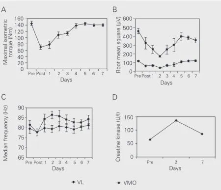
Figure 1A-C shows the results of torque and EMG measurements. Immediately after the eccentric exercise, there was a decrease in maximal isometric torque, as well as in the RMS and MDF values of both VL and VMO muscles. The maximal isometric torque of the quadriceps muscle decreased by 52% compared to its pre-exercise level and quickly started to recover, continually improving until reaching its pre-exercise level between the 4th and 5th day (Figure 1A). The MDF val-ues showed a similar behavior (Figure 1C) demonstrating a small initial decrease of 3 and 5% in VL and VMO, respectively. The RMS measurements also showed a 48 and 30% decrease of VL and VMO muscles, respectively, but, in contrast to the other experiments, they continued to decrease up to the 2nd day (Figure 1B).
When the recovery period was analyzed (days 1 to 7), a different behavior of maxi-mal isometric torque, RMS and MDF was observed. The maximal isometric torque of the quadriceps gradually recovered to pre-injury levels by the 5th day (Figure 1A). Although the largest reduction in RMS for both VMO and VL muscles occurred on the 2nd day after injury (63 and 66%, respec-tively), the VL muscle recovered the pre-injury values of RMS by the 7th day, while VMO did not recover them until the 7th day (Figure 1B).
The decrease of isometric torque, RMS and MDF, identified in the quadriceps muscle immediately after the eccentric exercise, in-dicates a failure in the mechanism of muscle fiber contraction, probably associated with muscle fiber fatigue. The difference in RMS between VMO and VL during the regenera-tion period cannot be easily explained, but one possibility that could be considered is that VMO muscle fibers were injured by the eccentric exercise more than VL fibers. Un-fortunately, it was not possible to compare the extent of injury between VMO and VL. It has been reported that RMS represents the number of active motor units during
muscle contraction, and is usually described as a measurement of skeletal muscle activity (12). As the maximal isometric torque is also a measurement of muscle activity, reduction in both isometric torque and RMS, as dem-onstrated in the present study, may be asso-ciated with muscle injury. Previous studies using eccentric exercise to induce injury in the quadriceps muscle also reported a de-crease in both muscle force (4) and RMS (13).
Immediately after injury, MDF decreased for both muscles (Figure 1C). A decrease in MDF after eccentric exercise was also ob-served in other studies (14,15). The decrease in MDF could be attributed to decreased conduction velocity of the active muscle fibers (16,17). Decreased muscle fiber con-duction velocity has been associated with proton accumulation (16). Generally, MDF may decrease more if the blood lactate con-centration is high, although changes were also observed in the absence of blood lactate (18). Other factors could also be associated with the decrease in MDF, such as impair-ment of the excitation-contraction coupling (18) and the selective injury of the fast twitch fibers after eccentric exercise (2), which is compensating for an increased activity of the slow twitch fibers trying to maintain force output (14). However, during the recovery period, an increase of both RMS and MDF was observed suggesting that after fatigue associated to muscle fiber injury, more muscle activation was needed to achieve the same relative torque level as pre-exercise level.
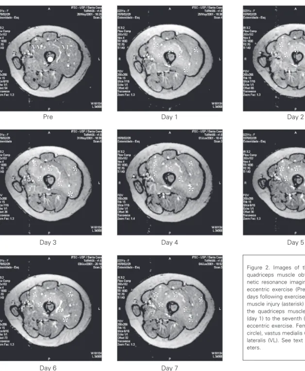
Figure 2. Images of the contralateral quadriceps muscle obtained by mag-netic resonance imaging (MRI) before eccentric exercise (Pre) and on the 7 days following exercise. Large areas of muscle injury (asterisk) are observed in the quadriceps muscle from the first (day 1) to the seventh (day 7) day after eccentric exercise. Femoral bone (Pre, circle), vastus medialis (VM) and vastus lateralis (VL). See text for MRI param-eters.
MRI showed large areas of muscle in-jury, predominantly located in the deep re-gions of the quadriceps. Observe the hyper-intense regions in the images from the 1st to the 7th day after injury (Figure 2) compared to the reference image obtained before exer-cise. The lesions formed diffuse areas of
muscle injury in all regions of the quadriceps muscle, although the deep regions of the muscle appeared to be more affected. Due to the diffuse character of the lesions and the low contrast of the images obtained it was not possible to compare the incidence of injury between VL and VMO, although it
Pre Day 1 Day 2
Day 3 Day 4 Day 5
could be seen that both muscles were af-fected.
In conclusion, the results of this study demonstrate that bouts of eccentric exercise decrease the isometric torque of the
quadri-ceps muscle, as well as the EMG signals of VMO and VL muscles and these changes are recovered within one week, despite the pres-ence of signals of muscle regeneration.
References
1. Prou E, Guevel A, Benezet P & Marini JF (1999). Exercise-induced muscle damage: Absence of adaptive effect after a single session of eccentric isokinetic heavy resistance exercise. Journal of Sports Medicine and Physical Fitness, 39: 226-232.
2. Lieber RL & Fridén J (1999). Mechanisms of muscle injury after eccentric contraction. Journal of Science and Medicine in Sport, 2: 253-265.
3. Clarkson PM (1992). Exercise-induced muscle damage - animal and human models. Medicine and Science in Sports and Exercise, 24: 510-511.
4. Brown S, Day S & Donnelly A (1999). Indirect evidence of human skeletal muscle damage and collagen breakdown after eccentric muscle actions. Journal of Sports Sciences, 17: 397-402.
5. Rinard J, Clarkson PM, Smith LL & Grossman M (2000). Response of males and females to high-force eccentric exercise. Journal of Sports Sciences, 18: 229-236.
6. Berry CB, Moritani T & Tolson H (1990). Electrical activity and soreness in muscles after exercise. American Journal of Physical Medicine and Rehabilitation, 69: 60-66.
7. Thompson D, Nicholas CW & Williams C (1999). Muscular soreness following prolonged intermittent high-intensity shuttle running. Jour-nal of Sports Sciences, 17: 387-395.
8. Nosaka K & Clarkson PM (1996). Changes in indicators of inflamma-tion after eccentric exercise of the elbow flexors. Medicine and Science in Sports and Exercise, 28: 953-961.
9. McHugh MP, Connolly DAJ, Eston RG & Gleim GW (2000). Electro-myographic analysis of exercise resulting in symptoms of muscle damage. Journal of Sports Sciences, 18: 163-172.
10. Foley JM, Javaraman RC, Prior BM, Pivarnik JM & Meyer RA (1999). MR measurements of muscle damage and adaptation after eccen-tric exercise. Journal of Applied Physiology, 87: 2311-2318. 11. DeLuca CJ (1997). The use of surface electromyography in
biome-chanics. Journal of Applied Biomechanics, 13: 135-163.
12. Basmajian JV & De Luca CJ (1985). Muscles Alive: Their Function Revealed by Electromyography. 5th edn. Williams & Wilkins, Balti-more.
13. Hortobágyi T, Houmard J, Fraser D, Dudek R, Lambert J & Tracy J (1998). Normal forces and myofibrillar disruption after repeated ec-centric exercise. Journal of Applied Physiology, 82: 492-498. 14. Linnamo V, Newton RU, Häkkinen K, Komi PV, Davie A, McGuigan
M & Triplett-McBride T (2000). Neuromuscular responses to explo-sive and heavy resistance loading. Journal of Electromyography and Kinesiology, 10: 417-424.
15. Linnamo V, Bottas R & Komi PV (2000). Force and EMG power spectrum during and after eccentric and concentric fatigue. Journal of Electromyography and Kinesiology, 10: 293-300.
16. Lindström L, Magnusson R & Petersen I (1970). Muscular fatigue and action potential conduction velocity changes studies with fre-quency analysis of EMG signals. Electromyography, 4: 341-356. 17. Merletti R, Rainoldi A & Farina D (2001). Surface electromyography
for noninvasive characterization of muscle. Exercise and Sport Sci-ences Reviews, 29: 20-25.