ABSTRACT
The association of diabetes insipidus and adipsia after craniopharyngioma surgery has high morbidity. Hypernatremia can be caused by adipsia and be aggravated by diabetes insipidus. Rhabdomyolysis rarely occurs. Case report:This is the first report of a diabetic patient with craniopharyngioma who developed diabetes insipidus and adipsia after surgery, evolving with severe hypernatremia that caused considerable rhabdomyolysis. Conclu-sion: The importance of the evaluation of muscle integrity when under hypernatremic states is pointed out. Although adipsia may have a simple solution through volunteer water ingestion, serious consequences such as repeated severe hypernatremia episodes and intense rhabdomyolysis with high morbidity could occur, if adipsia is not diagnosed. (Arq Bras Endocrinol Metab 2007;51/7:1175-1179)
Keywords: Rhabdomyolysis; Adipsia; Hypernatremia; Craniopharyn-gioma; Diabetes insipidus
RESUMO
Rabdomiólise Grave Devido a Hipernatremia Adípsica após Cirurgia de Craniofaringioma.
A associação de diabetes insipidus e adipsia após cirurgia de craniofarin-gioma implica em alta morbidade. Hipernatremia pode desenvolver-se devido a adipsia e ser agravada por diabetes insipidus. Rabdomiólise rara-mente ocorre. Descrição do caso:Esta é a primeira descrição de paci-ente diabético com craniofaringioma que desenvolveu diabetes insipidus e adipsia após a cirurgia, evoluindo com hipernatremia grave e conse-qüente rabdomiólise maciça. Conclusão:Ressalta-se a necessidade de avaliar a integridade muscular na vigência de estados hipernatrêmicos. Apesar de apresentar solução simples, como ingestão voluntária de água, pode haver sérias conseqüências se o diagnóstico de adipsia não é rea-lizado, como episódios repetidos de hipernatremia grave com rabdo-miólise intensa e elevada morbidade. (Arq Bras Endocrinol Metab 2007;51/7:1175-1179)
Descritores: Rabdomiólise; Adipsia; Hipernatremia; Craniofaringioma; Diabetes insipidus
D
IABETES INSIPIDUS CAN BE ASSOCIATEDwith craniopharyngioma, lead-ing to increased morbidity. Adipsia or hypodipsia rarely occurs as a hypothalamic lesion. The association of these two osmolarity dysfunctions causes a significantly higher morbidity (1). Hypernatremia can emerge from adipsia and be aggravated by diabetes insipidus. Clinical manifesta-tions include dehydration, muscular weakness, behavioural disturbances,apresentação de caso
DENISEE. ZANTUT-WITTMANN HERALDO MENDES GARMES ANITADENARDOPANZAN MARCELO DEOLIVEIRALIMA MARIATEREZAMATIASBAPTISTA
Endocrinology Division, Faculty of Medical Sciences, State University of Campinas (Unicamp), Campinas, SP.
delirium, lethargy, and coma. Rhabdomyolysis is a rare occurrence in hyperosmolarity states, and can progress with acute renal insufficiency and myoglobinuria. Muscular weakness and pain are marked symptoms and the diagnosis is confirmed through the elevation of creatine phosphokinase (CK) (2).
We report herein a diabetic patient with cranio-pharyngioma who developed diabetes insipidus and adipsia, as a complication of surgical resection of a suprasellar tumour, leading to a hypothalamic lesion, presenting repeated dehydration episodes with severe hypernatremia and at least one episode of massive rhabdomyolysis.
CASE REPORT
Male, 24 years, began his follow up in August 1997, presenting pubertal and growth delay, excessive weight gain and headache. Height = 1.63 m; weight = 74 kg; pubertal stage G2P2 (Tanner); bone age = 14 years (chronological age = 17 years, 10 months).
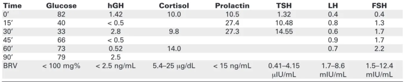
He presented amaurosis of the right eye and temporal hemianopsia of the left eye. Pituitary insuffi-ciency was revealed in the functional evaluation, except for the normal prolactin and thyrotropin response and free thyroxin levels (table 1). Magnetic resonance imaging (MRI) of sella turcica showed a heterogeneous suprasellar expansive lesion, predomi-nantly T1 hypointense and T2 hyperintense, with cys-tic areas and calcifications, displacing the 3rdventricle
and columns of the posterior fornix, pressing the optic nerve and gyrating laterally the optic tracts, with no sella turcica invasion (figure 1). Cerebral arteriography was in accordance to a suprasellar expansive mass. Lab-oratory findings were normal, except for hyper-glycemia (113 to 114 mg/dL), leading to the diagno-sis of diabetes mellitus. On November 1998, he was
submitted to a surgical transfrontal tumour resection. The anatomopathological diagnosis was craniopharyn-gioma. He developed diabetes insipidus and hyperna-tremia (Na = 173 mEq/L; reference values – RV: 133–145). He was normokalemic (K= 3.4 mEq/L, RV= 3.3–5.1). He started on intranasal Desmo-pressin–DDAVP (30 µg/day). Pituitary function was totally insufficient.
MRI (March/2002) showed gyrus rectus atro-phy, slight hypothalamic irregularity, free optic nerves, no identification of pituitary stalk, and no signs of recurrence. Bone age = 15 years (chronological age = 19 years, 01 month); Visual fields: lower lateral quad-rantopsia of the right eye and lateral hemianopsia of the left eye.
He was admitted 6 times at the Emergence Unit (January/1999 through August/2003), present-ing weakness and muscular pain, dehydration, hyper-natremia (158 to 173 mEq/L), hyperglycemia (126; 101; 116; 120; 224 mg/dL) and once with hyper-glycemic ketosis (980 mg/dL, Na = 161 mEq/L, K = 4.3 mEq/L). He was always normokalemic. In Sep-tember/2003, he referred to intense and generalized muscular pain weakness for the past two days and denied any thirst sensation. Laboratory findings: CK = 13,775 IU/L (RV < 170), Na = 165 mEq/L, K = 3.6 mEq/L, glucose = 224 mg/dL, Calcium (Ca) = 8.9 mg/dL (RV = 8.6–10.0), inorganic phosphorus (Pi) = 4.1 mg/dL (RV = 2.7–4.5), urea = 74 mg/dL (RV < 49), creatinine = 1.48 mg/dL (RV < 1.2). A CK peak was observed 24 hours after admission (CK = 21,261 IU/L, Na = 171 mEq/L, K = 4.2 mEq/L), and an acute non-oliguric renal insufficiency ensued as a com-plication. Daily CK (figure 2), natremia, and renal function were determined. The diagnosis was acute myopathy secondary to adipsic hypernatremia as a consequence of cranipharyngioma surgery. The treat-ment was based on natremia correction with
intra-Table 1.Pituitary functional evaluation before craniopharyngioma surgery.
Time Glucose hGH Cortisol Prolactin TSH LH FSH
0’ 82 1.42 10.0 10.5 1.32 0.4 0.4
15’ 40 < 0.5 27.4 10.48 0.8 1.3
30’ 33 2.8 9.8 27.3 14.55 0.6 1.7
45’ 66 < 0.5 0.9 1.7
60’ 73 0.52 14.0 0.7 2.2
90’ 79 2.5
BRV < 100 mg% < 2.5 ng/mL 5.4–25 µg/dL < 15 ng/mL 0.41–4.15 1.7–8.6 1.5–12.4
Figure 1. Magnetic resonance imaging of sella turcica showing a suprasellar expansive lesion in contact with the encephalic trunk, accom-plishing the 3rdventricle.
Figure 2. Graph representing serum creatine phosphokinase (CK) and sodium (Na) during the 6thhospital admission of the patient.
mEq/L IU/L
September/October/2003 180
160
140
120
100
80
60
40
20
0
13 15 17 19 21 23 25 27 29 1 3 5
25000
20000
15000
10000
5000
0
Na
venous hydration and on oriented and assisted water intake. After the reduction of CK levels and normali-sation of renal function, the patient was instructed to maintain continuous DDAVP use and regular and periodic water ingestion. Glucose control was attained with NPH and regular insulin and metformin.
Post-surgical visual field alterations remained unchanged. The muscle integrity follow up was per-formed through periodic CK, Na, and K serum deter-minations. His treatment includes daily prednisone, L-thyroxine, NPH and regular insulin, and monthly testosterone propionate.
He is now asymptomatic, although he has pre-sented some mild to moderate episodes of dehydration and consequences, resolved with outpatient treatment — as in June/2004: CK = 569 IU/L, Na = 147 mEq/L, K = 3.2 mEq/L, creatinine clearance = 113 mL/min/1.72 m2; glucose = 102 mg/dL, HbA1c = 5.8% (RV =
3.9–6.1) and in February/2006: CK = 1,258 IU/L, Na = 146 mEq/L, K = 3.4 mEq/L, creatinine = 0.94 mg/dL; glucose = 84 mg/dL, HbA1c = 6.5%.
DISCUSSION
Hypernatremia secondary to dehydration or to exces-sive saline administration is rare in healthy young adults. In hospitalised patients, hypernatremia is usual-ly secondary to erroneous intravenous administration of sodium (3). Only rarely do patients with cranio-pharyngioma develop adipsia or hypodipsia secondary to a tumour located in the hypothalamic region. How-ever, it is more common following surgery, occurring in approximately 20% of the cases (4). These alter-ations can lead to hypernatremic states that can unchain severe dehydration episodes, as well as behav-ioural disturbances, delirium, lethargy, coma, and muscular weakness due to rhabdomyolysis (1,2). Rhabdomyolysis is a rare osmolarity disturbance com-plication, characterised by muscular weakness and pain. Significant CK levels confirm the diagnosis. Acute renal failure and myoglobinuria may accompany the clinical picture. Immediate consequences of mus-cle destruction include hyperkalemia (increasing the risk of fatal heart arrhythmia) and hypocalcemia. Acute renal insufficiency is the result of renal vasoconstric-tion, intratubular myoglobin deposivasoconstric-tion, and nephro-toxicity caused by heme containing proteins (5). Hypokalemia, hypophosphatemia, and hyponatremia are uncommon causes of rhabdomyolysis (5).
Rhab-myopathy has been reported only a few times and the association with renal impairment is much more rare (2,8). One of the explanations for the development of muscle lesion by hypernatremia could be the inhibi-tion of Na-K transporter in the cytoplasm membrane (1). In patients whose myopathy was associated to hypernatremia (160 to 180 nmol/L), CK levels ranged from 500 to 60,000 UI/L and the occurrence of acute renal insufficiency was not rare. In some cases there was an association with germinoma, pinealoma, optic glioma or cerebral ischaemia. Diabetes insipidus was present in only a few cases (2,6,8,9). In this case, in addition to dehydration, the patient complained of intense pain and muscular weakness. Although he pre-sented severe dehydration and hyperglicemia, he did not mention thirst, information essential in order to establish the diagnosis of adipsia. Hypernatremia aggravated by dehydration and hyperglycemia can explain the aetiology of myopathy and acute renal insufficiency. The latter is probably due to myoglobin toxicity resulting from the rhabdomyolisis process after dehydration. It is relevant to highlight that the patient was euthyroid, normokalemic, normocalcemic, and normophosphatemic during rhabdomyolisis, hence the episodes were not precipitated by such metabolic disturbances. Muscle lesion was confirmed by extremely elevated CK levels, with a peak of 21,261 IU/L about 24 h after the admission. In such acute episodes, the treatment for dehydration corrected other hydro-electrolytic disturbances. Life long treat-ment of adipsia is needed to maintain a normal hydro-electrolytic balance, through systematic stimulated and regular water ingestion (1). Additionally, a periodic evaluation of sodium and CK serum levels is obligato-ry in these patients due to the frequent oligosympto-matic occurrence of mild or moderate dehydration and consequent rhabdomyolysis. This is the first report of a diabetic patient with craniophryngioma who devel-oped diabetes insipidus and adipsia as a complication of a hypothalamic tumour surgery, associated with hyperglycemia, contributing to the hyperosmolarity, evolving with hypernatremia and hydro-electrolytic imbalance that provoked severe rhabdomyolysis and acute renal insufficiency.
REFERENCES
1. Macias Batista A, Martinez Martins FJ, de Plabos Velasco PL. Diabetes insipidus and adpsic hypernatremia in a patient with craniopharyngioma. An Med Interna 1999;16:87-8. 2. Hiromatsu K, Kobayashi T, Fujii N, Itoyama Y, Goto I, Murakami
J. Hypernatremic myopathy. J Neurol Sci 1994;122:144-7. 3. Ofran Y, Lavi D, Opher D, Weiss TA, Elinav E. Fatal voluntary
salt intake resulting in the highest ever documented sodium plasma level in adults (255 mmol L-1): a disorder linked to
female gender and psychiatric disorders. J Int Med 2004;256:525-8.
4. Smith D, Finucane F, Phillips J, Baylis PH, Ficacane J, Tormey W, et al. Abnormal regulation of thirst and vasopressin secre-tion following surgery for craniopharyngioma. Clin
Endocrinol (Oxf) 2004;61(2):273-9.
5. Lane R, Phillips M. Rhabdomyolisis. BMJ 2003;327:115-6. 6. Kung AWC, Pun KK, Lam KSL, Yeung RTT. Rhabdomyolisis
associated with cranial diabetes insipidus. Postgrad Med J 1991;67:912-3.
7. Singhal PC, Abramovici M, Ayer S, Desroches L. Determinants of rhabdomyolysis in the diabetic state. Am J
Nephrol 1991;11:447-50.
8. Asahara H, Maruyama S, Motomura S, Tamura K, Mioshi T. A case of severe hypernatremia complicated with rhabdomy-olysis. Rinsho Shinkeigaku 1998;38:301-4.
9. Acquarone N, Garibotto G, Pontremoli R, Gurreri G. Hyperna-tremia associated with severe rhabdomyolysis. Nephron 1989;51:441-2.
Endereço para correspondência:
Denise Engelbrecht Zantut-Wittmann Disciplina de Endocrinologia Departamento de Clínica Médica Faculdade de Ciências Médicas
Universidade Estadual de Campinas (Unicamp) Caixa Postal 6111
Rua Tessália Vieira de Camargo 126 13084-971 Campinas, SP
Fax: (19) 3521-7408