Article
Printed in Brazil - ©2017 Sociedade Brasileira de Química0103 - 5053 $6.00+0.00*e-mail: [email protected]
A Novel Nanofibrous Film Chemosensor for Highly Selective and Sensitive Optical
Signaling of Zn
2+Chen Zhou* and Heng Liu
Research Center for Nanotechnology, Changchun University of Science and Technology, 130022 Changchun, People’s Republic of China
A novel nanofibrous film chemosensor was fabricated by copolymerization and electrospinning, which exhibited signaling behaviors for Zn2+. Upon addition of Zn2+, the sensor film exhibited
obvious fluorescence enhancement in a linear fashion. Owing to a larger surface area and high permeability, the selectivity and sensitivity of the nanofibrous film for Zn2+ were satisfactory and
achieving a limit of detection of 1.95 × 10⁻⁵ mol L-1. Moreover, this material could serve as an
adsorbent for Zn2+, as the adsorption capacity was 11.45 mg of Zn2+ ions per gram of nanofibrous
film.
Keywords: electrospinning, nanofibrous film, zinc ions, fluorescent sensor, adsorbent
Introduction
It is well known that Zn2+ is actively involved in
numerous physiological processes, such as brain function and pathology, gene transcription, immune function and mammalian reproduction.1,2 Zn2+ is the second most
abundant transition metal ion in human body after iron, so development of metal ion chemosensors for Zn2+ has
received considerable attention.3 Fluorescence detection
techniques have become powerful tools for sensing and imaging of trace amounts of metal ions because of their simplicity, sensitivity and real-time monitoring with fast response time.4 Nevertheless, single fluorescent sensor
molecule is not acceptable in practical application, especially in separation, removal and enrichment of target species.5,6 To introduce the solid substrate in preparing
fluorescent sensor could overcome the above problem effectively.7 In order to achieve the objective, we intend to
utilize electrospinning technology to fabricate nanofibrous film, which guaranteed the probes to be loaded in the solid substrate, furthermore, its porous network structure can greatly improve the diffusion velocity of interiors inside nanofibrous film.8
Electrospinning has been developed since 1934, it is an effective way for fabricating various porous composite nanofibers with high specific surface area and mechanical strength. With the development of nanotechnology,
it is increasingly concerned in many scientific fields such as chemical engineering, materials science and pharmacology.9-11 Several electrospun fibrous membrane
chemosensors for specific analytes (such as metal ions, anions, poisonous gas, etc.) were designed and synthesized in recent years, which were featured with large specific surface area, high porosity and good interconnectivity.12-19
However, according to the previous literatures, nanofibrous film for sensing Zn2+ was rarely reported. So synthesize
a functionalized electrospun nanofibrous film with high sensitivity and selectivity toward Zn2+ is very valuable.
Herein we reported a novel fluorescent nanofibrous film via copolymerization and electrospinning (Scheme 1). Upon addition of Zn2+, a striking fluorescence enhancement
was detected. The nanofibrous film also presented very excellent affinity for Zn2+ among other metal ions,
meanwhile the good adsorption of Zn2+ will expand its
application prospects.
Experimental
Synthesis of 2-hydroxy-4-acryloyloxy-benzaldehyde (HAB)20
2,4-Dihydroxybenzaldehyde (1.40 g, 10 mmol), hydroquinone (0.50 g, 4.5 mmol) and triethylamine (1.65 mL, 12 mmol) were dissolved in 200 mL 2-butanone. Then acryloyl chloride (12 mmol) in 30 mL 2-butanone was added dropwise into the mixture with constant stirring at
room temperature (25 °C) and kept stirring for 2 hours until precipitate appeared. The precipitate was filtered off and dried with anhydrous sodium sulfate. The crude 2-hydroxy-4-acryloyloxy-benzaldehyde (HAB) was recrystallized from ethanol. The product was collected as colorless oily liquid 1.02 g. Yield of pure product: 52.37%. 1H nuclear magnetic
resonance (NMR) (300 MHz, CDCl3) d6.26 (s, 1H, vinyl-H),
7.00 (m, 1H, vinyl-H), 7.01 (m, 1H, vinyl-H), 7.45 (t, 1H, J 3.3 Hz, Ph-H), 7.62 (d, 1H, J 3.3 Hz, Ph-H), 8.09 (s, 1H, Ph-H), 9.05 (s, 1H, carbonyl-H), 11.15 (s, 1H, OH).
Synthesis of salicylaldehyde-hydrazine
Salicylaldehyde (5.00 g, 41 mmol) in ethanol (50 mL) was added dropwise to the solution of hydrazine hydrate (5.00 g, 80 mmol) in ethanol at room temperature, then kept stirring for 12 h at room temperature. After removal of the solvent, the crude product was purified by column chromatography (silica gel, CH2Cl2/EtOH, 5:1). It was
produced 3.70 g of white crystals. Yield of pure product: 66.38%. 1H NMR (300 MHz, CDCl
3) d1.61 (s, 2H, NH2),
6.89 (m, 1H, Ph-H), 6.96 (t, 1H, J 3.6 Hz, Ph-H), 7.13 (m, 1H, Ph-H), 7.24 (m, 1H, Ph-H), 7.90 (s, 1H, Ar−H), 11.07
(s, 1H, OH).
Synthesis of poly (MMA-co-HAB)
HAB (1.90 g, 10 mmol), methyl methacrylate (MMA) (2.00 g, 20 mmol) and AIBN (0.02 g, 10 mmol) were dissolved in 10 mL DMF and introduced into a dry polymerization tube. The solution was deoxygenated by purging with N2 gas for 5 min. The tube was sealed and
placed in a regulated thermostat bath at 70 °C for 24 h. The obtained poly (MMA-co-HAB) was transparent and
colorless, then dissolved in CHCl3 (20 mL) and precipitated
with CH3OH (200 mL). After successive dissolving and
precipitation for 5 times, poly (MMA-co-HAB) was filtrated and dried under vacuum at 50 °C. Yield of pure product: 2.31 g (59.23%).
Synthesis of poly (MMA-co-Sal)
Poly (MMA-co-HAB) (0.35 g) reacted with salicylaldehyde-hydrazine (1.00 g, 7.3 mmol) in DMF (10 mL) at room temperature for 12 hours. The obtained poly (MMA-co-Sal) was pale yellow. It was dissolved in CHCl3 (20 mL) and precipitated with CH3OH (200 mL).
After successive dissolving and precipitation for 4 times, the poly (MMA-co-Sal) was filtrated and dried under vacuum at 70 °C. Yield of pure product: 0.73 g (54.07%).
1H NMR (300 MHz, CDCl
3) d2.51 (s, mH, CH), 3.17 (m,
3nH, methoxy-H), 3.34 (s, 2H, CH2), 4.12 (s, 3nH, CH3),
6.82 (d, 1H, J 3.3 Hz, Ph-H), 6.96 (m, 1H, Ph-H), 6.98 (m, 1H, Ph-H), 7.00 (m, 1H, Ph-H), 7.43 (m, 1H, Ph-H), 7.70 (d, 1H, J 2.7 Hz, Ph-H), 7.96 (s, 1H, Ph-H), 9.02 (s, 2H, Ar−H), 11.14 (s, 2H, OH).
Preparation of electrospinning precursory solution and film
at about 0.5 mL h-1 by the microinfusion pump. The
electrospinning was performed at 25 °C.
Results and Discussion
FTIR investigations of poly (MMA-co-Sal) film
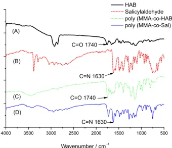
To confirm that fluorophore moiety was successfully grafted onto poly (MMA-co-HAB), Fourier transformed infrared spectroscopy (FTIR) was used. Figure 1 exhibited the FTIR spectra of (A) monomer HAB, (B) salicylaldehyde-hydrazine, (C) poly (MMA-co-HAB) and (D) poly (MMA-co-Sal) film. The characteristic bands of (A) and (C) at 1740 cm-1 (aldehyde C=O) confirmed that
aldehyde groups of HAB were successfully introduced into poly (MMA-co-HAB). But this band did not present in the spectra of (D), indicating that aldehyde and salicylaldehyde-hydrazine generated Schiff base salicylaldehyde-salicylaldehyde-hydrazine structure in the poly (MMA-co-Sal) film. Simultaneously, (B) and (D) exhibited a strong band at 1630 cm-1, which was
ascribed to C=N in the salicylaldehyde-hydrazine molecule. It was worth noting that the band of (D) at 1720 cm-1 might
be ascribed to N−N in the hydrazine molecule. Therefore,
we confirmed that salicylaldehyde-hydrazine group was successfully introduced into the nanofibrous film.
Morphologies of poly (MMA-co-Sal) nanofibers
Figure 2 shows the typical scanning electron microscope (SEM) images of the poly (MMA-co-Sal) nanofibrous film under different scales. It can be found that the poly (MMA-co-Sal) film was composed of numerous, randomly oriented nanofibers. The surface of the poly (MMA-co-Sal) (15 wt.%) nanofibrous film did not show any serious cracks or degradation under the optimized conditions. The average diameter (D) of nanofibers can be estimated in the following equation:
(1)
where n stands for the number of the nanofibers in SEM images, X stands for the diameter of each nanofiber, B is the scale bar, and L refers to the length of the scale bar. Therefore, the average diameter of poly (MMA -co-Sal) nanofibers is 1.26 µm as analyzed from SEM images (Figure 2b). This network structure of electrospun film provides a surface area-to-volume ratio roughly 1 to 2 orders of magnitude higher than that of known continuous thin films.21 This unique porous structure
could greatly accelerate the targets to diffuse close to the sensing elements and increase the complexation efficiency.22
Response of poly (MMA-co-Sal) nanofibrous film to Zn2+ ions
With the synthesis complete, the optical property of the poly (MMA-co-Sal) nanofibrous film in the presence of Zn2+ was investigated. Evidence for ion interaction with
the nanofibrous film was first sought using fluorescence spectroscopy, the fluorescence titration experiments were conducted under the condition of acetonitrile-Tris buffer solution at pH 7.1. The poly (MMA-co-Sal) nanofibrous Figure 1. FTIR spectra of (A) HAB; (B) salicylaldehyde; (C) poly (MMA-co-HAB); (D) poly (MMA-co-Sal).
film alone did not exhibit fluorescence at 504 nm [V(CH3CN):V(H2O), 9:1, pH 7.1, excited at 400 nm]. The
fluorescence intensity gradually enhanced upon the addition of Zn2+ from 0 to 200 µmol L-1, this “switch-on” process
could be observed under the irradiation of ultraviolet lamp (Figure 3). The increasing fluorescence intensity of the nanofibrous film depending on the concentration of Zn2+ was in a linear manner as illustrated in Figure 4
(R = 0.99255), which indicated that poly (MMA-co-Sal) nanofibrous film had potential application for quantitative determination of Zn2+, and the limit of detection (LOD)
could reach to 1.95 × 10⁻⁵ mol L-1 by calculation from this
linear relationship (based on LOD = KSb1/S, where K = 3;
Sb1 is the standard deviation of the blank solution; S is the
slope of the calibration curve).
Selective and competitive experiments
To gain insight into the selectivity of the fluorometric behavior of poly (MMA-co-Sal) nanofibrous film for Zn2+,
various common metal ions in environmental and biological interest were introduced to investigate their impact on the fluorescence response of poly (MMA-co-Sal). In selectivity experiments the fluorometric behavior of poly (MMA-co-Sal) was investigated upon addition of several metal ions such as K+, Al3+, Ni2+, Ca2+, Mg2+, Mn2+,
Fe3+, Cd2+, Pb2+, Hg2+, Cr3+, Cu2+, Co2+ (100 µmol L-1) in
acetonitrile-Tris buffer solution [V(CH3CN):V(H2O), 9:1,
pH 7.1, excited at 400 nm]. As shown in Figure 5 (black bar), only Zn2+ to the nanofibrous film caused a remarkable
fluorescence enhancement at 504 nm in the emission spectra, the introduction of other metal ions of K+, Al3+,
Ni2+, Ca2+, Mg2+, Mn2+, Fe3+, Cd2+, Pb2+, Hg2+, Cr3+, Cu2+,
Co2+ slightly affected the fluorescence. So it was clearly
indicated that our proposed nanofibrous film exhibited high selectivity to Zn2+ ions. In order to further test the
interference of other common cations in the determination of Zn2+, the competition experiments were performed:
the nanofibrous film were conducted with 30 µmol L-1
Zn2+ to induce fluorescence enhancement before mixing
300 µmol L-1 K+, Al3+, Ni2+, Ca2+, Mg2+, Mn2+, Fe3+, Cd2+,
Pb2+, Hg2+, Cr3+, Cu2+, Co2+. The fluorescence intensity
of the mixed system at 504 nm is shown in Figure 5 (red bar), all of the fluorescence intensity exhibited still enhancement, so the experimental results indicated that these ions showed no obvious interference for detecting Zn2+ in the fluorescence change at 504 nm. Thus the
fluorometric analysis above had proven that poly (MMA -co-Sal) nanofibrous film could serve as an outstanding sensitive and selective fluorescent sensor for Zn2+ in our
prospective. Figure 3. Fluorescent emission spectra of the nanofibrous film in
the presence of Zn2+ (0-200 µmol L-1) at different concentrations in
acetonitrile-water solution (9:1, v/v, 0.1 M Tris-HCl buffer at pH 7.10) (λex = 400 nm, λem = 504 nm).
Figure 4. Normalized response of the fluorescent signal to changing Zn2+
(0-200 µmol L-1) concentrations (λex = 400 nm, λem = 504 nm).
Adsorption kinetics of Zn2+ ions onto poly (MMA-co-Sal)
nanofibrous film
The adsorption and separation of Zn2+ was rarely
reported in previous studies about Zn2+ sensors. So in this
work, we comparative-deeply discussed the adsorptive and separable properties of poly (MMA-co-Sal) nanofibrous film in removing Zn2+ ions from solution.
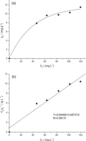
The nanofibrous film was cut to the size of 1.0 × 2.5 cm (about 8.97 mg in weight), and it was taken into the aqueous solution in various concentrations of Zn2+
for 24 hours.23 The relationship between equilibrium
adsorption amounts and equilibrium concentrations was demonstrated in Figure 6a. It was clearly depicted that the adsorption increased significantly with the increase in Zn2+ concentration until the saturation concentration
(100 mg L-1). As was illustrated in Scheme 2, the initial
increase in Zn2+ adsorption was due to the unsaturated
chelating sites being gradually occupied with the increasing Zn2+ concentration, when the chelating sites
became saturated, the equilibrium adsorption amounts leveled off as well.24 The concentration of Zn2+ left in
aqueous solution was inspected by inductively coupled plasma mass spectrometry (ICP-MS). The Langmuir adsorption equation was given to analyze experimental adsorption equilibrium data of Zn2+:25
(2)
where qe is the equilibrium quantity of the metals ions
adsorbed onto poly (MMA-co-Sal) nanofibrous film (mg g-1), C
e is the equilibrium concentration (mg L-1),
qm (mg g-1) and KL (L mg−1) are the Langmuir constants
related to the saturation adsorption capacity and binding energy, respectively.26 As was shown in Figure 6b, the
Langmuir Ceqe-1 versus Ce plot was in a good linear
relationship (R2 > 0.98). Based on the assumption of
the Langmuir theory that adsorption occurs at particular homogeneous sites within the adsorbent, this implies that one chelating sites is occupied by one metal ion exclusively, then no further adsorption occurs at that location. Thus, the adsorption that happened on the poly (MMA-co-Sal) nanofibrous film was monolayer.27,28 The
linear fit of the Ceqe-1 versus Ce plot was illustrated in
Figure 6b, relevant data indicated that Zn2+ ion adsorption
accorded with the Langmuir isotherm, and the values of KL and qm in Table 1 were calculated from the equation
in Figure 6b. The adsorption capacity was 11.45 mg of Zn2+ ions per gram of poly (MMA-co-Sal) nanofibrous
film (Table 1).
Scheme 2. Schematic illustration for preparation of poly (MMA -co-Sal) nanofiber fluorescent sensors by electrospinning for Zn2+ ions with
enhanced detection sensitivity.
Table 1. The Langmuir constants for Zn2+ on poly (MMA-co-Sal)
nanofibrous film
Metal ion KL / (L mg-1) qm / (mg g-1) R2
Zn2+ 0.135 11.45 0.98131
KL: related to the binding energy; qm: related to the saturation adsorption
capacity. Figure 6. (a) Adsorption isotherm and (b) Langmuir plot of Zn2+ on the
Conclusions
In conclusion, we synthesized a novel fluorescent nanofibrous film for sensing and adsorbing Zn2+ with
high selectivity and sensitivity.The limit of detection of the nanofibrous film for Zn2+ was calculated to be
1.95 × 10⁻⁵ mol L-1. Moreover, the adsorption capacity was
about 11.45 mg of Zn2+ ions per gram of the nanofibrous
film. So we expect that our sensing material to be a prospect in biomedical and environmental applications for the detection of Zn2+ in the future.
Supplementary Information
Supplementary information is available free of charge at http://jbcs.sbq.org.br as a PDF file.
Acknowledgments
The authors gratefully acknowledge the support of the Youth Science Foundation of Changchun University of Science and Technology (XQNJJ-2016-11) and the Natural Science Foundation of Inner Mongolia (No. 2015BS0202).
References
1. Fraker, P. J.; King, L. E.; Annu. Rev. Nutr. 2004, 24, 277. 2. Xu, Z.; Chem. Commun. 2012, 48, 4764.
3. Silva, A. P.; Gunaratne, H. Q.; Gunnlaugsson, T.; Huxley, A. J. M.; McCoy, C. P.; Rademacher, J. T.; Rice, T. E.; Chem. Rev. 1997, 97, 1515.
4. Tarkeshwar, G.; Anup, K.; Analyst 2011, 136, 4127.
5. Ito, H.; Matsuoka, M.; Ueda, Y.; Takuma, M.; Kudo, Y.; Lguchi, K.; Tetrahedron 2009, 65, 4235.
6. Kim, Y. R.; Kim, H. J.; Kim, J. S.; Kim, J. S.; Kim, H.; Adv. Mater. 2008, 20, 4428.
7. Wang, W.; Li, Y. P.; Sun, M. D.; Zhou, C.; Zhang, Y.; Li, Y. X.; Yang, Q. B.; Chem. Commun. 2012, 48, 6040.
8. Doshi, J.; Reneker, D. H.; J. Electrost. 1993, 35, 151.
9. Sun, L.; Yu, X.; Sun, M. D.; Wang, H. G.; Xu, S. F.; Dixon, J. D.; Wang, Y. A.; Li, Y. X.; Yang, Q. B.; Xu, X. Y.; J. Colloid Interface Sci. 2011, 358, 73.
10. Reneker, D.; Yarin, A.; Fong, H.; Koombhongse, S.; J. Appl. Phys. 2000, 87, 4531.
11. Shin, Y.; Hohman, M.; Brenner, M.; Rutledge, G.; Polymer 2001, 42, 9955.
12. Wang, X.; Drew, C.; Lee, S. H.; Senecal, K. J.; Kumar, J.; Samuelson, L. A.; Nano Lett. 2002, 2, 1273.
13. Onguna, M. Z.; Ertekin, K.; Gocmenturka, M.; Ergun, Y.; Suslu, A.; Spectrochim. Acta, Part A 2012, 90, 177.
14. Zhang, Y.; He, X.; Li, J.; Miao, Z.; Huang, F.; Sens. Actuators, B 2008, 132, 67.
15. Ding, B.; Yamazaki, M.; Shiratori, S.; Sens. Actuators, B 2005, 106, 477.
16. Zhou, C.; Xiao, N.; Li, Y. P.; Can. J. Chem. 2014, 92, 1092. 17. Song, X.; Liu, L.; Sens. Actuators, A 2009, 154, 175. 18. Nezhadali, A; Es’haghi, Z.; Bahar, S.; Banaei, A.; Shiran, J. A.;
J. Braz. Chem. Soc. 2016, 27, 99.
19. Wang, X.; Ding, B.; Sun, M.; Yu, J.; Sun, G.; Sens. Actuators, B 2010, 144, 11.
20. Wang, W.; Wang, X. L.; Yang, Q. B.; Fei, X. L.; Sun, M. D.; Song, Y.; Chem. Commun. 2013, 49, 4833.
21. Wang, X.; Drew, C.; Lee, S. H.; Senecal, K. J.; Kumar, J.; Samuelson, L. A.; Nano Lett. 2002, 11, 1273.
22. Wang, W.; Wang, X. L.; Yang, Q. B.; Fei, X. L.; Sun, M. D.; Song, Y.; Chem. Commun. 2013, 49, 4833.
23. Haider, S.; Park, S. Y.; J. Membr. Sci. 2009, 328, 90.
24. Ramazan, C.; Cengiz, S.; Mehmet, S.; Sep. Purif. Technol. 2006, 49, 107.
25. Saeed, K.; Haider, S.; Oh, T. J.; Park, S. Y.; Membr. Sci. 2008, 322, 400.
26. Deng, S.; Bai, R.; J. Colloid Interface Sci. 2003, 260, 265. 27. He, Z. Y.; Nie, H. L.; White, C. B.; Bioresour. Technol. 2008,
99, 7954.
28. Hsu, T. C.; J. Hazard. Mater. 2009, 171, 995.
Submitted: December 22, 2016