A contribution to the diagnosis of the small dentigerous
cyst or the paradental cyst
Contribuição para o diagnóstico do pequeno cisto
dentígero ou do cisto paradentário
José Hum ber to DAMANTE* Raul Ne grão FLEURY**
DAMANTE, J. H.; FLEURY, R. N. A con tri bu tion to the di ag no sis of the small dentigerous cyst or the paradental cyst.
Pesqui Odontol Bras, v. 15, n. 3, p. 238-246, jul./set. 2001.
The aim of this study was to ver ify the re la tion ship be tween the ra dio graph i cally mea sured width of the pericoronal space (PS) and the mi cro scopic fea tures of the fol li cle in or der to con trib ute to the di ag no sis of small dentigerous cysts and paradental cysts. One hun dred and thirty unerupted teeth (UT) and thirty-five par tially erupted teeth (PET) were radiographed and ex tracted. The ra dio graphic anal y sis con sisted of mea sur ing the width of the PS. The re sults of the ra dio graphic anal y sis were com pared with those of the histopathologic ex am i na tion of the den tal fol li cle. The width of the PS ranged from 0.1 to 5.6 mm. The most fre quently ob served lin ing of the fol li cles was a re duced enamel ep i the lium (REE) (68.4%) in UT and a hyperplastic strat i fied squamous ep i the lium (HSSE) (68.5%) in PET. In flam ma tion was pres ent in 36.1% of the UT and in 82.8% of the PET. There was a sta tis ti cally sig nif i cant as so ci a tion be tween the pres -ence of strat i fied squamous ep i the lium (SSE) and PS en large ment for UT (p < 0.05). There was a ten dency of as so ci a tion be tween in flam ma tion and PS en large ments in PET and, pos si bly, in UT, de spite the ab sence of sta tis ti cal sig nif i -cance. Sur gi cally, we did not de tect bone cav i ta tion or luminal cys tic con tents in pericoronal spaces smaller than 5.6 mm. We sug gest that the first ra dio graphic di ag no sis for a PS en large ment, in most of the rou tine clin i cal cases, should be of “in flam ma tion of the fol li cle”. The hy poth e sis of “dentigerous cyst” or “paradental cyst” is sug gested as a sec ond di ag no sis. The fi nal dif fer en tial di ag no sis be tween a small dentigerous or a paradental cyst and a pericoronal fol li cle de pends on clin i cal and/or sur gi cal find ings, such as the pres ence of bone cav i ta tion and cys tic con tent. UNITERMS: Tooth, unerupted; Dentigerous cyst.
INTRO DUC TI ON
Af ter the for ma tion of enamel, the crown of the tooth is sur rounded by the re duced enamel or gan and by ectomesenchyma. These two structures form the pericoronal fol li cle, which can be the or i -gin of sev eral types of dis eases dur ing or after odontogenesis. Hamartomas and odon to genic cysts or tu mors have been reported13,14,15,16,18,19,23, 24,29,34,
. Ab nor mal pericoronal fol li cles have also been as so ci ated with some syndromes28.
The dentigerous cyst is a le sion fre quently as so ci ated with unerupted teeth (UT). In the past, how -ever, many cysts con sid ered to be dentigerous turned out to be in flam ma tory paradental cysts1,5,31
or nor mal follicular vari a tions er ro ne ously di ag -nosed as cysts9,10,13,19,24,30
.
The patho logic pro cesses as so ci ated with the pericoronal fol li cle can be early iden ti fied by ra dio
-graphs that show an en large ment of the pericoronal space (PS). Ex act mea sure ments such as 2.0 mm, 2.5 mm and 5.0 mm in width have been used as ra dio graphic pa ram e ters for the di ag -no sis of dentigerous cysts since 19618,26,27,33
. In 1965 STAN LEY et al.34,35
dem on strated that in UT the re duced enamel ep i the lium (REE) pre dom i -nated in pa tients up to 22 years of age. The REE is then trans formed into a strat i fied squamous ep i -the lium (SSE) with -the nor mal ag ing of -the fol li cle. This in for ma tion re sulted in con fu sion and, some -times, in the misdiagnosis of small dentigerous cysts 2,4,11,19,20,22,25,30
. How ever, in 1987, ra dio graphic and mi cro scopic cri te ria were ex cluded from the di ag no sis of small dentigerous cysts10
. It was stated that the di ag no sis de pended on clin i cal and/or sur gi cal cri te ria such as the pres ence of bone cav i ta tion and luminal cys tic con tents. Re cent re ports have sup ported this con clu sion, em
pha siz ing the fact that the mi cro scopic fea tures of pericoronal fol li cles and dentigerous cysts are iden ti cal, with no pos si bil ity of differentiation9,13,18,19,24,30.
The pur pose of the pres ent study was to ver ify the re la tion ship be tween the ra dio graph i cally mea sured width of the PS and the mi cro scopic fea -tures of the fol li cles of unerupted teeth (UT) and par tially erupted teeth (PET) by in ves ti gat ing the role of in flam ma tion and the re la tion ship be tween this space and the type of ep i the lium. The study was car ried out in or der to con trib ute to the di ag -no sis of small patho log i cal PS en large ments such as those that oc cur with small dentigerous cysts and paradental cysts.
MA TE RI AL AND MET HODS
One hun dred and thirty UT and 35 PET were radiographed and ex tracted. Teeth were con sid -ered par tially erupted when any por tion of the crown was clin i cally ex posed in the mouth. The pericoronal fol li cles were sub mit ted to mi cro scopic eval u a tion. Most of the pa tients were in the sec ond and third de cades of life (51.5% and 41.2%, re -spec tively). The teeth most fre quently af fected were lower third mo lars (n = 84) and up per third mo lars (n = 38), fol lowed by ca nines (n = 21), pre mol ars (n = 17) and eumorphic su per nu mer ary teeth (n = 5). The pa tients were ran domly se lected among those re ferred to the sur gery clinic by den tists or by the de part ments of the School of Den -tistry of Bauru.
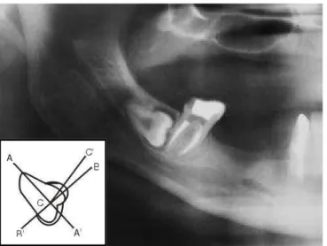
The wid est re gion of the PS was mea sured in periapical ra dio graphs or in pan oramic ra dio
-graphs. All pan oramic ra dio graphs were taken with the same X-ray ma chine (Ortopantomograph OP-3 - Palomex OY-Fin land & Siemens Corp-West Ger many). The con tours of the tooth and of the pericoronal space were traced on ultraphan pa per. Two per pen dic u lar lines (A-A’ and B-B’) were drawn on the im age of the tooth, one pass ing through the long axis and the other, through the cen ter of the crown (Fig ure 1). Starting from the in -ter sec tion of the two lines, a ruler (C-C’) was moved to the wid est point of the PS, where the mea sure ment was car ried out with a cal i per rule. The wid -est point was cho sen be cause THOMA36 has de
-scribed a lat eral form of dentigerous cyst. Im age mag ni fi ca tion in the pan oramic ra dio graphs was cor rected us ing a re duc tion of 0.5 mm, ac cord ing to LARA22.
The teeth were care fully ex tracted by means of a rou tine tech nique and care was taken to pre serve the fol li cle as much as pos si ble. The sur geon was al ways look ing for pericoronal bone cav i ta tion and luminal cys tic con tents. In some cases, when the ra dio graphic exam sug gested the pos si bil ity of a cys tic le sion, an as pi ra tion bi opsy was car ried out be fore the sur gery.
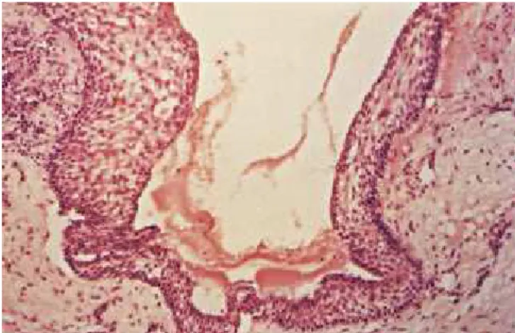
The fol li cles were mac ro scop i cally cut into semiserial sec tions, and 15 sec tions per spec i men were stained with hematoxylineosin. The ep i the -lium was iden ti fied as re duced enamel ep i the -lium (REE) when the su per fi cial cells were cy lin dri cal (Fig ure 2) or cuboidal (Fig ure 3), re sem bling typ i cal or re duced ameloblasts. When the su per fi cial cells were flat tened, the ep i the lium was iden ti fied as strat i fied squamous ep i the lium (SSE) (Fig ure 4) re
-FIGURE 1 - Met hod uti li zed for me a su ring the pe ri co ro -nal spa ce. Width = 2 mm.
gard less of the num ber of lay ers or the or ga ni za tion of the basal layer. Hy per pla sia was con sid ered to be pres ent when the num ber of lay ers ex ceeded twenty cell rows. The pres ence or ab sence of in flam ma tion, clas si fied as mild, mod er ate or in tense, was eval u -ated on the ectomesenchymal wall (Fig ure 5).
In or der to as so ci ate the dif fer ent ob served char ac ter is tics, the chisquare and Fisher tests were ap -plied. In flam ma tion de grees were com bined and con sid ered as be ing pres ent or ab sent. The width of the PS was di vided into two groups (nor mal and en -larged). En large ment was con sid ered when the mea sured width was greater than 3 mm.
RE SULTS
The ra dio graphic width of the PS of the spec i
-mens ranged from 0.1 mm to 5.6 mm; it ranged from 1 mm to 3 mm in most cases (86.6%) (Chart 1).
The REE was the most fre quent type of ep i the lium in the fol li cles of UT (68.4%) and in flam ma tion was pres ent in 36.1% of the cases. We also de -tected SSE in 20% of the cases, hyperplastic strat i fied squamous ep i the lium (HSSE) in 12.3%, and the ab sence of ep i the lium (AE) in 13% (Ta bles 1 and 2). In some fol li cles, two types of ep i the lium were pres ent or there was trans for ma tion of REE into SSE (Fig ure 6). In the fol li cles of PET, the most fre quent type of ep i the lium was the HSSE (68.5%), and in flam ma tion was pres ent in 82.8% of cases. We also de tected SSE in 17.1% of these fol li cles, REE in 5.7%, and AE in 11.4% (Ta bles 3 and 4). The sta tis ti cal anal y sis dem on strated no sig nif i
-FIGURE 3 - Re du ced ena mel epit he li um with cu bo i dal su per fi ci al cells and stra ti fi ca ti on of the un derl ying la -yers. Note the pre sen ce of a mild mo no nu cle ar in fil tra te on the con nec ti ve tis sue wall. Une rup ted to oth. Pe ri co -ro nal spa ce of 3.3. mm (H.E., X 160).
FIGURE 4 - Stra ti fi ed squa mous epit he li um with in ter -cel lu lar ede ma. Par ti ally erup ted to oth (H.E., X 100).
FIGURE 5 - Hyper plas tic stra ti fi ed squa mous epit he li um with nu me rous epit he li al cris tae. The con nec ti ve tis -sue wall is in ten sely in fla med. Par ti ally erup ted to oth. Pe ri co ro nal spa ce of 1.1 mm (H.E., X 40).
cant as so ci a tion be tween in flam ma tion and width of the PS for both UT and PET (p > 0.05). No sig nif i cance was ei ther ob served be tween the type of ep i -the lium and -the width of -the PS (p > 0.05) ex cept for the SEE in UT (p < 0.05).
The main find ings of this study are sum ma rized be low:
1. The ra di o grap hi cally me a su red pe ri co ro nal spa ce (PS) width ran ged from 1 mm to 3 mm in
most une rup ted (UT) and par ti ally erup ted te -eth (PET). The se me a su re ments cor res pond to nor mal fol li cles.
2. The pe ri co ro nal fol li cles of UT were most fre -quently li ned by re du ced ena mel epit he li um (REE). Tho se of PET were li ned by hyper plas tic stra ti fi ed squa mous epit he li um (HSSE).
3. PS en lar ge ments from 3.0 mm to 5.6 mm were fre quently re la ted to the pre sen ce of hyper plas -tic stra ti fi ed squa mous epit he li um (HSSE) with dif fe rent de gre es of hyper pla sia, and in flam ma -ti on of con nec -ti ve -tis sue.
DIS CUS SI ON
The re la ti ons hip bet we en pe ri co ro nal spa ce, in flam ma ti on and li ning epit he li um
The dis tri bu tion of the sam ple (Chart 1) shows that the ma jor ity of the PS widths ranged from 1 mm to 3 mm. In our study, we de fined as nor mal widths those smaller than 3 mm. The def i ni tion was based on the dis tri bu tion of our sam ple and on the con sulted lit er a ture which de scribes nor -mal pa ram e ters from 1.0 mm to 5.0 mm8,26,27,33.
The o retically, in flam ma tion should not be ex -pected in fol li cles of UT but it was pres ent in 36.1%
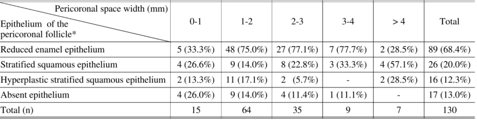
TABLE 1 - Re la ti ons hip bet we en the epit he li um of the pe ri co ro nal fol li cle and the pe ri co ro nal spa ce en lar ge ment in une rup ted te eth.
Pe ri co ro nal spa ce width (mm)
Epit he li um of the pe ri co ro nal fol li cle*
0-1 1-2 2-3 3-4 > 4 Total
Re du ced ena mel epit he li um 5 (33.3%) 48 (75.0%) 27 (77.1%) 7 (77.7%) 2 (28.5%) 89 (68.4%)
Stra ti fi ed squa mous epit he li um 4 (26.6%) 9 (14.0%) 8 (22.8%) 3 (33.3%) 4 (57.1%) 26 (20.0%)
Hyper plas tic stra ti fi ed squa mous epit he li um 2 (13.3%) 11 (17.1%) 2 (5.7%) - 2 (28.5%) 16 (12.3%)
Absent epit he li um 4 (26.0%) 9 (14.0%) 4 (11.4%) 1 (11.1%) - 17 (13.0%)
To tal (n) 15 64 35 9 7 130
*In some ca ses the re was du pli city of epit he li a. n - num ber of ca ses.
TABLE 2 - Re la ti ons hip bet we en in flam ma ti on of the pe ri co ro nal fol li cle and pe ri co ro nal spa ce en lar ge ment in une -rup ted te eth.
Pe ri co ro nal spa ce width (mm)
Inflam ma ti on
0-1 1-2 2-3 3-4 > 4 Total
Pre sent 6 (40.0%) 24 (37.5%) 9 (25.7%) 3 (33.3%) 5 (71.4%) 47 (36.1%)
Absent 9 (60.0%) 40 (62.5%) 26 (74.3%) 6 (66.7%) 2 (28.6%) 83 (63.9%)
Total (n) 15 64 35 9 7 130
n - num ber of ca ses.
of them (Ta ble 2). That can be ex plained by two hy poth e sis: the first one is phys i o log i cal. The erupt ing pro cess oc curs with an in flam ma tion orig i nat -ing from the pen e tra tion of oral an ti gens into the wider intercellular spaces of the ep i the lial cells of the re duced enamel or gan and oral ep i the lium. Many teeth could be erupt ing, al though this pro cess would not be com pleted. The sec ond hy poth e -sis is that many UT may com mu ni cate with the oral en vi ron ment through a periodontal pocket of an ad ja cent tooth. We de tected a few such cases us ing a periodontal probe, but these teeth were still clas si fied as UT.
In PET, in flam ma tion was pres ent in 82.8% of the cases (Ta ble 4). Some fol li cles may not have been to tally re moved dur ing the sur gery, which ex -plains the pres ence of cases with out in flam ma tion. The sur gi cal re moval of fol li cles is never com plete, es pe cially in the ar eas at tached to the oral mu -cosa. Unremoved parts con tinue to be at tached to the sur gi cal wounds.
By di vid ing the sam ple into teeth with nor mal and teeth with en larged PS, we could cal cu late from Ta bles 2 and 4 that in flam ma tion was pres -ent in 34% of the UT with nor mal PS and in 50% of
the UT with en larged PS. For PET, in flam ma tion was pres ent in 79% of the nor mal spaces and in 100% of the en larged ones. Al though there was no sig nif i cant as so ci a tion be tween such pa ram e ters (p > 0.05), the data sug gest that the larger the PS, the greater the prob a bil ity of ex ist ing in flam ma tion, par tic u larly for PET. That al lows us to spec u -late that in flam ma tion may be a de ter mi nant of the en large ment of the PS, de spite the fact that in flam -ma tion was oc ca sion ally pres ent in fol li cles whose PS var ied from 0.1 to 5.6 mm, in the gen eral sam -ple. Prob a bly, the as so ci a tion is stron ger for PET be cause tooth erup tion gen er ally oc curs along with an in flam ma tion pro cess. This is sup ported by our find ings in which only 20% of the nor mal PS had no in flam ma tion and in flam ma tion was pres -ent in 100% of the en larged pericoronal spaces in PET. On the con trary, one could ar gue that in flam ma tion is not caus ing en large ment of the PS be -cause it was pres ent in 79% of the PET with PS con sid ered nor mal (< 3.0 mm).
Al though our data do not sta tis ti cally sup port such as so ci a tion, we be lieve that in flam ma tion is de ter mi nant in the wid en ing of PS, mainly in PET, as it can be de picted from Ta bles 2 and 4.
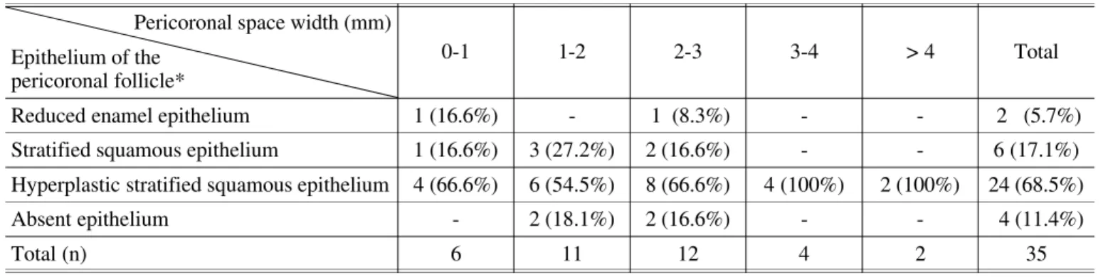
TABLE 3 - Re la ti ons hip bet we en the epit he li um of the pe ri co ro nal fol li cle and the pe ri co ro nal spa ce en lar ge ment in par ti ally erup ted te eth.
Pe ri co ro nal spa ce width (mm)
Epit he li um of the pe ri co ro nal fol li cle*
0-1 1-2 2-3 3-4 > 4 Total
Re du ced ena mel epit he li um 1 (16.6%) - 1 (8.3%) - - 2 (5.7%)
Stra ti fi ed squa mous epit he li um 1 (16.6%) 3 (27.2%) 2 (16.6%) - - 6 (17.1%)
Hyper plas tic stra ti fi ed squa mous epit he li um 4 (66.6%) 6 (54.5%) 8 (66.6%) 4 (100%) 2 (100%) 24 (68.5%)
Absent epit he li um - 2 (18.1%) 2 (16.6%) - - 4 (11.4%)
Total (n) 6 11 12 4 2 35
*In some ca ses the re was du pli city of epit he lia. n - num ber of ca ses.
TABLE 4 - Re la ti ons hip bet we en in flam ma ti on of the pe ri co ro nal fol li cle and pe ri co ro nal spa ce en lar ge ment in par ti -ally erup ted te eth.
Pe ri co ro nal spa ce width (mm)
Inflam ma ti on
0-1 1-2 2-3 3-4 > 4 Total
Pre sent 4 (66.6%) 8 (72.7%) 11 (91.6%) 4 (100%) 2 (100%) 29 (82.8%)
Absent 2 (33.3%) 3 (27.3%) 1 (8.4%) - - 6 (17.2%)
Total (n) 6 11 12 4 2 35
The re la tion ship be tween the lin ing ep i the lium, in flam ma tion and the PS width, was poor for UT (Ta bles 1 and 2), but was very clear for PET (Ta bles 3 and 4), where the REE al most dis ap peared, be ing sub sti tuted by HSSE. That sug gests the ex is tence of a re la tion ship be tween in flam ma tion, ep i the lial changes, par tic u larly hy per pla sia, and en larged PS; how ever, the chisquare anal y sis was sig nif i cant only for the pres ence of SSE and for PS en -large ment in UT.
Ac cord ing to Ta ble 1, it is also in ter est ing to note that REE was pres ent in the ma jor ity of the fol li cles (68.4%) that were not in di rect con tact with the oral cav ity. Ep i the lial lin ing was ab sent in 13% of the cases (Ta ble 1), but when it was pres -ent, it was mostly dis con tin u ous. That re sults from the ameloblastic ad her ence to the enamel cu -ti cle, which de tached from parts of the spec i men dur ing the sur gi cal treat ment. The ab sence of ep i the lium in the mi cro scopic ex am i na tion of the fol li -cle has al ready been men tioned in the literature4,35
and it does not de pend on the sur gi cal care at the time of re moval.
The pres ence of SSE in pericoronal fol li cles has al ready been men tioned by some authors2,4,6,35. Ac
-cord ing to ADELSPERGER et al.2
and STAN LEY et al.35,SEE ap pearsin fol li cles af ter the in di vid ual
reaches 21 or 22 years of age, re spec tively.
CONSOLARO6
showed that there was, with ag ing, a ten dency of trans for ma tion – REE into SEE – but, any of the epithelia could be pres ent in any age af ter nor mal erup tion. We do know that this trans for ma tion nor mally oc curs be cause, af ter amelogenesis, the ameloblasts turn from co lum -nar into cuboidal cells and then be come flat tened (Fig ures 2, 3 and 6). The re main ing lay ers are formed by the outer enamel or gan ep i the lial cells and stra tum intermedium.
We can not ex plain the sig nif i cance of the as so -ci a tion be tween SEE and PS en large ment in UT. We be lieve it was a ca sual find. We do not be lieve that the trans for ma tion of REE into SSE causes en large ment of the PS by it self. A re cently pub -lished study showed squamous metaplasia of REE with out changes in the PS2. We agree with those
au thors who de scribed the SSE as a nor mal part of the follicle4,6,35. We fol low the hy poth e sis that the
re la tion ship be tween in flam ma tion, HSSE and PS en large ment rep re sents a pro cess that ends with the cys tic trans for ma tion of the fol li cle. Some pro -spec tive stud ies have shown cys tic trans for ma tion
of the follicles16
. How ever, the risk of this trans for -ma tion re -mains un known.
The re la ti ons hip bet we en pe ri co ro nal spa ce en lar ge ment and the di ag no sis of
den ti ge rous cyst
It is im por tant to know the ac tual in ci dence of dentigerous cysts to rec om mend a pru dent man -age ment ther apy for UT. The lack of cri te ria for the di ag no sis of small dentigerous cysts dis torts the sta tis ti cal val ues, in creas ing ex ist ing doubts. MOURSHED26 found a 1.44% in ci dence of
dentigerous cysts in a ra dio graphic ex am i na tion of unerupted teeth. KNIGHTS et al.20
, in a mi cro -scopic study, found dentigerous cysts in 44.70% of UT. On the other hand, KIM; ELLIS19 found that the
most com mon histopathological mis take was to de fine pericoronal fol li cles as dentigerous cysts be cause of in ad e quate in ter pre ta tion of the lin ing ep -i the l-ium. EISENBERG11
em pha sized the im por -tance of in ter pret ing the ra dio graphic and clin i cal data when deal ing with os se ous pa thol ogy. Like -wise, SCIUBBA30
crit i cized the re sults ob tained by KNIGHTS et al.20 be cause they were based only on
mi cro scopic stud ies. He pointed out the lack of cri -te ria for de ci sion when one de pends on only one method of di ag no sis. In a let ter to the ed i tor, KNIGHTS et al.20 re plied that they con sid ered the
trans for ma tion of the REE into SSE as patho log i -cal. That con tra dicts STAN LEY; DIEHL34 for whom
the metaplasia of the REE oc curs with the ag ing pro cess, fol lowed by a de creas ing in ci dence of cysts and tu mors. In our opin ion, and in spite of these con clu sions, the lit er a ture con tin ues to re -port misdiagnoses of dentigerous cyst17
.
Dur ing the 70’s and 80’s, sev eral researchers4,12,22,25
came to the con clu sion that ra di -og ra phy alone was in suf fi cient to di ag nose small dentigerous cysts. Oth ers failed to es tab lish cor -rect di ag no ses when they chose the histopathological di ag no sis based only on the lin -ing ep i the lium. They as sumed that the pres ence of the SSE was an in di ca tion of cyst. It is im pos si ble to ac cept that the lin ing ep i the lium may char ac ter -ize a dentigerous cyst for three main rea sons. First, SSE was de scribed in the pericoronal fol li cles in clas si cal pa pers and was con sid ered a nor -mal vari a tion of the ag ing process35
the two en ti ties 9,10,13,30
. Third, the very def i ni tion of cyst in cludes bone cav i ta tion and luminal cys tic contents31. The histopathological di ag no sis of a
cyst may only be valid in con di tions sim i lar to those pre sented by AL-TALABANI; SMITH3
, where all the cyst com po nents were pres ent. Our in ves ti -ga tion showed us that pericoronal spaces up to 5.6mil li me terwide pre sented nei ther bone cav i ta -tion nor luminal cys tic con tents and, there fore, they were not cysts. They were rather en large -ments caused by a sum ma tion of fac tors such as in flam ma tion and ep i the lial trans for ma tion as dis -cussed above.
In 1995, DALEY; WYSOCKI9
pro posed that the sur gi cal cri te ria of bone cav i ta tion and luminal cys tic con tents were the only trust wor thy cri te ria to dis tin guish be tween dentigerous cyst and fol li -cles with radiolucent ar eas larger than 4 mm. This work9 re in forced the con clu sions al ready men
-tioned in 198710
. Ac cord ing to SHEAR31
,the def i ni -tion of a cyst no lon ger in cludes the pres ence of a lin ing. Our data sup port the be lief that clin i cal and/or sur gi cal cri te ria are nec es sary to con firm a di ag no sis of cyst in small PS en large ments.
Ac cord ing to EISENBERG11, the sub ject is of in
ter est only for ac a demic dis cus sion since the sur -gi cal treat ment is the same for fol li cles and small dentigerous cysts, de spite the fact that some in -sur ance car ri ers fa vor the di ag no sis of dentigerous cyst for an ar bi trary re im burse ment. In a re cent pre sen ta tion about this theme32 we dis cussed this
di ag no sis with some sci en tists, who have also men tioned in sur ance prob lems. The eco nomic fac -tor is men tioned be cause there are coun tries where over 50% of the ex pense in oral and maxillofacial sur gery oc cur as a re sult of the ex -trac tion of unerupted third mo lars.
The re la ti ons hip bet we en pe ri co ro nal spa ce en lar ge ment and the di ag no sis of pa ra den tal cyst
The fig ures which ex press the in ci dence of dentigerous cysts have be come in ac cu rate af ter CRAIG7
(1976) first de scribed the paradental cyst as a spe cific en tity. Sev eral cases of paradental cysts have been la beled as dentigerous. Ad di -tionally, a num ber of epidemiologic stud ies have also be come in ac cu rate af ter the fi nal sep a ra tion of in flam ma tory cysts in the new WHO classification21
.
STAN LEY et al 35
, men tioned some histological char ac ter is tics of radicular cysts in unerupted third mo lar fol li cles. They most cer tainly de scribed paradental cysts. There was ep i the lial hy per pla sia, in tense in flam ma tion, cho les terol crys tals, macrophages and for eign body type gi ant cells. This pic ture has also been men tioned in AMÊNDOLA’s work4
. Both works have only shown a lack of clin i cal and sur gi cal cri te ria to de scribe small in flam ma tory cysts.
Based on these re ports from the lit er a ture, we have di vided our sam ple into UT and PET, and most of the teeth were third mo lars, the main site of oc cur rence of paradental cysts1,5,31.
None of the cases we have stud ied have ful -filled the di ag nos tic cri te ria of paradental or dentigerous cysts, de spite a pericoronal radiolucency of up to 5.6 mm. There are con sis -tent ra dio graphic fea tures of a paradental cyst that should be rec og nized: the cyst is small, lo -cated on the distobuccal as pect of the tooth1
and its im age is of ten cov ered by the radiopacity of the crown. Some times, the sur geon can only dis cover the cav ity and its con tents when he el e -vates the flap. The dis sec tion of the cyst is eas ier than that of a fol li cle be cause the walls are thicker. Pericoronal in fil tra tion of an es thetic should be avoided be cause the so lu tion may press the fol li cle lead ing to the misdiagnosis of a paradental cyst. We there fore sug gest the fol low ing pro to col for the fi nal di ag no sis of PS en large ments: in most cases of UT or PET, the ra di ol o -gist should choose in flam ma tion as the pri mary di ag no sis and dentigerous or paradental cyst as a sec ond di ag no sis. The cli ni cian and/or sur -geon should ob serve the pres ence or ab sence of bone cav i ta tion and its luminal cys tic con tents which will dif fer en ti ate the pericoronal fol li cle from the dentigerous cyst or the paradental cyst. The ma te rial must be thor oughly ex am ined mi -cro scop i cally. The pa thol o gist, when in ter pret ing clin i cal, sur gi cal and ra dio graphic data, will con -firm or re fute the di ag no sis. Only the pa thol o gist will be able to rule out the ex is tence of keratocysts, hamartoplasias, in cip i ent ameloblastomas or other le sions as so ci ated with pericoronal fol li cles.
CON CLU SI ONS
en lar ge ment in PET and pos sibly in UT but, the data were not sta tis ti cally sig ni fi cant.
2. In most of the rou ti ne cases of PS en lar ge ment, the first ra di o grap hic di ag no sis should be “in -flam ma ti on of the fol li cle”. The hypot he sis of “den ti ge rous cyst” or “pa ra den tal cyst” is sug -ges ted as a se cond di ag no sis.
3. The fi nal dif fe ren ti al di ag no sis bet we en small den ti ge rous cyst or pa ra den tal cyst and pe ri co
ro nal fol li cle de pends on cli ni cal and/or sur gi cal fin dings, such as the pre sen ce of bone ca vi -ta ti on and cystic con tent.
ACKNOW LED GMENTS
The au thors thank Dr. José Roberto Pereira Lauris, Dr. Eliete Neves da Silva, Dr. Ricardo Marins de Carvalho and Dr. Carla Andreotti Damante for their help.
DAMANTE, J. H.; FLEURY, R. N. Con tri bu i ção para o di ag nós ti co do pe que no cis to den tí ge ro ou do cis to pa ra den tá rio.
Pes qui Odon tol Bras, v. 15, n. 3, p. 238-246, jul./set. 2001.
Foi pro pó si to des te es tu do ve ri fi car a re la ção en tre a lar gu ra do es pa ço pe ri co ro ná rio (EP) me di da ra di o gra fi ca men te e os as pec tos mi cros có pi cos do fo lí cu lo. O ob je ti vo foi con tri bu ir com o di ag nós ti co de pe que nos cis tos den tí ge ros e cis tos pa ra den tá ri os. Cen to e trin ta den tes nãoirrompidos (DNI) e trin ta e cin co den tes par ci al men te ir rom pi dos (DPI) fo ram ra di o gra fa dos e ex tra í dos. O es tu do ra di o grá fi co con sis tiu na me di ção da lar gu ra do EP se gui da pelo exa me mi -cros có pi co do fo lí cu lo. A lar gu ra do EP va ri ou de 0,1 a 5,6 mm. O re ves ti men to mais f re qüen te men te ob ser va do em DNI foi o epi té lio re du zi do do es mal te (ERE = 68,4%). Em DPI foi o epi té lio pa vi men to so es tra ti fi ca do hi per plá si co (EPEH = 68,5%). Infla ma ção es ta va pre sen te em 36,1% dos DNI e 82,8% dos DPI. Hou ve uma as so ci a ção es ta tis ti ca men te sig ni fi can te en tre a pre sen ça de epi té lio pa vi men to so es tra ti fi ca do (EPE) com o alar ga men to do es pa ço pe ri co ro ná rio em DNI (p < 0,05). Hou ve uma ten dên cia da in fla ma ção es tar as so ci a da com o alar ga men to do EP em DPI. Em es -pa ços pe ri co ro ná ri os me no res que 5,6 mm não fo ram de tec ta dos ca vi da de ós sea e con te ú do cís ti co ci rur gi ca men te. Na ma i o ria dos ca sos clí ni cos de ro ti na com alar ga men to do EP, su ge ri mos que o pri me i ro di ag nós ti co ra di o grá fi co deva ser “fo lí cu lo in fla ma do”. “Cis to den tí ge ro” ou “cis to pa ra den tá rio” deve ser su ge ri do como se gun do di ag nós ti co. O di ag -nós ti co di fe ren ci al fi nal en tre um pe que no cis to den tí ge ro ou cis to pa ra den tá rio e um fo lí cu lo pe ri co ro ná rio de pen de rá de acha dos clí ni cos e/ou ci rúr gi cos de ca vi da de e con te ú do.
UNI TER MOS: Den te não-erupcionado; Cis to den tí ge ro.
BI BLI O GRAP HIC RE FE REN CES
1. ACKERMANN, G.; COHEN, M. A.; ALTINI, M. The pa ra den -tal cyst: a cli ni co pat ho lo gic study of 50 ca ses. Oral Surg Oral Med Oral Pat hol, v. 64, p. 308-312, 1987. 2. ADELSPERGER, J. et al. Early soft tis sue pat ho sis as so ci a
ted with im pac ted third mo lars wit hout pe ri co ro nal ra -di o lu cency. Oral Surg, v. 89, n. 4, p. 402-406, Apr. 2000.
3. ALTALABANI, N. G.; SMITH, C. J. Expe ri men tal den ti ge rous as pects and ena mel hi po pla sia, the ir pos si ble sig ni fi can ce in ex pla i ning the pat ho ge ne sis of hu man den -ti ge rous cysts. J Oral Pat hol, v. 9, p. 82-91, 1980. 4. AMÊNDOLA, C. H. P. Estu do ra di o grá fi co e his to pa to ló
gi co do es pa ço e mem bra nas pe ri co ro ná ri as de ter -ceiros mo la res in fe ri o res não-ir rom pi dos. Ba u ru, 1983. Tese Fa cul da de de Odon to lo gia de Ba u ru, Uni -ver si da de de São Pa u lo.
5. BOHAY, R. N.; WEINBERG, S.; THORNER, P. S. The pa ra den tal cyst of the man di bu lar per ma nent first mo lar: re -port of a bi la te ral case. ASDC J Dent Child, v. 59, p. 361-365, 1992.
6. CONSOLARO, A. Caracterização microscópica de folículos pericoronários de dentes não-irrompidos e parcialmente irrompidos – sua relação com a idade.
Bauru, 1987. Tese - Faculdade de Odontologia de Bauru, Universidade de São Paulo.
7. CRAIG, G. T. The pa ra den tal cyst: a spe ci fic in flam ma tory odon to ge nic cyst. Br Dent J, v. 141, p. 9-14, 1976. 8. DACHI, S. F.; HOWELL, F. V. A sur vey of 3.874 rou ti ne
full-mouth ra di o graphs. II: a study of im pac ted te eth.
Oral Surg, v. 14, p. 1165-1169, 1961.
9. DALEY, T. D.; WYSOCKI, G. P. The small den ti ge rous cyst: a di ag nos tic di lem ma. Oral Surg Oral Med Oral Pat hol Oral Ra di ol Endod, v. 79, p. 77-81, 1995.
10. DAMANTE, J. H. Estudo dos folículos pericoronários de dentes não-irrompidos e parcialmente irrompidos. Inter-relação clínica, radiográfica e microscópica. Bauru, 1987. Tese (Livre-Docência) - Faculdade de Odontologia de Bauru, Universidade de São Paulo. 11. EISENBERG, E. Dis cus si on. Den tal fol li cu lar tis sue: mi
-sin ter pre ta ti on as odon to ge nic tu mors. J Oral Ma xil lo -fac Surg, v. 51, p. 767-768, 1993.
13. FUKUTA, Y.; TOTSUKA, M.; TAKEDA, Y.; YAMAMOTO, H. Pat ho lo gi cal study of the hyper plas tic den tal fol li cle. J Ni hon Univ Sch Dent, v. 33, p. 166-173, 1991. 14. GARDNER, D. G. The cen tral odon to ge nic fi bro ma: an at
-tempt at cla ri fi ca ti on. Oral Surg, v. 50, p. 425-432, 1980.
15. GARDNER, D. G.; RADDEN, B. Mul ti ple cal cif ying hyper -plas tic den tal fol li cles. Oral Surg Oral Med Oral Pat hol Oral Ra di ol Endod, v. 79, p. 603-606, 1995.
16. GIROD, S. C.; GERLACH, K. L.; KRUEGER, G. Cysts as so -ci a ted with long-standing im pac ted third mo lars. Int J Oral Ma xil lo fac Surg, v. 22, p. 110-112, 1993. 17. GLOSSER, J. W.; CAMPBELL, J. H. Pat ho lo gic chan ge in
soft tis su es as so ci a ted with ra di o grap hi cally “nor mal” third mo lar im pac ti ons. Br J Oral Ma xill ofac Surg, v. 37, p. 259-260, 1999.
18. HIRSHBERG, A.; BUCHNER, A.; DAYAN, D. The cen tral odon to ge nic fi bro ma and the hyper plas tic den tal fol li cle: study with Pi cro si ri us red and po la ri zing mi cros copy. J Oral Pat hol Med, v. 25, p. 125-127, 1996.
19. KIM, J.; ELLIS, G. L. Den tal fol li cu lar tis sue: mi sin ter pre -ta ti on as odon to ge nic tu mors. J Oral Ma xil lo fac Surg, v. 51, p. 762-768, 1993.
20. KNIGHTS, E. M.; BROKAW, W. C.; KESSLER, H. P. The in -ci den ce of den ti ge rous cysts as so -ci a ted with a ran dom sam pling of une rup ted third mo lars. Gen Dent, v. 39, p. 96-98, 1991.
21. KRAMER, I. R. H.; PINDBORG, J. J.; SHEAR, M. The WHO his to lo gi cal typing of odon to ge nic tu mours. Can cer, v. 70, p. 2988-2994, 1992.
22. LARA, H. R. O. Contribuição ao estudo da correlação en -tre a imagem radiográfica e o aspecto histológico dos sacos pericoronários normais e dos cistos dentígeros. Porto Alegre, 1982. Tese - Faculdade de Odontologia de Porto Alegre, Universidade Católica do Rio Grande do Sul.
23. LEIDER, A. S.; EVERSOLE, L. R.; BARKIN, M. E. Cystic ame lo blas to ma: a cli ni co pat ho lo gic analy sis. Oral Surg Oral Med Oral Pat hol, v. 60, p. 624-630, 1985. 24. LUKINMAA, P. L.; HIETANEN, J.; ANTTINEN, J. et al. Con
-ti guous en lar ged den tal fol li cles with his to lo gic fe a tu res
re sem bling the WHO type of odon to ge nic fi bro ma. Oral Surg Oral Med Oral Pat hol, v. 70, p. 313-317, 1990. 25. MOREIRA DÍAZ, E.; RUIZ, M. D. V.; ALONSO, L. R. R.
Estudio de la correlación en tre la imagem radiográfica y el aspecto hístico del saco pericoronário de los terceros molares retenidos. Rev Cubana Estomat, v. 14, p. 137-144, 1977.
26. MOURSHED, F. A. Ro ent ge no grap hic study of den ti ge rous cysts. I: inci den ce in a po pu la ti on sam ple. Oral Surg, v. 18, p. 47-53, 1964.
27. MOURSHED, F. A. Ro ent ge no grap hic study of den ti ge rous cysts. II: role of ro ent ge no grams in de tec ting den ti ge -rous cyst in the early sta ges. Oral Surg, v. 18, p. 54-61, 1964.
28. ROBERTS, M. W.; BLAKEY, G. H.; JACOWAY, J. R. et al. Enlar ged den tal fol li cles, a fol li cu lar cyst, and ena mel hypo pla sia in a pa ti ent with Lowe syndro me. Oral Surg Oral Med Oral Pat hol, v. 77, p. 264-265, 1994. 29. SANDLER, H. J.; NERSASIAN, R. R.; CATALDO, E. et al.
Mul ti ple den tal fol li cles with odon to ge nic fi bro ma-like chan ges (WHO type). Oral Surg Oral Med Oral Pat hol, v. 66, p. 78-84, 1988.
30. SCIUBBA, J. J. Eva lu a ting den ti ge rous cysts. Gen Dent, v. 39, p. 313-315, 1991. Let ter.
31. SHEAR, M. Cysts of the oral re gi ons. 3. ed. Oxford : Wright PSG, 1992.
32. SILVA, E. N.; FLEURY, R. N.; CHINELLATO, L. E. M. et al. Re la ti on bet we en the width of the pe ri co ro nary spa ce and some mi cros co pic as pects of the fol li cle. J Dent Res, v. 76, p. 854. 1997. (Spe ci al Issue)
33. STANLEY, H. R.; ALATTAR, M.; COLLET, W. K. et al. Pat ho -lo gi cal se quel of “ne glec ted” im pac ted third mo lars. J Oral Pat hol, v. 17, p. 113-117, 1988.
34. STANLEY, H. R.; DIEHL, D. L. Ame lo blas to ma po ten ti al of fol li cu lar cysts. Oral Surg, v. 20, p. 269-278, 1965. 35. STANLEY, H. R.; KROGH, H.; PANNKUK, E. Age chan ges in
the epi te li al com po nents of fol li cles (den tal sacs) as so ci -a ted with im p-ac ted third mo l-ars. Oral Surg, v. 19, p. 128-139, 1965.
36. THOMA, K. H. The cir cun fe ren ci al den ti ge rous cyst. Oral Surg, v. 18, p. 368-371, 1964.