Arq Neuropsiquiatr 2007;65(4-A):1007-1009
1007
BILATERAL PTOSIS AND SUPRANUCLEAR
DOWNGAZE PARALYSIS
Péricles Maranhão-Filho
1,2, João Carlos S. Campos
1, Marco A. Lima
1ABSTRACT - The purpose of this article is to highlight an uncommon combination of supranuclear down-ward gaze paralysis with bilateral eyelid ptosis in a 53-years-old man with a radiation induced midbrain tu-mor and to discuss the aspects regarding the centers and pathways that mediate supranuclear vertical gaze movements.
KEY WORDS: midbrain, rostral intersticial medial longitudinal fasciculus nucleus, downgaze paralysis, eye-lid ptosis, tumor.
Ptose bilateral e paralisia supranuclear do olhar conjugado para baixo
RESUMO - O objetivo deste artigo é ressaltar uma rara condição caracterizada por paralisia supranuclear do olhar conjugado para baixo associada a ptose palpebral bilateral em um homem de 53 anos, causada por tumor mesencefálico radio-induzido, e discutir os aspectos relacionados ao controle supranuclear dos mo-vimentos oculares verticais.
PALAVRAS-CHAVE: mesencéfalo, núcleo rostral intersticial do fascículo longitudinal medial, paralisia da mi-rada conjugada para baixo, ptose palpebral, tumor.
1Department of Neurosurgery, Brazilian National Cancer Institute, Rio de Janeiro RJ, Brazil; 2Department of Neurology, Federal
University of Rio de Janeiro, Rio de Janeiro RJ, Brazil.
Received 9 March 2007, received in fi nal form 31 May 2007. Accepted 1 August 2007.
Dr. Péricles Maranhão-Filho - Avenida Canal de Marapendi 1680 / 1802 - 22631-050 Rio de Janeiro RJ - Brasil. E-mail: pmaranhaofi lho @gmail.com
The structures and pathways that control the ver-tical eye movements are complex. In 1883, Henri Par-inaud described three types of vertical gaze paraly-sis1, which are: upgaze (most frequent); both upward and downward gaze simultaneously; and downgaze (uncommon). All three types may also be associated with convergence paralysis2 and lid retraction3. Down-gaze paralysis caused by a tumor has been rarely re-ported in the literature1,4-6.
We describe a patient with bilateral eyelid ptosis, downgaze voluntary saccades, and pursuit paralysis due to a radiation induced midbrain tumor. We also discuss the aspects regarding the nuclei and pathways that mediate supranuclear vertical gaze movements.
CASE
A 53-year-old man underwent resection of a left cerebel-lar hemangioma and ventriculoperiteal shunting with ad-juvant local fi eld radiation therapy when he was fourteen years old. In May 2003, he started complaining of diplopia and developed ptosis on the left eye, which progressed in a few months to a bilateral ptosis. The symptoms and signs were not affected by eye movements, and did not fl uctuate over the day. His general examination was unremarkable. By forcible frontal muscle contraction, we could observe a
discrete left exotropia in primary eye position. He was not able to make downward saccades spontaneously or on com-mand, and could not follow downward moving objects with smooth-pursuit eye movements. Upward and horizontal sac-cades and pursuit movements (Fig 1), as well as the horizon-tal and vertical doll’s head maneuvers were intact. The orbic-ularis oculi strength was normal. Convergence was absent. Pupils were reactive to the light and symmetric. No other ab-normalities were observed in the neurological examination.
The magnetic resonance imaging (MRI) (Fig 2) showed a midbrain lesion. A stereotaxic biopsy of this lesion revealed an anaplasic astrocytoma (WHO grade III).
Despite the treatment, the patient’s neurological condi-tion worsened, and he died 25 months after diagnosis.
Her sister gives us the written consent for this publication.
DISCUSSION
Arq Neuropsiquiatr 2007;65(4-A)
1008
Bilateral ptosis and supranuclear downgaze paralysis Maranhão-Filho et al.
Taking into account both the clinicopathologi-cal fi ndings and the results of experimental studies1,3 we have a clearer impression that slightly different lesions in these three structures are responsible for central up versus downgaze paralysis1,3,7. The riMLF is the principal pre-motor structure for the generation of voluntary saccades in both vertical directions. Cir-cumscribed and symmetric lesions in his efferent way result in downgaze paralysis1,4,8. The pre-motor areas controlling smooth pursuit movements are less clear3.
The riMLF receives afferent innervation from re-gions controlling eye movements, such as the para-median pontine reticular formation and vestibular nuclei, and sends efferent bilaterally to the oculo-motor nuclei1.
There is a separation between the burst neurons in the riMLF for pathways of upgaze and downgaze movements2,3. Upgaze prenuclear neurons send bilat-eral projections to the elevator muscles crossing
with-in the oculomotor nuclear complex. In contrast, axons that mediate downgaze project ipsilaterally to the de-pressor muscles2,8. In our patient, although there was no pathologic study, we can conjecture an involve-ment of this second riMLF efferent axons by the tumor. Another aspect to be considered is the bilat-eral ptosis presented by this patient. Eyelid move-ments are intimately coordinated with vertical eye movements3,9. Neural coordination of vertical gaze and lid position is due to a synkinesis between the vertical acting extraocular muscles and the levator
palpebrae muscle7. There are some situations
asso-ciating brainstem lesions and bilateral ptosis. Lesions to pathways coursing in the fl oor of the third ven-tricle and in the rostral midbrain, or in the central caudal nucleus (CCN), that is the small subgroup of the oculomotor nucleus containing levator motor neurons, may cause bilateral ptosis3. Destruction of the periaqueductally gray matter (PAG), can also lead to ptosis9. More rarely, bilateral ptosis associated with ophthalmoplegia may occur by an isolated lesion of an unilateral nuclear oculomotor complex10. Although there have been several indications that some central structures may be involved, the precise mechanism and pathways responsible by central bilateral ptosis until now remain unclear3.
In respect to the tumor, even without knowing the fi rst radiation therapy schedule and dosage, we can speculate that the anaplastic glioma was radia-tion induced. Radiaradia-tion therapy has important late effects on the CNS. Prominent among these effects are the radiation necrosis of nervous tissue11,12 and the appearance of tumors, which may be present as benign13,14 or malignant15,16. Most of these are
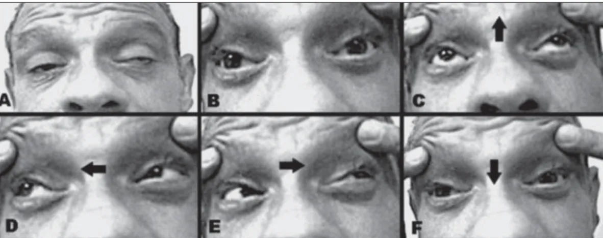
dose-Fig 1. Eye movements (images taken from video). Bilateral ptosis with compensatory contraction of the fron-tal muscle and slightly left exotrophy in inert looking (A). Eyelid forcibly opened and symmetric pupils (B). Up-ward, right, and left eye movements (arrows indicate direction) are not limited (C,D,E). There was no move-ment down upon command (F) (or saccadic – not show).
Arq Neuropsiquiatr 2007;65(4-A)
1009
Bilateral ptosis and supranuclear downgaze paralysis Maranhão-Filho et al.
dependent and occur within the original radiation treatment fi eld. They are usually meningiomas17, sar-comas18, or gliomas19, but all types of nervous system neoplasms can occur20,21. Children receiving prophylac-tic irradiation for acute lymphaprophylac-tic leukemia have 22 times higher chance of developing CNS WHO grade II, III, and IV astrocytomas in the fi rst fi ve to 10 years after radiation therapy19,20.
Similar to the Parinaud’s syndrome1, our patient presented with a vertical defi cit of voluntary saccades and pursuit movements with preservation of oculo-cephalic refl exes. These aspects represent the fi nger-print of the supranuclear nature of the process. How-ever, our patient developed bilateral ptosis (no eyelid retraction) associated with downgaze paralysis (not up gaze paralysis). The convergence was impaired and pupillary reaction remained normal, which may or not occur in Parinaud’s syndrome.
In conclusion, in light of the current knowledge and based on the neuro-ophthalmologic clinical exam-ination and imaging aspects, we can assume the pos-sibility of the involvement of the efferent riMLF con-nections and the central caudal nucleus, or the PAG, in a patient with midbrain radiation induced tumor.
Acknowledgement – The authors are in debit with Pro-fessor U. Buetnner for his critical review and manuscript suggestions and Péricles Maranhão Neto for his technical assistance.
REFERENCES
1. Pierrot-Deseiligny CH, Chain F, Gray F, Serdaru M, Escourolle R. Lher-mit e F. Parinaud syndrome: electro-oculographic and anatomical anal-yses of six vascular cases with deductions about vertical gaze organiza-tion in the premotor structures. Brain 1982;105: 667-696.
2. Pearce JMS. Parinaud’s syndrome: historical note. J Neurol Neurosurg Psychiatry 2005;76:99.
3. Schmidtke K, Büt ner-Ennever JA. Nervous control of eyelid function: a review of clinical, experimental and pathological data. Brain 1992; 115:227-247.
4. Büt ner-Ennever JA, Büt ner U, Cohen B, Baumgartner G. Vertical gaze paralysis and the rostral intersticial nucleus of the medial longitudinal fasciculus. Brain 1982;105:125-149.
5. Johkuraa K, Komiyamaa A, Hasegawab O, Kuroiwab Y. Downgaze pal-sy and bilateral ptosis due to a thalamomesencephalic lesion. J Neurol Sci 1998;161:176-179.
6. Moncayo J, Bogousslavsky J. Vertebro-basilar syndromes causing ocu-lo-motor disorders. Curr Opin Neurol 2003;16:45-50.
7. Leigh JR., Zee DS. The neurology of eye movements. 3.Ed. New York: Oxford University Press, 1999.
8. Roongroj B, Gordon T P, John LR. A hypothetical scheme for the brain-stem control of vertical gaze [Medical Hypothesis]. Neurology 2000; 54:1985-1993.
9. Büt ner-Ennever JA, Acheson JF, Büt ner U, et al. Ptosis and supranu-clear downgaze paralyis. Neurology 1989;39:385-389.
10. Maranhão-Filho PA, Pires MEP. Metastasis to the unilateral oculomotor nucleus complex: case report. Arq Neuropsiquiatr 2006;64:520-522. 11. Kong L, Lu JJ, Hu C, Guo X, Wu Y, Zhang Y. The risk of second
prima-ry tumors in patients with nasophaprima-ryngeal carcinoma at er defi nitive radiotherapy. Cancer 2006;107:1287-1293.
12. Modan B, Mart H, Baidatz D, Steinitz R, Levin SG. Radiation-induced head and neck tumours. Lancet 1974;1:277-279.
13. Benbassat CA, Olchovsky D.Prolactinoma and other head and neck tu-mors at er scalp irradiation. J Surg Oncol 1992;50:270-273.
14. Juven Y, Sadetzki S. A possible association between ionizing radiation and pituitary adenoma: a descriptive study. Cancer 2002;95:397-403. 15. Chung CK, Stryker iA, Cruse R, Vannuci R, Towfighi.
Gliob l a s t o m a m u l t i f o r m e f o l l o w i n g p r o p h y l a c t i c c r a n i -al irradiation and intrathec-al methotrexate in a child with a c u t e l y m p h o c y t i c l e u k e m i a . C a n c e r 1 9 8 1 ; 4 7 : 2 5 6 3 - 2 5 6 6 . 16. Shore RE, Albert RE, Pasternack BS. Follow-up study of patients treat-ed by X-ray epilation for Tinea capitis; resurvey of post-treatment ill-ness and mortality experience. Arch Environ Health 1976;31:21-28. 17. Spallone A, Gagliardi FM, Vagnozzi R. Intracranial meningiomas
relat-ed to external cranial irradiation. Surg Neurol 1979;12:153-159. 18. Yap J, Chuba PJ, Thomas R, et al. Sarcoma as a second malignancy at er
treatment for breast cancer. Int J Radiat Oncol Biol Phys 2002;52:1231-1237. 19. Bruce J, Allen W, Senatus PB. Astrocytoma. eMedicine. ht p://www.
emedicine.com/med/topic2693.htm. August 7, 2006.
20. Neglia JP, Meadows AT, Robison LL, et al. Second neoplasms at er acute lymphoblastic leukemia in childhood. N Engl J Med 1991;325:1330-1336. 21. Salvati M, Polli FM, Caroli E, Frati A, Missori P, Delfi ni R.