Physical exercise mobilizes endothelial progenitor cells (EPCs) to peripheral blood. However, this effect seems to depend on exercise characteristics, such as duration and intensity.
The aim of this systematic review was to verify the impact of a single bout of aerobic exercise on the mobilization of EPCs in healthy individuals, and the potential mechanisms involved.
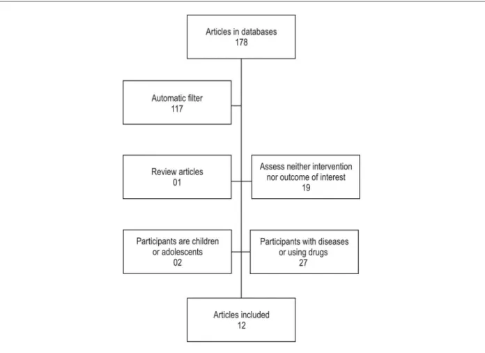
The bibliographic search was conducted on the following electronic databases in May 2011: SciELO, LILACS, Cochrane, ClinicalTrials.gov, SPORTDiscus and Medline. Of the 178 articles initially identified, 12 met the inclusion criteria and were classified regarding quality according to the PEDro scale.
The magnitude and duration of the EPC mobilization response were higher after long/ultralong duration exercises, and they are correlated with vascular endothelial growth factor (VEGF) plasma levels. The EPC mobilization peak in response to a maximal or submaximal single bout of exercise lasting up to one hour occurs immediately after the exercise or within the first hour after it. One possible mechanism is nitric oxide (NO) bioavailability. The individuals’ age and exercise intensity seem to interfere with the EPC mobilization response.
Long/ultralong duration exercises promote more pronounced EPC mobilization as compared with maximal or submaximal exercises. The mechanisms involve VEGF release in long/ ultralong duration exercises and NO bioavailability in maximal or submaximal exercises lasting less than one hour.
Introduction
Endothelial progenitor cells (EPCs), described in 1997 by Asahara et al1, constitute a heterogeneous population of circulating cells in peripheral blood1. Their origin is found in multiple precursors, such as hemangioblasts1,
non-hematopoietic precursors, monocytic cells2, or tissue-resident stem cells3. Endothelial progenitor cells play an important role in vascular repair and new vessel formation, because of their capacity to proliferate, migrate, differentiate in vivo and in vitro into endothelial cells1, and incorporate into the preexisting endothelium2,4. Thus, phenotypically, they have morphofunctional characteristics of both hematopoietic and mature endothelial cells5.
Endothelial progenitor cells are rare, representing approximately 0.01% to 0.0001% of the mononuclear fraction in peripheral blood6. However, several stimuli, such as physical exercise, can mobilize them from bone marrow, temporarily increasing their number in peripheral circulation7-9. In the peripheral circulation, EPCs secret pro-angiogenic factors, such as vascular endothelial growth factor (VEGF)10 and granulocyte colony stimulating factor (G-CSF)11, which are capable of paracrine stimulation of neovasculogenesis and angiogenesis1.
The regular practice of physical exercise contributes to an approximate 30% reduction in mortality due to cardiovascular diseases12. Because the reduction in classical cardiometabolic risk factors, such as hyperlipidemia, hypertension and insulin resistance, explains only approximately 40% of the exercise-induced reduction in mortality13, variables directly related to the endothelium could explain why and how physical exercise prevents and decreases the progression of disease, and reduces cardiovascular mortality14.
One of the possible mechanisms involved in that process is EPC mobilization to peripheral blood9. However, different exercise types, durations and intensities can promote distinct responses, and, thus, alter the bioavailability and functionality of EPCs. Thus, the impact of a bout of aerobic exercise on EPC mobilization in healthy individuals and the possible mechanisms involved in the process were systematically reviewed.
Methods
Selection of the studies
Bibliographic search was performed in May 2011 by two independent examiners (JFRS and NGR) by use of the following electronic databases: SciELO, Cochrane, LILACS, ClinicalTrials.gov, SPORTDiscus, and Medline. The studies were assessed by use of all or part of the following descriptors in English or their Portuguese correspondents: (“endothelial progenitor” OR CD34+KDR+ OR CD34+VEGFR2+ OR sca-1_flk-1 OR CD133+VEGFR2+ OR CD133+KDR+ OR AC133+VEGFR2+ OR AC133+KDR+ OR
Keywords
Exercise; endothelial progenitor cells; cell mobilization.
Mailing Address: Antonio Claudio Lucas da Nóbrega •
Laboratório de Ciências do Exercício - Instituto Biomédico, Universidade Federal Fluminense - Rua Professor Hernani Pires de Melo, 101 - Sala 106 - 24210-130, Niterói, RJ, Brazil
E-mail: anobrega@id.uff.br
Manuscript received June 03, 2011; revised manuscript received July 13, 2011; accepted July 18, 2011.
Mobilization of Endothelial Progenitor Cells with Exercise in Healthy
Individuals: a Systematic Review
Jemima Fuentes Ribeiro da Silva
1, Natália Galito Rocha
1, Antonio Claudio Lucas da Nóbrega
1,2Programa de Pós-Graduação em Ciências Cardiovasculares - Universidade Federal Fluminense1; Departamento de Fisiologia e Farmacologia -
“circulating angiogenic cells” OR “blood derived progenitor cells” OR “circulating progenitor”) AND (exercise OR “aerobic fitness” OR training OR “physical activity” OR “physical fitness” OR “sports activities” OR “sports medicine” OR marathon OR athletes OR cyclists OR runners OR ergometer OR endurance OR treadmill).
From all electronic databases searched, 178 studies were identified. After applying the terms NOT disease and NOT review to the search, 56 studies were selected in the Medline database, four in the ClinicalTrials.gov database, two in the SciELO database, two in the Cochrane database, one in the SPORTDiscus database, and none in the LILACS database. Of those studies, two were found in two different databases (Medline and Cochrane), and one was found in three different databases (Medline, ClinicalTrials. gov, and SPORTDiscus).
Through manual analysis, studies with the following characteristics were excluded: children or adolescents as participants (n = 02); participants affected by disease, inflammation, or metabolic change, and those on drugs (n = 27); studies assessing neither the intervention nor the outcome of interest, that is, if the intervention was not one single physical exercise bout, or the cell population analyzed did not correspond to the EPC profile, according to inclusion criteria (n = 19); and review studies (n = 01) (Figure 1).
When the title and abstract of the study suggested its potential eligibility for inclusion, a copy of the complete text was obtained and the study was classified according to the following five inclusion criteria: 1. year of publication: from 1997, date of the first publication about EPCs; 2. study designs: cohort, cross-sectional, clinical trials, controlled clinical trials, and randomized controlled clinical trials; 3. study population: healthy adults (humans or animals); 4. intervention: one aerobic physical exercise bout; and 5. outcome of interest: quantitative assessment of EPCs, characterized as positive for CD133 (AC133) or CD34 (sca-1) and VEGFR2 (KDR or flk-1), or endothelial cell colony forming units (EC-CFUs)15.
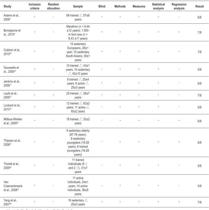
The quality of the studies was assessed by use of the PEDro scale16, according to which studies with a score equal to or higher than four are classified as of high quality. The criteria adopted were as follows: 1. well-defined inclusion criteria; 2. random allocation into groups; 3. description of the demographic characteristics and
size of the sample (≥ eight individuals); 4. blind (blind participants
or examiners or result analyses); 5. sufficient method (when the exercise protocol was described in details or the cell analysis was performed by use of at least one quantitative or qualitative method); 6. measurements (were obtained in more than 85% of the individuals initially allocated into groups for at least one key-result); 7. statistical analysis (at least one result of the statistical
comparisons between groups or moments was described); 8. regression analysis (to assess the association between the intervention and outcome variables) (Table 1).
Data extraction
The following data were obtained from each publication: name of the first author and year of publication; population data, demography and size of the sample; blood sample collection time; variables studied and the method through which they were analyzed; the protocol, intensity and volume of exercise; and the results of the study, with correlation between intervention and outcome.
Characteristics of the participants
The exercise training level of the participants ranged from non-active or sedentary17,18 to athletes who finished a 246-km foot race in up to 36 hours19. Thus, in the present systematic review, a classification based on the volume of exercise training of the participants of all studies was elaborated to standardize and classify the individuals into only three levels of physical fitness as follows: sedentary group, participants whose volume of aerobic training was lower than or equal to once a week, with 20-minute bouts each; active group, those that practiced aerobic exercise twice or three times a week, with 20-minute bouts each; trained group, participants whose volume of aerobic training was greater than or equal to four times a week, with 30-minute bouts each.
Table 1 – Methodological quality of the studies
Study Inclusion
criteria
Random
allocation Sample Blind Methods Measures
Statistical analysis
Regression
analysis Result
Adams et al.,
20087 ⁺ ⁻
68 trained ♂, 57±6
years ⁺ ⁺ ⁺ ⁺ ⁻ 6/8
Bonsignore et
al., 20108 ⁺ ⁻
Marathon (n = 9,44
±12 years);
1,500-m foot race (n =
8,43 ±11 years)
⁺ ⁺ ⁺ ⁺ ⁺ 7/8
Cubbon et al.,
201026 ⁺ ⁻
15 sedentary Europeans, 28±1 year; 15 sedentary South-Asians, 30±1
years
⁺ ⁺ ⁺ ⁺ ⁺ 7/8
Goussetis et
al., 200919 ⁺ ⁻
10 trained ♂, 43±1 years; 10 sedentary
♂, 42±10 years ⁻ ⁺ ⁺ ⁺ ⁻ 5/8
Jenkins et al.,
200917 ⁺ ⁻
8 trained ♂, 25±4 years; 8 active ♂,
25±3 years ⁺ ⁺ ⁺ ⁺ ⁻ 6/8
Laufs et al.,
200527 ⁺ ⁺
25 trained ♂, 28±7
years ⁺ ⁺ ⁺ ⁺ ⁻ 7/8
Lockard et al.,
201018 ⁺ ⁻
12 trained ♂, 62±2 years; 11 active ♂,
65±2 years ⁻ ⁺ ⁺ ⁺ ⁻ 5/8
Möbius-Winkler
et al.,200922 ⁺ ⁻
18 trained ♂, 32±2
years ⁻ ⁺ ⁺ ⁺ ⁺ 6/8
Thijssen et al.,
200621 ⁺ ⁻
8 sedentary elderly
(67-76 years);
8 sedentary
youngsters (19-28 years); 8 trained youngsters (18-28
years)
⁻ ⁺ ⁺ ⁺ ⁺ 6/8
Thorell et al.,
200925 ⁺ ⁻
11 trained
individuals (9 ♀ and 2 ♂), 31±7
years
⁻ ⁺ ⁺ ⁺ ⁻ 5/8
Van Craenenbroeck et al., 200823
⁺ ⁻
11 active individuals, 24±1
years; 14 active individuals, 36±9
years
⁻ ⁺ ⁺ ⁺ ⁻ 5/8
Yang et al.,
200724 ⁺ ⁻
16 sedentary ♂,
25±3 years ⁺ ⁺ ⁺ ⁺ ⁺ 7/8
Exercise characteristics
The response of EPCs to exercise can vary according to the type of protocol used7,8,20. Thus, the following data were obtained from each study: intensity, volume, duration, and frequency of physical exercise training; percentage values of maximal heart rate, maximal oxygen consumption (VO2) and anaerobic threshold of participants; authors’ description of the exercise protocol used, enabling the classification of the exercises into the following three categories: 1. long/ultralong duration exercise (half-marathon, marathon or ultramarathon training); 2. maximal intensity exercise, usually progressive and reaching maximal effort and/or maximal VO2 in 5 to 20 minutes. e.g. typical examples are the conventional exercise test or cardiopulmonary exercise test (CPET); and 3. Submaximal intensity exercise, usually using a percentage of the anaerobic threshold and/or maximal VO2 as reference, and with maximal duration of one hour. In the present study, exercise intensity ranged from 80% to 100% of the anaerobic threshold or exceeded 75% of the maximal VO2.
Results
The 12 studies included were classified as of high quality and were as follows: seven cross-sectional studies; four longitudinal studies; and one study with both designs. In all studies included, the methodological procedures adopted in dimensioning and selecting the samples, the measurements of the variables, and the ethical aspects were sufficiently described. The composition of the samples (total of 286 participants) varied in age, from 18 to 80 years, and physical condition, from sedentary to trained (tab. 1).
Most studies reported significant effects of different types of exercises on the mobilization of EPCs to peripheral blood. However, neither the volume of training nor the physical condition of the participants has influenced the number of EPCs prior to exercise17,18,21. In addition, after one single bout of submaximal exercise, the number of EPCs increased in the blood stream of the participants with a higher volume of training17, suggesting that EPC mobilization requires an acute disturbing stimulus in the organism.
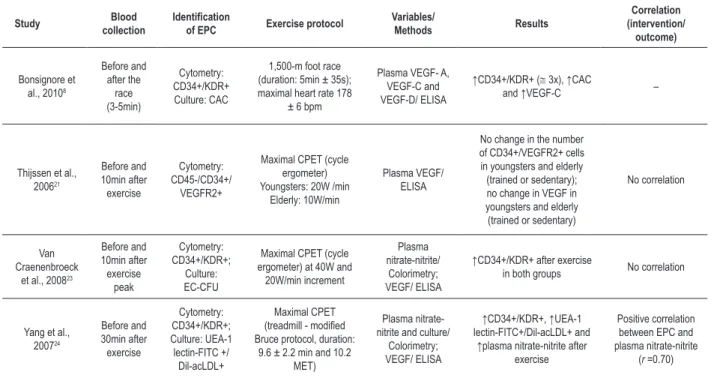
Tables 2a-2c show specific information about the studies included, which were classified as: 1. long/ultralong duration exercise (n = 4 studies, tab. 2a); 2. maximal intensity exercise (n = 4 studies, tab. 2b); 3. submaximal intensity exercise (n = 5 studies, tab. 2c).
Long/ultralong duration exercise
An increase in the number of CD34+/KDR+ cells or EC-CFUs after long/ultralong duration exercise (marathon/ultra-distance running) was observed in three of the four studies assessed (tab. 2a). An elevation in inflammatory markers, mainly interleukin-6, was observed after exercise in the studies assessed8,19,22.
In trained individuals, the number of CD34+/KDR+ cells increased approximately twice at the end of a marathon (p < 0.005), while the number of circulating angiogenic cells (CAC) increased approximately three times (8.5 ± 1.9 vs. 30.2 ± 4.6 cells/1x106 mononuclear cells; p < 0.0001), with
a return to baseline levels on the next morning8 (10.9 ± 1.8 cells/1x106 mononuclear cells). In addition, in trained men, the number of EC-CFUs increased by 11-fold (44.5 ± 2.5 vs. 494.5 ± 27.9/mL) at the end of an ultra-distance foot race (246 km) in up to 36 hours, and remained increased for 48 hours (428.5 ± 31.5/mL; p < 00001) after the end of the race19. In addition, in trained runners, the number of CD34+/ VEGFR2+ cells did not change after a marathon7 (117.0 ± 8.0 vs. 128.0 ± 9.0 cells/mL; p = 0.33). One exercise bout on the cycle ergometer at 70% of the anaerobic threshold revealed a 5.5-fold increase in CD34+/KDR+ cells (p < 0.001) and a 3.5-fold increase in CD133+/KDR+ cells from 210 minutes of exercise (p < 0.001) in a time-dependent way, with return to baseline levels after 24 hours22. The VEGF-C levels increased on the day following a marathon and were greater than those of maximal exercise (p < 0.05)8. Ten minutes after exercising on the cycle ergometer22, VEGF plasma levels showed a 1.9-fold maximal increase (79.8 ± 15.4 pg/mL vs. 132.6 ± 32.1 pg/mL; p < 0.05), which significantly correlated with the number of CD133+/KDR+ cells (r = 0.67; p = 0.0045)22. However, Adams et al7have reported a reduction in VEGF plasma levels after a marathon (48.9 ± 8.0 vs. 34.0 ± 7.5 pg/ mL; p < 0.05), which did not correlate with the number of CD34+/VEGFR2+ cells.
Maximal intensity exercise
Four studies assessed the effect of one single bout of maximal exercise on the number of EPCs8,21,23,24 (tab. 2b).
Yang et al24 have reported a significant increase in CD34+/ KDR+ cells (p < 0.05) and UEA-1 lectin-FITC+/Dil-acLDL+ cells (p < 0.05) 30 minutes after a CPET on the treadmill. In trained individuals, the number of CD34+/KDR+ cells has increased three times (p < 0.01) after the end of a 1,500-m foot race8. The levels of CAC have also increased significantly after a 1,500-m foot race8 (p < 0.005). Similar results have been shown by Van Craenenbroeck et al23, who have reported a 76% increase in CD34+/KDR+ cells in young individuals aged 24 ± 1 years (15.4 ± 10.7 vs. 27.2 ± 13.7 cells/mL; p = 0.01) and a 69% increase in adults aged 36 ± 9 years (30.9 ± 14.6 vs. 52.5 ± 42.6 cells/mL; p = 0.03); however, no significant change was observed in the percentage of EC-CFUs (11.9 ± 10.9 vs. 9.0 ± 8.3; p = 0.2). Only Thijssen et al21 have reported no change in the number of EPCs after CPET in trained and sedentary young and elderly individuals (p > 0.05).
Most studies have shown no significant change in VEGF plasma levels after one single bout of maximal exercise21,23,24; however, Bonsignore et al8 have reported an increase in VEGF-C plasma levels (7.3 ± 1.8 vs. 8.8 ± 1.7 pg/mL; p < 0,001) after a 1,500-m foot race (tab. 2b).
According to the study by Yang et al24, an increase in nitric oxide (NO) plasma levels has been observed 30 minutes after a treadmill CPET (p < 0.05). In addition, linear regression analysis has revealed a positive correlation (r = 0.70; p < 0.05) between EPC increase and NO levels24.
Table 2b – Results of studies using maximal intensity exercise protocols
Study Blood
collection
Identiication
of EPC Exercise protocol
Variables/ Methods Results Correlation (intervention/ outcome) Bonsignore et al., 20108
Before and after the race (3-5min) Cytometry: CD34+/KDR+ Culture: CAC
1,500-m foot race (duration: 5min ± 35s);
maximal heart rate 178
± 6 bpm
Plasma VEGF- A, VEGF-C and VEGF-D/ ELISA
↑CD34+/KDR+ (@ 3x), ↑CAC
and ↑VEGF-C –
Thijssen et al., 200621 Before and 10min after exercise Cytometry: CD45-/CD34+/ VEGFR2+
Maximal CPET (cycle
ergometer) Youngsters: 20W /min
Elderly: 10W/min
Plasma VEGF/
ELISA
No change in the number of CD34+/VEGFR2+ cells in youngsters and elderly
(trained or sedentary);
no change in VEGF in youngsters and elderly
(trained or sedentary)
No correlation
Van Craenenbroeck
et al., 200823
Before and 10min after exercise peak Cytometry: CD34+/KDR+; Culture: EC-CFU
Maximal CPET (cycle
ergometer) at 40W and 20W/min increment
Plasma
nitrate-nitrite/ Colorimetry; VEGF/ ELISA
↑CD34+/KDR+ after exercise
in both groups No correlation
Yang et al., 200724 Before and 30min after exercise Cytometry: CD34+/KDR+; Culture: UEA-1 lectin-FITC +/ Dil-acLDL+ Maximal CPET
(treadmill - modiied
Bruce protocol, duration:
9.6 ± 2.2 min and 10.2 MET)
Plasma
nitrate-nitrite and culture/
Colorimetry; VEGF/ ELISA
↑CD34+/KDR+, ↑UEA-1 lectin-FITC+/Dil-acLDL+ and
↑plasma nitrate-nitrite after
exercise
Positive correlation
between EPC and
plasma nitrate-nitrite
(r =0.70)
EPC – endothelial progenitor cells; (+) – positive; CD34 – cluster of differentiation 34; VEGFR2 – receptor 2 of the vascular endothelial growth factor; VEGF – vascular endothelial growth factor (types A, C, D); ELISA – enzyme-linked immunosorbent assay; KDR – kinase insert domain receptor of the vascular endothelial growth factor;
CAC – circulating angiogenic cell; EC-CFU – endothelial cell colony forming unit; UEA-1-FITC – Ulex Europaeus agglutinin type 1- conjugated to luorescein isothiocyanate;
Dil-acLDL – 1,1’-dioctadecyl-3,3’,3’-tetramethylindocarbocyanine-labelled acetylated low-density lipoprotein; CPET – cardiopulmonary exercise test; MET –metabolic equivalent; W – watts; bpm – beats per minute; CD45 – cluster of differentiation 45.
Table 2a – Results of studies using long/ultralong duration exercise protocols
Study Blood collection Identiication
of EPC Exercise protocol
Variables/ Methods Results Correlation (intervention/ outcome) Adams et al., 2008 7
Before and immediately after the
marathon
Cytometry: CD34+/ VEGFR2+
Dusseldorf Marathon 2006, duration @ 4h
Plasma VEGF
and EGF/ ELISA
No change in the number of CD34+/VEGFR2 + cells and
↓VEGF after the marathon No correlation
Bonsignore et al., 2010 8
2-3 days before,
immediately after,
and 18-20h after the
marathon Cytometry: CD34+/KDR+; Culture: CAC Palermo International Marathon 2005,
duration @ 3h30min
Plasma VEGF-A, VEGF-C and VEGF-D/ ELISA
↑CD34+/KDR+
(@ 2x), ↑CAC (3x) immediately after and
↑VEGF-C (18-20h after)
–
Goussetis et al., 2009 19
Before, immediately after and 48h after the
foot race
Culture:
EC-CFU
246-km ultradistance race (Spartathlon),
duration @ 32h8min
– ↑EC-CFU (11x) immediately after and 48h after the race –
Möbius-Winkler et
al., 2009 22
Before, during (5, 10, 15, 30, 60, 90, 120, 150, 180, 210, 240min)
and after exercise (30,
60, 120, 1,440min)
Cytometry: CD34+/KDR+ and CD133+/
KDR+
Cycle ergometer test at 70% of the AT
for 4h
Plasma VEGF/
ELISA
↑CD34+/KDR+ (5,5x), ↑CD133+/KDR+ (3,5x) and
↑VEGF after 10min
Correlation between CD133+/ KDR+ cells and VEGF (r = 0.67)
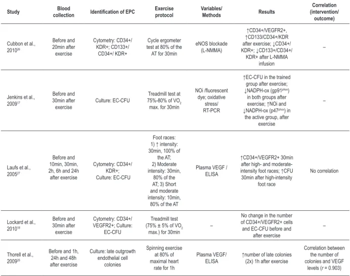
(tab. 2c), three studies17,18,27 have used a protocol based on a 400-m foot race or a treadmill race for 30 minutes and exercise intensity between 68% and 82% of maximal VO2.
Although Lockard et al18 have found no alteration in the number of CD34+/VEGFR2+ cells (p > 0.05) and EC-CFUs (p > 0.05) 30 minutes after an exercise with intensity of 75% ± 5% of maximal VO2, most studies do not seem to support that finding. Jenkins et al17 have shown that, in trained men, EC-CFUs increased significantly 30 minutes after submaximal exercise (p = 0.02). Moderate or intense exercise (80% and 100% of the anaerobic threshold, respectively) of same duration has increased the number of circulating EPCs (CD34+/VEGFR2+ and UEA-1 lectin-FITC+/Dil-acLDL+ cells), with a peak response between 10 and 30 minutes after the end of the exercise bout (p < 0.01). The number of EC-CFUs has also increased 30 minutes
after high-intensity running27 (p < 0.05). In addition, one cycle ergometer exercise bout, lasting 30 minutes and at 80% of the anaerobic threshold26, caused an increase in the number of CD34+/KDR+ and CD133+/CD34+/KDR+ cells in European Caucasians (85.4 ± 1.3% and 73.9 ± 9.1%; respectively) and in Asians (53.2 ± 6.9% and 48.3 ± 8.8%; respectively) 20 minutes after the end of the exercise bout. In another study25, one single bout of spinning exercise, with mean duration of 44.3 ± 3.4 minutes and heart rate ranging from 77% to 95% of maximal heart rate, could double the number of late outgrowth endothelial cell colonies, another type of EPC colony. In addition, VEGF levels have shown a significant correlation with the number of late outgrowth endothelial cell colonies (r = 0.903)25. The effects of the single bout of spinning exercise have decreased over time with return to baseline levels after 48 hours.
Table 2c – Results of the studies using submaximal intensity exercise protocols lasting less than one hour
Study Blood
collection Identiication of EPC
Exercise protocol Variables/ Methods Results Correlation (intervention/ outcome)
Cubbon et al., 201026 Before and 20min after exercise Cytometry: CD34+/ KDR+; CD133+/ CD34+/ KDR+ Cycle ergometer test at 80% of the AT for 30min
eNOS blockade
(L-NMMA)
↑CD34+/VEGFR2+, ↑CD133/CD34+/KDR after exercise; ↓CD34+/ KDR+; ↓CD133+/CD34+/
KDR+ after L-NMMA
infusion
–
Jenkins et al., 200917
Before and 30min after exercise
Culture: EC-CFU 75%-80% of VOTreadmill test at 2
max. for 30min
NOi /luorescent dye; oxidative
stress/
RT-PCR
↑EC-CFU in the trained group after exercise; ↓NADPH-ox (gp91phox)
in both groups after
exercise; ↑NOi and ↓NADPH-ox (p47phox) in
the active group, after
exercise
–
Laufs et al.,
200527
Before and 10min, 30min, 2h, 6h and 24h
after exercise
Cytometry: CD34+/
KDR+; Culture: EC-CFU
Foot races:
1) ↑ intensity:
30min, 100% of
the AT; 2) Moderate
intensity: 30min, 80% of the
AT; 3) Short
and moderate intensity: 10min,
80% of the AT
Plasma VEGF /
ELISA
↑CD34+/VEGFR2+ 30min after high- and moderate-intensity foot races; ↑CFU
30min after high-intensity
foot race
No correlation
Lockard et al., 201018 Before and 30min after exercise Cytometry: CD34+/ VEGFR2+; Culture: EC-CFU Treadmill test
(75% ± 5% of VO2
max.) for 30min –
No change in the number of CD34+/VEGFR2+ cells
and EC-CFU before and
after exercise
–
Thorell et al., 200925
Before and 1h, 24h and 48h after exercise
Culture: late outgrowth endothelial cell
colonies
Spinning exercise at 80% of maximal heart
rate for 1h
Plasma VEGF/
ELISA ↑number of late colonies (2x) 1h after exercise
Correlation between the number of colonies and VEGF
levels (r = 0.903)
In trained men, exercise has acutely reduced the messenger RNA (mRNA) levels of NADPH oxidase (gp91phox) in EC-CFUs17 (p = 0.02). Active men showed an increased production of intracellular NO (p = 0.004) and a reduced expression of the p47phox subunit of NADPH oxidase (p < 0.05), but with no difference for other oxidative stress markers17 (p > 0.05). The infusion of L-NMMA, an inhibitor of the endothelial nitric oxide synthase (eNOS), has reduced the number of EPCs (CD34+/KDR+: -3.3% vs. 68.4%; p < 0.001; CD133+/ CD34+/KDR+: 0.7% vs. 71.4%; p < 0.001). In addition, linear regression analysis has shown a positive correlation between flow-mediated vasodilation and CD34+/KDR+ (r = 0.41; p = 0.02) and CD133+/ CD34+/KDR+ cells (r = 0.39; p < 0.04)26.
Discussion
Recent studies15,24 have shown that the exercise-induced improvement in endothelial function is mainly due to the mobilization of EPCs to the peripheral circulation, where they participate in the processes of neovascularization and endothelial repair15. Thus, physical exercise is an important method to promote health for cardiovascular system in both healthy individuals28 and those with cardiovascular risk factors29,30.
Long/ultralong duration exercise
Marathons, half-marathons, and ultramarathons are models of induced inflammation that stimulate the increase in the circulating levels of pro- and anti-inflammatory mediators31, promoting the release, migration and differentiation of bone marrow stem cells32. That type of exercise also increases the plasma levels of cytokines and endothelial activation markers, providing an opportunity to assess the physiological mechanisms related to vascular repair, before an irreversible tissue damage has occurred33.
Long/ultralong duration exercises have shown to significantly increase the concentration of progenitor cells, especially the endothelial ones, and leukocytes8,19,22. Interleucin-6 plasma levels have increased during and after long/ultralong duration exercises, evidencing their inflammatory character8,19.
In addition, the highest VEGF plasma levels have been observed after that type of exercise. Elevated VEGF levels have been believed to associate with the presence of tissue hypoxia, favoring the stimulus of EPC mobilization and endothelial repair. Thus, that increase in VEGF seems to demonstrate a mechanism of physiological adaptation to long/ultralong duration exercise, suggesting the existence of a positive correlation between exercise intensity and the release of growth factors8.
Exercise duration, on its turn, seems to be associated with the permanence of the exercise effects on EPC. Protocols of extreme exercises19 have shown longer-lasting responses (up to 48 hours after the end of the exercise bout) than marathons do (up to 24 hours after the end of the exercise bout)8.
Finally, the difference in age between the populations studied is believed to explain the controversial results obtained, because age influences the number and function of EPCs34.
Maximal intensity exercise
Maximal intensity exercises are characterized by a large increase in vascular shear stress35, which can be considered a subacute effect of physical exercise28. Most results have shown that one single maximal exercise bout seems to increase the number of EPCs8,23,24, NO plasma levels24, and VEGF-C plasma levels8. However, a positive correlation has been found only between the increase in the number of EPCs and NO levels24. In addition, the magnitude of the increment used in exercise protocols seems to contribute to more significant results24.
Some studies using similar conditions of exercise and population profile21,23,24 have shown very different results for the number of EPCs at baseline condition. At least part of that heterogeneity is due to the absence of a standardized protocol to quantify, identify and analyze EPCs. Thus, comparing and consolidating the results of the different studies is difficult, and that constitutes the greatest methodological challenge to knowledge advance in that area.
Submaximal intensity exercise lasting less than one hour Successive submaximal intensity exercise bouts comprise the training; thus, the study of the physiological responses to that type of exercise can contribute to the understanding of the global process of endothelial adaptation to physical training36. In young individuals, submaximal intensity exercise has caused a significant increase in EC-CFUs, EPCs and late outgrowth endothelial cell colonies. In addition, circulating EPC levels have not varied in response to a greater intensity exercise of same duration27. However, a strenuous protocol, combining submaximal intensity and longer duration (approximately one hour), has shown a longer-lasting effect of exercise on EPCs, observed up to 48 hours after the end of exercise bout. In addition, that protocol has strongly correlated with VEGF plasma levels (r = 0.903)25.
Because of the large interindividual variability found, no significant difference in the number of EPCs or EC-CFUs has been observed after exercise in elderly athletes18.
Studies are controversial regarding the influence of physical condition on EPCs. Trained youngsters17 have shown a significant increase in EC-CFUs. However, an increase in CD34+/KDR+ or CD34+/VEGFR2+ cells has been observed in both sedentary26 and trained individuals27.
The major mechanism involved in EPC mobilization after performing a submaximal exercise seems to be associated with NO bioavailability. In active men, exercise has caused a reduction in the expression of NADPH oxidase, an important cause of oxidative stress on the cardiovascular system, and an increase in intracellular NO levels in EC-CFUs17. In sedentary individuals, exercise has caused a significant reduction in the mobilization of CD34+/VEGFR2+ cells after eNOS blockade with L-NMMA infusion26. Those data suggest that NADPH oxidase might be one of the mechanisms involved in the inhibition of intracellular NO production in EC-CFUs17.
endothelial shear stress, elevating intracellular calcium levels, and, consequently, activating eNOS. The activation of that enzyme, on its turn, promotes NO increase, metalloproteinase 9 activation, and the release of the soluble kit ligand, which is crucial for the mobilization of EPCs from the bone marrow to peripheral circulation26.
Despite the fact that an ischemic tissue is by definition a pathological tissue, and, thus, lacks in healthy individuals, long and ultralong duration exercises are models of physiological ischemia, because they cause changes in the anaerobic glycolytic metabolism, in addition to generating oxidative stress23. Oxygen reactive species are essential for hypoxia37. Hypoxia inducible factor-1 is activated after single exercise bouts38, stimulating the gene expression of important EPC mobilizing molecules, such as VEGF10. In the presence of VEGF, eNOS activation and NO increase occur, thus mobilizing EPCs from bone marrow26. In addition, NO is also the key molecule in shorter-duration exercises, and the increase in its availability seems to depend especially on two mechanisms: in the first, the activation of protein kinase 3 mediated by the increase in shear stress activates eNOS, which increases NO production39. In the second mechanism, exercise reduces NADPH expression, increasing NO bioavailability, and, consequently, EPC mobilization from the bone marrow occurs. Considering that, one can suggest that single exercise bouts of different intensities and durations mobilize EPCs to peripheral blood through different mechanisms; however, NO seems to be a molecule common to both mobilization pathways.
Conclusion
The mobilization of endothelial progenitor cells was more marked in long/ultralong duration exercises and seems to be associated with the vascular endothelial growth factor plasma levels. Maximal exercises and submaximal exercises lasting less than one hour cause a lower magnitude increase in the number of circulating endothelial progenitor cells, and their main mechanism of mobilization seems to be nitric oxide bioavailability.
Acknowledgements
This study was financially supported by the following institutions: Capes, Faperj, CNPq, Finep, and Labs D’Or.
Potential Conflict of Interest
No potential conflict of interest relevant to this article was reported.
Sources of Funding
This study was funded by Capes, Faperj, CNPq, Finep, and Labs D’Or.
Study Association
This article is part of the thesis of Doctoral submitted by Natália Galito Rocha, from Universidade Federal Fluminense.
References
1. Asahara T, Murohara T, Sullivan A, Silver M, van der Zee R, Li T, et al. Isolation of putative progenitor endothelial cells for angiogenesis. Science. 1997;275(5302):964-7.
2. Yoder MC, Mead LE, Prater D, Krier TR, Mroueh KN, Li F, et al. Redefining endothelial progenitor cells via clonal analysis and hematopoietic stem/ progenitor cell principals. Blood. 2007;109(5):1801-9.
3. Beltrami AP, Barlucchi L, Torella D, Baker M, Limana F, Chimenti S, et al. Adult cardiac stem cells are multipotent and support myocardial regeneration. Cell. 2003;114(6):763-76.
4. Shantsila E, Watson T, Lip GY. Endothelial progenitor cells in cardiovascular disorders. J Am Coll Cardiol. 2007;49(7):741-52.
5. Sauter B, Foedinger D, Sterniczky B, Wolff K, Rappersberger K. Immunoelectron microscopic characterization of human dermal lymphatic microvascular endothelial cells. Differential expression of CD31, CD34, and type IV collagen with lymphatic endothelial cells vs blood capillary endothelial cells in normal human skin, lymphangioma, and hemangioma in situ. J Histochem Cytochem. 1998;46(2):165-76.
6. Khan SS, Solomon MA, McCoy JP Jr. Detection of circulating endothelial cells and endothelial progenitor cells by flow cytometry. Cytometry B Clin Cytom. 2005;64(1):1-8.
7. Adams V, Linke A, Breuckmann F, Leineweber K, Erbs S, Krankel N, et al. Circulating progenitor cells decrease immediately after marathon race in advanced-age marathon runners. Eur J Cardiovasc Prev Rehabil. 2008;15(5):602-7.
8. Bonsignore MR, Morici G, Riccioni R, Huertas A, Petrucci E, Veca M, et al. Hemopoietic and angiogenetic progenitors in healthy athletes:
different responses to endurance and maximal exercise. J Appl Physiol. 2010;109(1):60-7.
9. Laufs U, Werner N, Link A, Endres M, Wassmann S, Jurgens K, et al. Physical training increases endothelial progenitor cells, inhibits neointima formation, and enhances angiogenesis. Circulation. 2004;109(2):220-6.
10. Asahara T, Takahashi T, Masuda H, Kalka C, Chen D, Iwaguro H, et al. VEGF contributes to postnatal neovascularization by mobilizing bone marrow-derived endothelial progenitor cells. EMBO J. 1999;18(14):3964-72.
11. Powell TM, Paul JD, Hill JM, Thompson M, Benjamin M, Rodrigo M, et al. Granulocyte colony-stimulating factor mobilizes functional endothelial progenitor cells in patients with coronary artery disease. Arterioscler Thromb Vasc Biol. 2005;25(2):296-301.
12. Kokkinos P, Myers J, Faselis C, Panagiotakos DB, Doumas M, Pittaras A, et al. Exercise capacity and mortality in older men: a 20-year follow-up study. Circulation. 2010;122 (8):790-7.
13. Shi L, Morrison JA, Wiecha J, Horton M, Hayman LL. Healthy lifestyle factors associated with reduced cardiometabolic risk. Br J Nutr. 2011;105(5):747-54
14. Seligman BG, Polanczyk CA, Santos AS, Foppa M, Junges M, Bonzanini L, et al. Intensive practical lifestyle intervention improves endothelial function in metabolic syndrome independent of weight loss: a randomized controlled trial. Metabolism. 2011 Jun 21. [Epub ahead of print].
16. Giacomini MK, Cook DJ. Users’ guides to the medical literature: XXIII. Qualitative research in health care A. Are the results of the study valid? Evidence-Based Medicine Working Group. JAMA. 2000;284(3):357-62.
17. Jenkins NT, Witkowski S, Spangenburg EE, Hagberg JM. Effects of acute and chronic endurance exercise on intracellular nitric oxide in putative endothelial progenitor cells: role of NAPDH oxidase. Am J Physiol Heart Circ Physiol. 2009;297(5):H1798-805.
18. Lockard MM, Witkowski S, Jenkins NT, Spangenburg EE, Obisesan TO, Hagberg JM. Thrombin and exercise similarly influence expression of cell cycle genes in cultured putative endothelial progenitor cells. J Appl Physiol. 2010;108(6):1682-90.
19. Goussetis E, Spiropoulos A, Tsironi M, Skenderi K, Margeli A, Graphakos S, et al. Spartathlon, a 246 kilometer foot race: effects of acute inflammation induced by prolonged exercise on circulating progenitor reparative cells. Blood Cells Mol Dis. 2009;42(3):294-9.
20. Bonsignore MR, Morici G, Santoro A, Pagano M, Cascio L, Bonanno A, et al. Circulating hematopoietic progenitor cells in runners. J Appl Physiol. 2002;93(5):1691-7.
21. Thijssen DH, Vos JB, Verseyden C, van Zonneveld AJ, Smits P, Sweep FC, et al. Haematopoietic stem cells and endothelial progenitor cells in healthy men: effect of aging and training. Aging Cell. 2006;5(6):495-503.
22. Mobius-Winkler S, Hilberg T, Menzel K, Golla E, Burman A, Schuler G, et al. Time-dependent mobilization of circulating progenitor cells during strenuous exercise in healthy individuals. J Appl Physiol. 2009;107(6):1943-50.
23. Van Craenenbroeck EM, Vrints CJ, Haine SE, Vermeulen K, Goovaerts I, Van Tendeloo VF, et al. A maximal exercise bout increases the number of circulating CD34+/KDR+ endothelial progenitor cells in healthy subjects. Relation with lipid profile. J Appl Physiol. 2008;104(4):1006-13.
24. Yang Z, Wang JM, Chen L, Luo CF, Tang AL, Tao J. Acute exercise-induced nitric oxide production contributes to upregulation of circulating endothelial progenitor cells in healthy subjects. J Hum Hypertens. 2007;21(6):452-60.
25. Thorell D, Borjesson M, Larsson P, Ulfhammer E, Karlsson L, DuttaRoy S. Strenuous exercise increases late outgrowth endothelial cells in healthy subjects. Eur J Appl Physiol. 2009;107(4):481-8.
26. Cubbon RM, Murgatroyd SR, Ferguson C, Bowen TS, Rakobowchuk M, Baliga V, et al. Human exercise-induced circulating progenitor cell mobilization is nitric oxide-dependent and is blunted in South Asian men. Arterioscler Thromb Vasc Biol. 2010;30(4):878-84.
27. Laufs U, Urhausen A, Werner N, Scharhag J, Heitz A, Kissner G, et al. Running exercise of different duration and intensity: effect on endothelial progenitor cells in healthy subjects. Eur J Cardiovasc Prev Rehabil. 2005;12(4):407-14.
28. Bousquet-Santos K, Soares PP, Nobrega AC. Subacute effects of a maximal exercise bout on endothelium-mediated vasodilation in healthy subjects. Braz J Med Biol Res. 2005;38(4):621-7.
29. Adams V, Lenk K, Linke A, Lenz D, Erbs S, Sandri M, et al. Increase of circulating endothelial progenitor cells in patients with coronary artery disease after exercise-induced ischemia. Arterioscler Thromb Vasc Biol. 2004;24(4):684-90.
30. Sandri M, Adams V, Gielen S, Linke A, Lenk K, Krankel N, et al. Effects of exercise and ischemia on mobilization and functional activation of blood-derived progenitor cells in patients with ischemic syndromes: results of 3 randomized studies. Circulation. 2005;111(25):3391-9.
31. Ostrowski K, Rohde T, Asp S, Schjerling P, Pedersen BK. Pro- and anti-inflammatory cytokine balance in strenuous exercise in humans. J Physiol. 1999;515 (Pt 1):287-91.
32. Aicher A, Zeiher AM, Dimmeler S. Mobilizing endothelial progenitor cells. Hypertension. 2005;45(3):321-5.
33. Margeli A, Skenderi K, Tsironi M, Hantzi E, Matalas AL, Vrettou C, et al. Dramatic elevations of interleukin-6 and acute-phase reactants in athletes participating in the ultradistance foot race spartathlon: severe systemic inflammation and lipid and lipoprotein changes in protracted exercise. J Clin Endocrinol Metab. 2005;90(7):3914-8.
34. Hoetzer GL, Van Guilder GP, Irmiger HM, Keith RS, Stauffer BL, DeSouza CA. Aging, exercise, and endothelial progenitor cell clonogenic and migratory capacity in men. J Appl Physiol. 2007;102(3):847-52.
35. Padilla J, Harris RA, Rink LD, Wallace JP. Characterization of the brachial artery shear stress following walking exercise. Vasc Med. 2008;13(2):105-11.
36. Nobrega ACL. The subacute effects of exercise: concept, characteristics, and clinical implications. Exerc Sport Sci Rev. 2005;33(2):84-7.
37. Schroder K, Kohnen A, Aicher A, Liehn EA, Buchse T, Stein S, et al. NADPH oxidase Nox2 is required for hypoxia-induced mobilization of endothelial progenitor cells. Circ Res. 2009;105(6):537-44.
38. Lundby C, Gassmann M, Pilegaard H. Regular endurance training reduces the exercise induced HIF-1alpha and HIF-2alpha mRNA expression in human skeletal muscle in normoxic conditions. Eur J Appl Physiol. 2006;96(4):363-9.